E. Murad, D-95615 Marktredwitz, Germany
E. Murad, D-95615 Marktredwitz, Germany
E. Murad, D-95615 Marktredwitz, Germany

Create successful ePaper yourself
Turn your PDF publications into a flip-book with our unique Google optimized e-Paper software.
Martian Phyllosilicates: Recorders of Aqueous Processes (2008) 7020.pdf<br />
57 Fe MÖSSBAUER SPECTROSCOPY: A TOOL FOR THE REMOTE CHARACTERIZATION OF<br />
PHYLLOSILICATES? E. <strong>Murad</strong>, D-<strong>95615</strong> <strong>Marktredwitz</strong>, <strong>Germany</strong> (emurad@yahoo.com)<br />
Introduction: The earliest Mössbauer study of<br />
phyllosilicates dates back to 1962, when Pollak et al.<br />
[1] published Mössbauer data on several biotites.<br />
Some years later Malden and Meads [2] carried out a<br />
detailed Mössbauer study of a commercial kaolin. By<br />
removing associated mica, these authors arrived at a<br />
set of “true” parameters for the pure clay-sized<br />
phyllosilicate kaolinite. Since then a plethora of papers<br />
on phyllosilicates has been published. The Mössbauer<br />
Minerals Handbook [3] lists 111 papers that make<br />
reference to biotite, 26 papers that refer to muscovite<br />
and 11 references to kaolinite.<br />
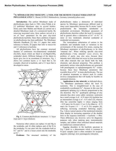
All phyllosilicates have the common structural<br />
features of continuous two-dimensional tetrahedral<br />
(Si,Al)O4 sheets which are liked to (Al,Mg)(O,OH)6<br />
octahedral sheets. Different linkages of the sheets to<br />
layers give rise to an assortment of structures. Fig. 1<br />
shows two common layers: a 1:1 layer that is, for<br />
example, observed in kaolinite, and a 2:1 layer that is<br />
developed in micas.<br />
OH,O<br />
Si<br />
Si<br />
Fe<br />
Fig. 1. Common phyllosilicate structures: 1:1 layer<br />
(top) and 2:1 layer (bottom).<br />
Numerous other cations can replace Al and Mg in<br />
the octahedra, and Al often replaces part of the Si in<br />
the tetrahedra. Fe can enter into the octahedra both as<br />
Fe 2+ and Fe 3+ and to a lesser extent into the tetrahedra<br />
as Fe 3+ .<br />
Problems: The structural similarity of the<br />
Al<br />
OH<br />
Al<br />
O<br />
Si<br />
O<br />
O<br />
OH<br />
phyllosilicates makes a distinction of individual<br />
species by Mössbauer spectroscopy difficult (and in<br />
many cases impossible), because the Fe atoms “sense”<br />
predominantly the immediate (tetrahedral or<br />
octahedral) environment. Mössbauer parameters of<br />
phyllosilicates therefore reflect the local Fe symmetry,<br />
and in general allow a distinction of Fe 2+ or Fe 3+ in<br />
more or less moderately distorted octahedral or<br />
tetrahedral environments.<br />
The isomorphous substitution of cations in the<br />
octahedra and tetrahedra leads to variations in the<br />
environments of the resonant (Fe) atoms, causing the<br />
Mössbauer parameters of phyllosilicates to be often<br />
“smeared out”. When studying specific clay-sized<br />
phyllosilicates (“clay minerals” sensu stricto), the<br />
problem of purity may also arise, because these<br />
minerals more often than not occur in close association<br />
with other minerals that can falsify both the bulk<br />
chemistry and physical properties. This problem is<br />
particularly serious when phyllosilicates are associated<br />
with paramagnetic or superparamagnetic Fe 3+ oxides,<br />
because these have Mössbauer parameters that<br />
resemble those of Fe 3+ in many phyllosilicates. The use<br />
of chemical treatments to remove such Fe oxides<br />
involves manipulations that will hardly be feasible on<br />
unmanned missions.<br />
Applications to clay minerals: as indicated above,<br />
57 Fe Mössbauer parameters reflect the local Fe<br />
symmetry. This is straightforward in the case of<br />
octahedrally-coordinated Fe 3+ , because in this case the<br />
quadrupole splitting (Δ) is directly proportional to the<br />
site distortion. The Fe 3+ quadrupole splittings of clay<br />
minerals thus show some subtle variations: nontronite<br />
has a small average Δ of ~ 0.35 mm/s [4], kaolinite has<br />
an intermediate average Δ of ~ 0.51 mm/s [5], whereas<br />
illite has somewhat higher Δ values ranging from<br />
~ 0.59 mm/s for Fe-rich (> 5 wt.-% Fe) illites to ~ 0.73<br />
mm/s for Fe-poor (
Martian Phyllosilicates: Recorders of Aqueous Processes (2008) 7020.pdf<br />
Transmission (%)<br />
100<br />
98<br />
96<br />
100<br />
99<br />
100<br />
98<br />
96<br />
94<br />
92<br />
Nontronite<br />
Kaolinite<br />
Illite<br />
-5 -2.5 0<br />
Velocity (mm/s)<br />
2.5 5<br />
Fig. 2. Room-temperature Mössbauer spectra of<br />
nontronite (top), kaolinite (center) and illite (bottom).<br />
A parameter which is readily revealed by<br />
Mössbauer spectroscopy is the Fe 2+ /Fe 3+ ratio, which<br />
can provide important clues regarding the redox<br />
environment of a sample. The determination of this<br />
ratio by Mössbauer spectroscopy is relatively<br />
straightforward: thus Fig. 2 shows the illite to contain<br />
a minor proportion of Fe 2+ , indicated by a resonant line<br />
at ~ 2.4 mm/s. Such a line is missing in the other two<br />
spectra, indicating Fe in these samples to be<br />
exclusively trivalent.<br />
Applications to natural clays of complex<br />
mineralogy on Earth and Mars: a basic difference<br />
between clay minerals s.s. (clay-sized phyllosilicates)<br />
and Fe oxides is that almost all of the former are<br />
paramagnetic to ~ 10 K, whereas many Fe oxides are<br />
magnetically ordered at 300 K and all are magnetically<br />
ordered at ~ 20 K [7]. Natural clays (and sediments<br />
and soils) are usually more or less complex mixtures of<br />
clay minerals, Fe oxides, and other (Fe-bearing and<br />
Fe-free) minerals. Their room-temperature Mössbauer<br />
spectra thus generally comprise three sets of<br />
components: (1) Fe 3+ doublets from paramagnetic clay<br />
minerals and other silicates, and superparamagnetic Fe<br />
oxides and related minerals (e.g. schwertmannite),<br />
(2) Fe 2+ doublets that, if present, would in most cases<br />
originate from clay minerals, and (3) Fe 3+ sextets from<br />
magnetically ordered Fe oxides. These features have<br />
been used to characterize clays from a variety of<br />
environments using Mössbauer spectroscopy in<br />
combination with other techniques such as X-ray<br />
diffraction, visible, infrared and Raman<br />
spectroscopies, selective chemical extraction<br />
procedures, optical and electron microscopies, and<br />
thermal analysis [8,9,10]. As an example, Fig. 3 shows<br />
how the selective removal of Fe oxides affects the<br />
room-temperature Mössbauer spectrum of a bauxite.<br />
This treatment not only allows a better characterization<br />
of the paramagnetic components (residual minerals,<br />
some or all of which could contribute to the Fe 3+<br />
doublet, have been identified by X ray diffraction as<br />
boehmite, kaolinite, anatase and crandallite), but also<br />
shows the residual hematite to be identical to that<br />
which has been extracted [11].<br />
Transmittance (%)<br />
100<br />
98<br />
96<br />
100<br />
99<br />
98<br />
BX-N<br />
3 x DCB 295 K<br />
-10 -5 0 5 10<br />
Velocity (mm/s)<br />
Fig. 3. Mössbauer spectra of a bauxite before (top)<br />
and after extraction with Na dithionite (bottom).<br />
A constraint on remote work, e.g. on Mars, is that<br />
only minimal sample manipulation will be possible.<br />
Although Mössbauer work has been instrumental in<br />
identifying a variety of Fe oxides on Mars [12,13,14],<br />
a combination of various techniques including<br />
Mössbauer spectroscopy holds promise for a more<br />
comprehensive characterization of clay mineralogy<br />
than Mössbauer spectroscopy alone [15,16].<br />
References: [1] Pollak et al (1962) phys. stat. sol.,<br />
2, 1653, [2] Malden P.J. and Meads R.E. (1967)<br />
Nature, 215, 844, [3] Stevens J.G. et al. (1988)<br />
Mössbauer Mineral Handbook, MEDC, Asheville,<br />
N.C., [4] Cardile C.M. & Johnston J.H. (1985) Clays<br />
Clay Min., 33, 295, [5] <strong>Murad</strong> E. & Wagner U. (1991)<br />
N. Jb. Min. Abh., 162, 281, [6] <strong>Murad</strong> E. & Wagner U.<br />
(1994) Clay Min., 29, 1, [7] <strong>Murad</strong> E. & Cashion J.<br />
(2004) Mössbauer Spectroscopy of Environmental<br />
Materials, Springer, [8] <strong>Murad</strong> E. & Wagner U.<br />
(1989) Hyp. Int., 45, 161, [9] <strong>Murad</strong> E. and Rojík P.<br />
(2005) Clay Min., 40, 427, [10] Bishop J.L. et al. (2007)<br />
Clays Clay Min., 55, 1, [11] <strong>Murad</strong> E. (200x) Min Eng.,<br />
18, 984, [12] Klingelhöfer G. et al. (2005) Hyp. Int., 166,<br />
549, [13], Morris R.V. et al. (2006) JGR, 111, E02S13,<br />
[14], Ming D.W. et al. (2006) JGR, 111, E02S12, [15]<br />
Bishop J.L. et al. (2004) IJA, 3, 275, [16] Lane M.D. et<br />
al. (2004) GRL, 31, L17902.