Pulmonary Wegener's Granulomatosis*
Pulmonary Wegener's Granulomatosis*
Pulmonary Wegener's Granulomatosis*

Create successful ePaper yourself
Turn your PDF publications into a flip-book with our unique Google optimized e-Paper software.
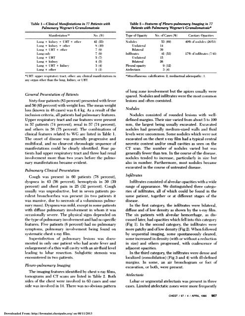
Table 1 -Clinical Manifestations in 77 Patients with<br />
<strong>Pulmonary</strong> Wegener’s Granulomatosis<br />
IoIamiifestatiouim* Nou . (%)<br />
Ltommg + kidney + (JET + other 42 (55)<br />
Lmong + kidney + outher 8 (10)<br />
Lung + URT + oother 7 (9)<br />
Lung ounl 7 (9)<br />
Lung + URT 5 (7)<br />
Lumig + kidney 4 (5)<br />
Lung + URT + kidney 3 (4)<br />
Lung + oother 1 (1)<br />
*URT: topper respiratoumy tract; (other: any clinical mamiifestations imi<br />
amm orgamm other timan time lumig, kidne; oor URT<br />
General Presentation of Patients<br />
Sixty-four patients (83 percent) presented with fever<br />
and 50 (65 percent) with weight boss. The mean weight<br />
loss (known in 48 cases) was 6.4 kg. As a result of the<br />
inclusion criteria, all patients had pulmoonary features.<br />
Upper respiratory tract and ear features were present<br />
in 57 patients (74 percent), renal in 57 (74 percent),<br />
amid others in 58 (75 percent). Tile combinations of<br />
clinical features related to WG are listed in Table 1.<br />
The onset oof disease was generally progressive and<br />
multifocal, and no clear-cut chronologic sequence of<br />
manifestations could be clearly identified. Four pa-<br />
tients had upper respiratory tract and three had renal<br />
involvement more than two) years before the pulmo-<br />
nary manifestations l)ecame evident.<br />
<strong>Pulmonary</strong> Clinical Presentation<br />
Cough was present in 60 patients (78 percent),<br />
dyspnea in 43 (56 percent), hemoptysis in 30 (39<br />
percent) and chest pain in 25 (32 percent). Cough<br />
usually was unproductive, but in seven patients pu-<br />
rulent hronchoorrhea -as present (in two patients it<br />
was massive, due to) necrosis of a voluminous pulmo-<br />
nary mass). Dyspnea was mild, except in some patients<br />
with diffuse pulmonary involvement in whom it was<br />
ooccasio)nally severe. The physical signs depended on<br />
the type o)fpulmonary involvement and had noo specific<br />
features. Five patients (6 percent) had no pulmonary<br />
symptoollls, pulmonary involvement being foound on<br />
systematic chest x-ray film.<br />
Superinfectioon of pulmoonary lesio)ns was doocu-<br />
mented in only one patient who had acute fever and<br />
enlargement ofa thin wall cavity with an air-fluid level<br />
leading to lobar resection. Subglottic stenosis was<br />
encountered in two patients.<br />
Pleuro-pulmonary Imaging<br />
The iinaglllg features identified by chest x-ray films,<br />
tomograms and CT scans are listed in Table 2. Both<br />
sides of the chest were involved in 63 cases and one<br />
side was involved in 14. There was no obvious pattern<br />
Downloaded From: http://hwmaint.chestpubs.org/ on 08/11/2013<br />
Table 2-Features ofPleuro-pulmonary lmaging in 77<br />
Patients with <strong>Pulmonary</strong> Wegener’s Granulomatosis5<br />
Type ofOpacity No. of Cases (%) Cavitary Opacities<br />
Nodules 53 (69) 49% of nodules (26/53)<br />
Unilateral 14<br />
Bilateral 39<br />
Infiltrates 41 (53) 17% ofinfiltrates (7/41)<br />
Unilateral 15<br />
Bilateral 26<br />
Pletoral opacity 9 (12)<br />
Atelectasis 3 (4)<br />
*Miscellaneoosis: calcification: 2; mediastinal adenopathy: 1.<br />
of lung zone involvement but the apices usually were<br />
spared. Nodules and infiltrates were the most commoon<br />
lesions and (often coexisted.<br />
Nodules<br />
Nodules consisted of rounded lesions with well-<br />
defined margins. Their size varied from about 5 to 100<br />
mm, the largest being usually excavated. Excavated<br />
nodules had generally medium-sized walls and fluid<br />
levels were uncommon. Some nodules which were no)t<br />
excavated on the chest x-ray film had a typical central<br />
necrotic content and/or small cavities as seen on the<br />
CT scan. The number of nodules varied but was<br />
generally fewer than ten. In the course ofthe disease,<br />
nodules tended to increase, particularly in size but<br />
also in number. Furthermore, most nodules became<br />
excavated in the course of untreated disease.<br />
Infiltrates<br />
Infiltrates consisted ofalveolar opacities with a wide<br />
range (Of appearance. We distinguished three categoo-<br />
ries of infiltrates, all of which could be found in the<br />
same patient, together or at different stages of the<br />
disease.<br />
In the first category, the infiltrates were bilateral,<br />
diffuse and of low density as shown by the x-ray film.<br />
The six patients with alveolar hemorrhage, as dis-<br />
cussed later, had opacities which fell into this category<br />
(Fig 1). In the second category, the infiltrates were<br />
more patchy and oflow density (Fig 2). When foollowed<br />
by sequential imaging, some spontaneously cleared,<br />
some increased in density (with or without a reductiooml<br />
in size) and others progressed, with coalescence oof<br />
adjacent opacities.<br />
In the third category, the infiltrates were dense and<br />
localized (consolidation) (Fig 3 and 4) with ill-defined<br />
margins. In some, an air bronchogram or foci oof<br />
excavation, or both, were present.<br />
Atelectasis<br />
Lobar or segmental atelectasis was present in tilree<br />
cases. Limited atelectatic zones were more frequently<br />
CHEST I 97 I 4 I APRIL 1990 907