Respiratory Rate during Acute Asthma*
Respiratory Rate during Acute Asthma*
Respiratory Rate during Acute Asthma*

Create successful ePaper yourself
Turn your PDF publications into a flip-book with our unique Google optimized e-Paper software.
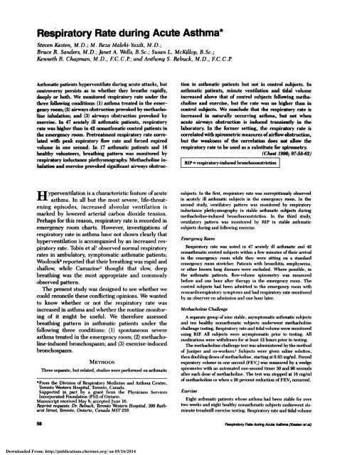
<strong>Respiratory</strong> <strong>Rate</strong> <strong>during</strong> <strong>Acute</strong> <strong>Asthma*</strong>Steven Kesten, M.D.; M. Reza Maleki-Yazdi, M.D.;Bnwe R. Sander M.D.; Janet A. Well B. Sc.; Susan L. McKillop, B. Sc.;Kenneth R. Chapman, M.D. , F. C. C.P; and Anthony S. Rebuck, M. D. , F.C. C.PAsthmatic patients hyperventilate <strong>during</strong> acute attacks, butcontroversy persists as to whether they breathe rapidly,deeply or both. We monitored respiratory rate under thethree following conditions: (1) asthma treated in the emergencyroom; (2) airways obstruction provoked by methacholineinhalation; and (3) airways obstruction provoked byexercise. In 47 acutely ill asthmatic patients, respiratoryrate was higher than in 42 nonasthmatic control patients inthe emergency room. Pretreatment respiratory rate correlatedwith peak expiratory flow rate and forced expiredvolume in one second. In 17 asthmatic patients and 16healthy volunteers, breathing pattern was monitored byrespiratory inductance plethysmography. Methacholine inhalationand exercise provoked significant airways obstructionin asthmatic patients but not in control subjects. Inasthmatic patients, minute ventilation and tidal volumeincreased above that of control subjects following methacholineand exercise, but the rate was no higher than incontrol subjects We conclude that the respiratory rate isincreased in naturally occurring asthma, but not whenacute airways obstruction is induced transiently in thelaberator In the former setting, the respiratory rate iscorrelated with spirometric measures ofairflow obstruction,but the weakness of the correlation does not allow therespiratory rate to be used as a substitute for spirometry.(Chest 19; 97:58-62)I RIP= respiratory-induced bronchoconstrictionH yperventilation is a characteristic feature of acuteasthma. In all but the most severe, life-threateningepisodes, increased alveolar ventilation ismarked by lowered arterial carbon dioxide tension.Perhaps for this reason, respiratory rate is recorded inemergency room charts. However, investigations ofrespiratory rate in asthma have not shown clearly thathyperventilation is accompanied by an increased respiratoryrate. Tobin et al’ observed normal respiratoryrates in ambulatory, symptomatic asthmatic patients;Woolcock2 reported that their breathing was rapid andshallow, while Camazine3 thought that slow, deepbreathing was the most appropriate and commonlyobservedpattern.The present study was designed to see whether wecould reconcile these conflicting opinions. We wantedto know whether or not the respiratory rate wasincreased in asthma and whether the routine monitoringof it might be useful. We therefore assessedbreathing pattern in asthmatic patients under thefollowing three conditions: (1) spontaneous severeasthma treated in the emergency room; (2) methacholine-inducedbronchospasm; and (3) exercise-inducedbronchospasm.METHODSThree separate, but related, studies were performed on asthmatic*From the Division of <strong>Respiratory</strong> Medicine and Asthma Centre,Toronto Western Hospital, Toronto, Canada.Supported in part by a grant from the Physicians ServicesIncorporated Foundation (PSI) of Ontario.Manuscript received May 8; accepted June 16.Reprint requests: Dt Rebuck, Toronto Western Hospital, 399 BathurstStreet, Toronto, Ontario, Canada M5T 2S8subjects. In the first, respiratory rate was surreptitiously observedin acutely ill asthmatic subjects in the emergency room. In thesecond study, ventilatory pattern was monitored by respiratoryinductance plethysmography in stable asthmatic subjects <strong>during</strong>methacholine-induced bronchoconstriction. In the third study,ventilatory pattern was monitored by RIP in stable asthmaticsubjects <strong>during</strong> and following exercise.EmergencyRoom<strong>Respiratory</strong> rate was noted in 47 acutely ill asthmatic and 42nonasthmatic control subjects within a few minutes of their arrivalin the emergency room while they were sitting on a standardemergency room stretcher. Patients with bronchitis, emphysema,or other known lung diseases were excluded. Where possible, inthe asthmatic patients, flow-volume spirometry was measuredbefore and one hour after therapy in the emergency room. Thecontrol subjects had been admitted to the emergency mom withnoncardiorespiratory symptoms and had respiratory rate monitoredby an observer on admission and one hour later.MethacholineChallengeA separate group ofnine stable, asymptomatic asthmatic subjectsand ten healthy nonasthmatic subjects underwent methacholinechallenge testing. <strong>Respiratory</strong> rate and tidal volume were monitoredusing RIP All subjects were asymptomatic prior to testing. Allmedications were withdrawn for at least 12 hours prior to testing.The methacholine challenge test was administered by the methodof Juniper and co-workers. Subjects were given saline solution,then doubling doses ofmethacholmne, starting at 0.03 mg/mI. Forcedexpiratory volume in one second (FEy,) was measured by a wedgespirometer with an automated one-second timer 30 and 90 secondsafter each dose ofmethacholine. The test was stopped at 16 mg/nilof methacholine or when a 20 percent reduction of FEV, occurred.ExerciseEight asthmatic patients whose asthma had been stable for overtwo weeks and eight healthy nonasthmatic subjects underwent sixminutetreadmill exercise testing. <strong>Respiratory</strong> rate and tidal volume58 <strong>Respiratory</strong> <strong>Rate</strong> <strong>during</strong> <strong>Acute</strong> Asthma (Kesten eta!)Downloaded From: http://publications.chestnet.org/ on 05/16/2014
were monitored using RIP. All medications were withdrawn for at500least 12 hours prior to testing.The exercise test was performed in an air.condifioned laboratoryunder stable environmental conditions. The treadmill speed (6to8.5 km/h) and inclination (8 to 13 percent) were adjusted so thatthe subjects achieved at least 80 percent oftheir predicted maximalheart rates by the end ofthe challenge. Exercise was stopped early‘; 400C. 300SSS #{149} r0.42p< 0.02if the patient could no longer continue because of dyspnea orfatigue. The FEy, was measured by a wedge spirometer with anautomated one-second timer before and serially upon terminationofthe exercise challenge. 200U-w0.SRespiratonj Inductance PlethysmographyIn studies 2 and 3, each subject had a suitably sized transducerinductance coil placed around the rib cage just below the axilla,and a second coil positioned at the umbilicus above the iliac crest.The location of the coils was marked and checked regularly toensure that their positions did not change. Theleast squares methodof calibration for RIP was used. Following the technique specifiedby the manufacturer, calibration of RIP (DC mode) was performedwith the subjects in both standing and supine postures. Rib cageand abdominal deflections from at least three representative breathsin each of the two postures were recorded on a multichannelrecorder and separate compartmental amplification factors werecalculated from simultaneous spirometric measures of tidal volumeusingsimultaneous equations. Calibration was verified by comparingthe sum of the deflections with tidal volume measured by spirometry.The calibration procedure was repeated if tidal volumemeasured by RIP and spirometry differed by more than ± 10percent. At the conclusion of the studies, tidal volume by RIP wasmeasured against tidal volume by spirometry. The trial data wererejected if there was a mean tidal volume difference between thetwo methods ofgreater than 10 percent.EmergencyRoomRESULTSForty-seven acutely ill asthmatic patients were enteredinto this study. Seventeen were men and 30were women (mean age, 30 years, range 16 to 65 years)and had presented to the emergency room for treatmentof asthma. Technically satisfactory values forspirometry, taken within a few minutes of arriving inthe emergency room, were obtained in 27 of 47patients, while satisfactory spirometry postbronchodilatortherapy was obtained in 26 patients. Peakexpiratory flow rate (PEFR) was obtained in 31 of 47patients. At the time of presentation, the mean initialFEY1 (1.43 ± 0.49 L) was 43 ± 16 percent of predictedvalue, calculated for age, sex and height.5 Accordingly,these patients may be viewed, as a group, as havinghad an episode ofasthma ofmoderate severity. Amongpatients for whom initial spirometry was obtained,there were inverse correlations between respiratoryrate and several indices of airflow: FEY1 (r = - 0.45,p
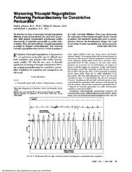
‘Up>0.2P>O.2DASTHMATICS0-..I-.CONTROLS6:‘U16 p.(O.OO1F’ )0! p)O.2p>O.2p0.2Bassllne Last doseMETHACHOLINECHALLENGETESTFicuax 3. <strong>Respiratory</strong> rate, minute ventilation andtidal volume before and after exercise and before andafter methacholine inhalation fur asthmatic and nonasthmaticcontrol subjects. Bars represent ± 1 SEM.percent. All asthmatic subjects had a PC2O of lessthan 8 mg/mi, whereas all control subjects had a PC2Oexceeding 8 mg/ml of methacholine. In three controlsubjects, FEY1 fell between 22 and 26 percent afterinhaling 16 mg/ml ofmethacholine. In these subjects,respiratory rate changed 1 breath/minute or lessbetween baseline and completion ofthe challenge. Inasthmatic patients but not in control subjects, overallYE and YT were increased by methacholine (after lastdose: VE=11.1±3.5 vs 7.6±2.1 Llmin, pO.2). Following exercise, FEY1 fell a mean of25 percent for asthmatics and 2 percent for controlsubjects. Following exercise, YE and Y’r were bothsignfficantly higher throughout the postexercise periodin asthmatics than in normal subjects. When FEY1 inasthma patients was at its lowest (15 minutes postexercise,2.22±0.49 L), YE and YT were both more thandouble that of control subjects (YE = 14.60 vs 7.24Lfmin, p
gency room patients exposed to a similarly stressfulenvironment. One might also postulate that hypoxemiais responsible for the observed increase in respiratoryrate. However, most of our asthmatic patientswere given supplemental oxygen in the emergencyroom and it is unlikely that they were hypoxemic atthe time of our assessments. Moreover, there is evidencethat the mild hypoxemia of acute asthma haslittle effect on ventilation and no measurable effect onrespiratory rate. Freedman studied a group of asthmaticswith a mean FEY1 of 43 ± 16 percent ofpredicted and a mean oxygen saturation of 89 ± 5percent.7 While supplemental oxygen increased meanoxygen saturation to 95 ± 2 percent, respiratory rateremained unchanged (22 ± 1 and 21 ± 1 breaths!min,respectively).Is respiratory rate simply an indicator ofthe severityof airflow obstruction? If this were so, the differencein respiratory rate between asthmatic patients seen inthe emergency room and asthmatics studied in thelaboratory would simply be a consequence of moresevere airflow obstruction observed in the formersetting. Although both emergency room and laboratory-studiedasthmatic patients were symptomaticwith wheezing and dyspnea, mean FEY1 was lower inthe former (1.42±0.50 L vs a mean postmethacholineFEY1 of2. 12 ± 0.53 L). This explanation is not entirelysatisfactory for three reasons. First, Chadha et al8 havemonitored respiratory rate with inductive plethysmographyin a group of asthmatic subjects while theyprovoked more severe airways obstruction with methacholinethan we did in the current study. Despite amean FEY1, just 45 percent of predicted and comparableto that seen in our emergency room patients,they observed no increase in respiratory rate. Second,the inverse correlations we observed between pretreatmentrespiratory rate and pretreatment airflowindices were little more than vague trends achievingstatistical significance by virtue of the relatively largenumber of subjects involved. Following treatment,respiratory rate remained abnormally high as comparedto control subjects, but any hint of correlationbetween respiratory rate and airflow measurementsdisappeared. Clearly, respiratory rate and measures ofFEY1 or PEFR are not redundant 1:1 correlates.Finally, the two asthmatic patients in our study whoshowed the most marked response to inhaled methacholinehad a decline in FEY1 of 49 percent (similarto pretreatment values we saw in the emergencyroom) but no change in respiratory rate.The airway obstruction induced in the laboratory instable asthmatics differed from that in the emergencyroom in duration of obstruction, severity of obstruction,and perhaps most importantly, in the degree ofaccompanying inflammation. The inflammatory responseand release of mediators likely contribute tothe alterations in breathing patterns and respiratorydrive. The increase in breathing frequency that wehave noted in the emergency room presentation ofasthma and the lack of any such change in thelaboratory implies that the inflammatory process,rather than the degree of bronchospasm, may beresponsible for changes in respiratory rate.’2At first glance, neither methacholine nor exercisewould appear to be responsible for inducing acuteinflammatory changes. Methacholine directly inducessmooth muscle contraction. However, we acknowledgethe possibility that methacholine could possibly stimulateirritant airway receptors by physical irritation ofthe mucosa, smooth muscle contraction in itself activatingirritant receptors, and stimulation ofa deflationreflex secondary to compression by contiguous hyperinflated Similarly, in asthmatic patients, exerciseresults in thermal changes in the airways leadingto sudden increases in blood supply to the bronchi,hyperemia and edema ofthe mucosa with subsequentairway narrowing,’4 changes which could arguably bedescribed as inflammatory. It remains unclear whatclinical outcome these laboratory findings have onirritant airway receptors. Nevertheless, the durationand degree of inflammation in the airways of anasthmatic subject presenting to an emergency roomand subsequent stimulation ofintrapulmonary irritantreceptors must exceed that which accompanies theacute, transient airflow limitation induced in thelaboratory by methacholine or exercise.Our observation of unchanged respiratory rate followingmethacholine inhalation appears to confirmearlier studies of ventilatory pattern following methacholineor histamine challenge in asthmatic subjects.<strong>Respiratory</strong> rate appears to be unchanged when bronchospasmis induced in this manner, but variouschanges in minute ventilation have been reported:some report an increase,8”3 others a decrease’5 andstill other observers report unchanged levels of ventilation.16,17Tobin et alh8 noted that the relief of bronchoconstrictionin stable asthmatic subjects was notaccompanied by any important changes in respiratoryrate or minute ventilation. Our findings in laboratoryinducedasthma are more similar to those of Chadhaet al8 in that we noted no change in respiratory rateand a significant increase in minute ventilation derivedmainly from an increased tidal volume. The reasonsfor such conflicting results among different researchgroups may be related to a variety of factors thatinclude the differing degrees of bronchoconstrictioninduced, the use of mouthpieces and nose clips bysome investigators,’9’2#{176} small sample size, or differentpopulations (healthy volunteers, COPD or asthmaticpatients). Our protocol was limited to asthmatic subjects,had a sample size larger than that reported inseveral of the other studies, and measured respiratoryCHEST I 97 I I I JANUARY, 1990 61Downloaded From: http://publications.chestnet.org/ on 05/16/2014