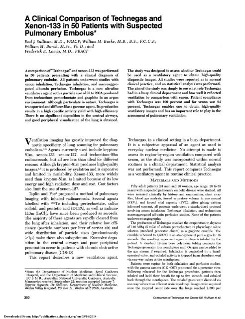
A Clinical Comparison of Technegas and Xenon-133 in 50 Patients ...
A Clinical Comparison of Technegas and Xenon-133 in 50 Patients ...
A Clinical Comparison of Technegas and Xenon-133 in 50 Patients ...

Create successful ePaper yourself
Turn your PDF publications into a flip-book with our unique Google optimized e-Paper software.
A <strong>Cl<strong>in</strong>ical</strong> <strong>Comparison</strong> <strong>of</strong> <strong>Technegas</strong> <strong>and</strong><br />
<strong>Xenon</strong>-<strong>133</strong> <strong>in</strong> <strong>50</strong> <strong>Patients</strong> with Suspected<br />
Pulmonary Embolus*<br />
Paulj Sullivan, M.D., FRACP; William M. Burke, M.B., B.S., F.C.C.P;<br />
William M. Burch, M. Sc., Ph.D.; <strong>and</strong><br />
Frederick E. Lomas, M.D., FRACP<br />
A comparison <strong>of</strong> "<strong>Technegas</strong>" <strong>and</strong> xenon-<strong>133</strong> was performed<br />
<strong>in</strong> <strong>50</strong> patients present<strong>in</strong>g with a cl<strong>in</strong>ical diagnosis <strong>of</strong><br />
pulmonary embolus. All patients underwent studies with<br />
xenon <strong>in</strong>halation, <strong>Technegas</strong> <strong>in</strong>halation, <strong>and</strong> macroaggregated<br />
album<strong>in</strong> perfusion. <strong>Technegas</strong> is a new ultraf<strong>in</strong>e<br />
ventilatory agent with a particle size <strong>of</strong> <strong>50</strong> to 200A produced<br />
from technetium pertechnetate <strong>and</strong> graphite <strong>in</strong> an argon<br />
environment. Although particulate <strong>in</strong> nature, <strong>Technegas</strong> is<br />
transported <strong>and</strong> diffuses like a gaseous agent. Its production<br />
results <strong>in</strong> a high specific activity yield with high efficiency.<br />
There is no significant deposition <strong>in</strong> the central airways,<br />
<strong>and</strong> good peripheral visualization <strong>of</strong> the lung is obta<strong>in</strong>ed.<br />
The study was designed to assess whether <strong>Technegas</strong> could<br />
be used as a ventilatory agent to obta<strong>in</strong> high-quality<br />
diagnostic images. All studies were reported as <strong>in</strong> normal<br />
cl<strong>in</strong>ical practice, <strong>and</strong> no statistical analysis was performed.<br />
The aim <strong>of</strong> the study was simply to see what role <strong>Technegas</strong><br />
had <strong>in</strong> a busy cl<strong>in</strong>ical department <strong>and</strong> how well it reflected<br />
ventilation by comparison with xenon. Patient compliance<br />
with <strong>Technegas</strong> was 100 percent <strong>and</strong> for xenon was 94<br />
percent. <strong>Technegas</strong> enables one to obta<strong>in</strong> high-quality<br />
ventilatory images <strong>and</strong> has an important role to play <strong>in</strong> the<br />
assessment <strong>of</strong> pulmonary ventilation.<br />
Ventilation imag<strong>in</strong>g has greatly improved the diagnostic<br />
specificity <strong>of</strong> lung scann<strong>in</strong>g for pulmonary<br />
embolism. 1-3 Agents currently used <strong>in</strong>clude krypton-<br />
81m, xenon-<strong>133</strong>, xenon-127, <strong>and</strong> technetium-99m<br />
radioaerosols, but all are less than ideal for different<br />
reasons. Although krypton-81m produces high-quality<br />
images,4'5 it is produced by cyclotron <strong>and</strong> is expensive<br />
<strong>and</strong> limited <strong>in</strong> availability <strong>Xenon</strong>-<strong>133</strong>, more widely<br />
used than krypton-81m, is limited because <strong>of</strong> its low<br />
energy <strong>and</strong> high radiation dose <strong>and</strong> cost. Cost factors<br />
also limit the use <strong>of</strong> xenon-127.<br />
Tapl<strong>in</strong> <strong>and</strong> Poe6 proposed a method <strong>of</strong> pulmonary<br />
imag<strong>in</strong>g with <strong>in</strong>haled radioaerosols. Several agents<br />
labelled with 99mTc <strong>in</strong>clud<strong>in</strong>g pertechnetate, sulfur<br />
colloid, <strong>and</strong> pentetic acid (DTPA), as well as <strong>in</strong>dium-<br />
113m (JnCl3), have s<strong>in</strong>ce been produced as aerosols.<br />
The majority <strong>of</strong> these agents are rapidly cleared from<br />
the lung after <strong>in</strong>halation, <strong>and</strong> their relative low efficiency<br />
(particle numbers per liter <strong>of</strong> carrier air) <strong>and</strong><br />
wide distribution <strong>of</strong> particle sizes (predom<strong>in</strong>antly<br />
>lI>) make them also suboptimum. Excessive deposition<br />
<strong>in</strong> the central airways <strong>and</strong> poor peripheral<br />
penetration occur <strong>in</strong> patients with chronic obstructive<br />
pulmonary disease (COPD).<br />
This report describes a new ventilation agent,<br />
*From the Department <strong>of</strong> Nuclear Medic<strong>in</strong>e, Royal Canberra<br />
Hospital, <strong>and</strong> the Department <strong>of</strong> Medic<strong>in</strong>e <strong>and</strong> <strong>Cl<strong>in</strong>ical</strong> Science,<br />
J. C. S. M. R., Australian National University Canberra, Australia.<br />
Manuscript received September 22; revision accepted January 7.<br />
Repr<strong>in</strong>t requests: Dr Sullivan, Department <strong>of</strong> Nuclear Medic<strong>in</strong>e,<br />
Woden Valley Hospital, PO Box 11, Woden ACT 2606, Australia<br />
300<br />
<strong>Technegas</strong>, <strong>in</strong> a cl<strong>in</strong>ical sett<strong>in</strong>g <strong>in</strong> a busy department.<br />
It is a subjective appraisal <strong>of</strong> an agent as used <strong>in</strong><br />
everyday nuclear medic<strong>in</strong>e. No attempt is made to<br />
assess its region-by-region ventilation compared with<br />
xenon, as the study was <strong>in</strong>corporated with<strong>in</strong> normal<br />
rout<strong>in</strong>es <strong>in</strong> a cl<strong>in</strong>ical department. Statistical analysis<br />
was not performed. This report compares <strong>Technegas</strong><br />
as a ventilatory agent <strong>in</strong> rout<strong>in</strong>e cl<strong>in</strong>ical practice.<br />
MATERIALS AND METHODS<br />
Fifty adult patients (24 men <strong>and</strong> 26 women; age range, 28 to 85<br />
years) with suspected pulmonary embolic disease were studied. All<br />
were assessed cl<strong>in</strong>ically by history <strong>and</strong> exam<strong>in</strong>ation, chest x-ray<br />
film, blood gas analysis, forced expiratory volume <strong>in</strong> one second<br />
(FEV,), <strong>and</strong> forced vital capacity (FVC). After giv<strong>in</strong>g written<br />
<strong>in</strong>formed consent, all patients underwent a st<strong>and</strong>ardized protocol<br />
<strong>in</strong>volv<strong>in</strong>g xenon <strong>in</strong>halation, <strong>Technegas</strong> <strong>in</strong>halation, <strong>and</strong> technetium<br />
macroaggregated album<strong>in</strong> perfusion studies. None <strong>of</strong> the patients<br />
underwent angiography<br />
The production <strong>of</strong> <strong>Technegas</strong> <strong>in</strong>volves the evaporation to dryness<br />
<strong>of</strong> 140 MBq (5 mCi) <strong>of</strong> sodium pertechnetate <strong>in</strong> physiologic sal<strong>in</strong>e<br />
solution (st<strong>and</strong>ard generator eluent) <strong>in</strong> a graphite crucible. The<br />
crucible is heated to 2,<strong>50</strong>0°C <strong>in</strong> an atmosphere <strong>of</strong> pure argon for 15<br />
seconds. The result<strong>in</strong>g vapor <strong>and</strong> argon mixture is <strong>in</strong>haled by the<br />
patient. A st<strong>and</strong>ard 12-mm bore polythene tub<strong>in</strong>g connects the<br />
<strong>Technegas</strong> generator to a mouthpiece unit. Oxygen can be added to<br />
the gas stream if required. Inhalation is controlled by a h<strong>and</strong>operated<br />
valve, <strong>and</strong> exhaled activity is trapped <strong>in</strong> an absorbent wad<br />
via one-way valves at the mouthpiece.<br />
<strong>Patients</strong> were sup<strong>in</strong>e for both <strong>in</strong>halation <strong>and</strong> perfusion studies,<br />
with the gamma camera (GE 400T) positioned for a posterior view<br />
Follow<strong>in</strong>g rehearsal for the <strong>Technegas</strong> procedure, patients then<br />
<strong>in</strong>haled <strong>and</strong> held their breath for up to five seconds <strong>and</strong> exhaled<br />
back through the mouthpiece. The exhaled gases were diverted via<br />
one-way valves to an efficient res<strong>in</strong> wood trap. Images were acquired<br />
once the <strong>in</strong>spired count rate over the lungs reached 2,<strong>50</strong>0 per<br />
<strong>Comparison</strong> <strong>of</strong> <strong>Technegas</strong> <strong>and</strong> <strong>Xenon</strong>-<strong>133</strong> (Sullivan et al)<br />
Downloaded From: http://publications.chestnet.org/ on 05/16/2014
second (equivalent to about 37 MBq [1 mCi] <strong>in</strong> the lung). Adequate<br />
counts were normally obta<strong>in</strong>ed <strong>in</strong> one or two <strong>in</strong>halations. Sicker<br />
patients <strong>of</strong>ten required several more breaths. Six planar views (100k)<br />
were performed: posterior; posterior obliques; anterior; <strong>and</strong> anterior<br />
obliques.<br />
<strong>Xenon</strong> imag<strong>in</strong>g <strong>in</strong> the posterior view was performed immediately<br />
prior to the <strong>Technegas</strong> study A s<strong>in</strong>gle-use disposable breath<strong>in</strong>g<br />
system (Atomic Products) was used. After <strong>in</strong>halations <strong>of</strong> approximately<br />
400 MBq <strong>of</strong> xenon-<strong>133</strong> (11 mCi), a st<strong>and</strong>ard protocol <strong>of</strong><br />
breath-hold<strong>in</strong>g, equilibration, <strong>and</strong> washout was performed. Perfusion<br />
imag<strong>in</strong>g (macroaggregated album<strong>in</strong>) was performed immediately<br />
after the <strong>Technegas</strong> study An adm<strong>in</strong>istered activity <strong>of</strong> 180<br />
MBq (5 mCi) to ma<strong>in</strong>ta<strong>in</strong> a pixel count ratio <strong>of</strong> about five times the<br />
<strong>Technegas</strong> activity was used. Multiple planar images identical with<br />
the <strong>Technegas</strong> sequence were collected.<br />
<strong>Technegas</strong> <strong>and</strong> xenon images were compared for technical quality<br />
<strong>and</strong> diagnostic value. The images were viewed <strong>and</strong> graded without<br />
digital process<strong>in</strong>g by two experienced nuclear medic<strong>in</strong>e physicians<br />
review<strong>in</strong>g all studies <strong>in</strong> three sessions <strong>in</strong> isolation from other data.<br />
Image quality was assessed on a scale <strong>of</strong> zero to five <strong>in</strong> terms <strong>of</strong><br />
<strong>in</strong>terpretability All studies were reported as high or low probability<br />
<strong>of</strong> pulmonary embolus or <strong>in</strong>determ<strong>in</strong>ate; however, for this report,<br />
only those diagnosed as a high probability <strong>of</strong> pulmonary embolus<br />
were <strong>in</strong>cluded as truly positive studies.<br />
RESULTS<br />
Thirty-n<strong>in</strong>e patients had a risk factor <strong>of</strong> prolonged<br />
bed rest or recent surgery Eleven had a past history<br />
<strong>of</strong> phlebitis or previous pulmonary embolus. Thirtyseven<br />
patients presented with dyspnea, <strong>and</strong> 32 had<br />
pleuritic chest pa<strong>in</strong>. There were 13 smokers <strong>and</strong> 12<br />
ex-smokers. Electrocardiograms were normal <strong>in</strong> 27<br />
patients <strong>and</strong> showed s<strong>in</strong>us tachycardia <strong>in</strong> 19. The chest<br />
x-ray film was normal <strong>in</strong> 14 patients <strong>and</strong> showed<br />
pleural effusions <strong>in</strong> 15 <strong>and</strong> atelectasis <strong>in</strong> six. Blood gas<br />
analysis showed an oxygen pressure <strong>of</strong> less than 80<br />
mm Hg <strong>in</strong> 37 patients, <strong>and</strong> 19 had an FEV1 <strong>of</strong> less<br />
than 1.20 L.<br />
Ten patients were diagnosed as hav<strong>in</strong>g a high<br />
probability <strong>of</strong> pulmonary embolism by the criterion <strong>of</strong><br />
the PIOPED trial.8 Diffuse match<strong>in</strong>g defects thought<br />
to be consistent with COPD were visible <strong>in</strong> 13 patients.<br />
Diagnostic images with <strong>Technegas</strong> were obta<strong>in</strong>ed <strong>in</strong><br />
49 patients. One patient showed marked central airway<br />
deposition with poor visualization <strong>of</strong> the peripheral<br />
areas <strong>of</strong> the lungs. This was due to technical problems<br />
with the prototype equipment.<br />
Three patients were unable to cooperate sufficiently<br />
to obta<strong>in</strong> a xenon study (Fig 1), <strong>and</strong> diagnostic images<br />
were obta<strong>in</strong>ed <strong>in</strong> 46 patients. One image was un<strong>in</strong>terpretable<br />
because <strong>of</strong> poor compliance by the patient.<br />
In a subjective comparison <strong>of</strong> the quality <strong>of</strong> the<br />
images, 37 studies were similar (Fig 2), ten had better<br />
images with <strong>Technegas</strong>, <strong>and</strong> three had better images<br />
with xenon. Diagnostically, <strong>Technegas</strong> was thought to<br />
be better <strong>in</strong> 21 studies, with this predom<strong>in</strong>antly due<br />
to the multiple images obta<strong>in</strong>ed.<br />
Of the ten patients diagnosed with pulmonary<br />
embolism, the extra views with <strong>Technegas</strong> enabled a<br />
more confident diagnosis <strong>in</strong> six (eg, Fig 3). Two <strong>of</strong> the<br />
ten patients with pulmonary embolism were diagnosed<br />
on the <strong>Technegas</strong> image but not with xenon. In both<br />
patients the scan's diagnosis was based on the summation<br />
<strong>of</strong> small subsegmental nonmatched defects.8<br />
The subsequent course <strong>of</strong> both patients validated the<br />
<strong>Technegas</strong> f<strong>in</strong>d<strong>in</strong>gs.<br />
Patient compliance with <strong>Technegas</strong> was 100 percent<br />
<strong>and</strong> was 94 percent with xenon. All patients who had<br />
previously undergone xenon or aerosol studies reported<br />
that a <strong>Technegas</strong> study was less dem<strong>and</strong><strong>in</strong>g.<br />
From a technical po<strong>in</strong>t <strong>of</strong> view, the <strong>Technegas</strong> study<br />
was easier to perform <strong>and</strong> caused less disruption to<br />
the patient's schedul<strong>in</strong>g <strong>and</strong> camera time.<br />
DiscusSION<br />
The important role <strong>of</strong> ventilation lung scann<strong>in</strong>g <strong>in</strong><br />
pulmonary embolism is well documented, <strong>and</strong> <strong>in</strong><br />
cooperative patients with relatively normal respiratory<br />
FicuRE 1. Anterior <strong>Technegas</strong> (left) <strong>and</strong> perfusion (right) views <strong>of</strong> 84-year-old man with carc<strong>in</strong>oma <strong>of</strong><br />
prostate <strong>and</strong> pulmonary embolus. Patient was unable to tolerate xenon study<br />
CHEST / 94 / 2 / AUGUST 1988 301<br />
Downloaded From: http://publications.chestnet.org/ on 05/16/2014
FIGURE 2. Posterior views with xenon-<strong>133</strong> (top), <strong>Technegas</strong> (center),<br />
<strong>and</strong> macroaggregated album<strong>in</strong> (bottom) perfusion from 37-year-old<br />
female diabetic whose study was with<strong>in</strong> normal limits.<br />
function, both radioactive gas <strong>and</strong> technetium aerosol<br />
systems can be expected to perform well; however,<br />
many patients seen <strong>in</strong> rout<strong>in</strong>e cl<strong>in</strong>ical practice suffer<br />
from COPD, dyspnea, or are otherwise distressed or<br />
enfeebled <strong>and</strong> unable to cooperate fully Three <strong>Xenon</strong><br />
studies <strong>in</strong> this series failed for such reasons. Us<strong>in</strong>g<br />
aerosols, such patients show deposition <strong>in</strong> the central<br />
airways with poor visualization <strong>of</strong> the peripheral areas<br />
<strong>of</strong> the lungs.<br />
With <strong>Technegas</strong>, compliance by patients is much<br />
higher because there is little need for breath-hold<strong>in</strong>g<br />
302<br />
or rebreath<strong>in</strong>g. Adequate lung images are obta<strong>in</strong>ed <strong>in</strong><br />
most patients with a s<strong>in</strong>gle breath because <strong>of</strong> the high<br />
production efficiency (up to 40 percent <strong>of</strong> <strong>in</strong>itial eluent<br />
as breathable activity <strong>in</strong> 4 L <strong>of</strong> argon). This high<br />
efficiency also allows dynamic <strong>in</strong>halation studies to be<br />
performed.7<br />
Alderson et al have compared xenon <strong>and</strong> radioaerosols<br />
<strong>in</strong> 81 patients. These <strong>in</strong>vestigators9 found that<br />
aerosols compared favorably with xenon-<strong>133</strong> <strong>and</strong> concluded<br />
that they were diagnostically equivalent. In<br />
our study <strong>Technegas</strong> appeared diagnostically superior<br />
to xenon-<strong>133</strong>. Whereas Alderson et a19 found more<br />
ventilation <strong>in</strong> dependent pulmonary regions with<br />
aerosols, we did not f<strong>in</strong>d this with <strong>Technegas</strong>, although<br />
ventilation did parallel perfusion <strong>in</strong> its distribution <strong>in</strong><br />
normal lungs.<br />
The distribution <strong>of</strong> particle size <strong>of</strong> <strong>Technegas</strong> is<br />
prov<strong>in</strong>g technically difficult to determ<strong>in</strong>e but is <strong>in</strong> the<br />
range <strong>of</strong> <strong>50</strong> to 200A, two orders <strong>of</strong> magnitude smaller<br />
than typical aerosols <strong>and</strong> smaller than produced by<br />
the new ultrasonic nebulizers. The half-life <strong>of</strong> <strong>Technegas</strong><br />
with<strong>in</strong> the lungs follows the physical half-life <strong>of</strong><br />
Tc-99m, <strong>and</strong> there is no evidence <strong>of</strong> clearance across<br />
the alveolar capillary membrane.<br />
Nonclearance <strong>of</strong> <strong>Technegas</strong> allows high-quality multiple<br />
planar <strong>and</strong> tomographic studies to be performed.7<br />
When <strong>Technegas</strong> <strong>and</strong> perfusion tomography are performed<br />
sequentially without mov<strong>in</strong>g the patient, identical<br />
ventilation <strong>and</strong> perfusion slices can be analyzed<br />
by computer for mismatch<strong>in</strong>g. This technique is impossible<br />
with xenon.<br />
There are three major reasons why <strong>Technegas</strong>produced<br />
ventilation images are more comparable to<br />
the perfusion studies than other current ventilatory<br />
agents, <strong>in</strong>clud<strong>in</strong>g xenon-<strong>133</strong>. First, similar pulmonary<br />
depths are imaged, <strong>and</strong> resolution is the same, s<strong>in</strong>ce<br />
both studies use 9omTc. Secondly, some m<strong>in</strong>or deposition<br />
<strong>in</strong> the central airways was seen only <strong>in</strong> the sickest<br />
patients, <strong>and</strong> <strong>of</strong>ten xenon-<strong>133</strong> studies cannot be performed<br />
<strong>in</strong> such patients. All xenon equilibrium images<br />
conta<strong>in</strong> activity <strong>in</strong> the conduct<strong>in</strong>g airways. Thirdly,<br />
visualization <strong>of</strong> the peripheral lung is excellent. The<br />
one exception <strong>in</strong> this series was caused by a technical<br />
fault <strong>in</strong> the apparatus.<br />
The other advantages <strong>of</strong> us<strong>in</strong>g<br />
aOmTc for both ventilation <strong>and</strong> perfusion studies are<br />
obvious, namely low cost, wide availability, ideal<br />
energy, <strong>and</strong> half-life.<br />
Provided that the follow<strong>in</strong>g perfusion study uses<br />
about five times the pulmonary activity <strong>of</strong> the ventilation<br />
study, there is no significant degradation <strong>of</strong> the<br />
perfusion image <strong>in</strong> analogue (film) record<strong>in</strong>g. This is<br />
readily calculated <strong>and</strong> obta<strong>in</strong>able with st<strong>and</strong>ard conventional<br />
tracer doses. For computer-assisted studies,<br />
it would be practical to use similar activities <strong>and</strong><br />
subtract the ventilation image.<br />
<strong>Technegas</strong> is cl<strong>in</strong>ically a ventilatory agent <strong>of</strong> high<br />
302<strong>Comparison</strong> <strong>of</strong> <strong>Technegas</strong> <strong>and</strong> <strong>Xenon</strong> -<strong>133</strong> (Sullivan et al)<br />
Downloaded From: http://publications.chestnet.org/ on 05/16/2014
FiCURE 3. Posterior projection with xenon- <strong>133</strong> (top left), <strong>Technegas</strong><br />
(upper right), <strong>and</strong> macroaggregated album<strong>in</strong> (middle left) perfusion<br />
<strong>and</strong> right posterior oblique projections with <strong>Technegas</strong> (lower right)<br />
<strong>and</strong> macroaggregated album<strong>in</strong> (bottom left) perfusion from 76-yearold<br />
woman with acute onset <strong>of</strong> dyspnea, pleuritic chest pa<strong>in</strong>, <strong>and</strong><br />
syncope. Right posterior oblique views assisted level <strong>of</strong> confidence<br />
<strong>in</strong> diagnosis <strong>of</strong> pulmonary embolism.<br />
quality It has dist<strong>in</strong>ct advantages over xenon-<strong>133</strong>. In<br />
<strong>in</strong>dependent trials, the transport <strong>of</strong><strong>Technegas</strong> through<br />
the lungs is <strong>in</strong>dist<strong>in</strong>ct from gaseous agents, <strong>and</strong> therefore<br />
although particulate <strong>in</strong> nature, <strong>Technegas</strong> may be<br />
compared to gaseous agents. The capacity to obta<strong>in</strong><br />
multiple views directly comparable with the perfusion<br />
study with excellent compliance <strong>and</strong> acceptance by<br />
patients are the pr<strong>in</strong>cipal advantages. High-quality<br />
diagnostic images can be rout<strong>in</strong>ely obta<strong>in</strong>ed with very<br />
little effort <strong>in</strong> even very ill patients. The equipment is<br />
compact <strong>and</strong> mobile.<br />
The ability to perform dynamic <strong>and</strong> tomographic<br />
studies <strong>of</strong> directly comparable ventilation <strong>and</strong> perfusion<br />
images may well open an important new role for<br />
computerized analysis.<br />
<strong>Technegas</strong> allows high-quality ventilation imag<strong>in</strong>g.<br />
Its ease <strong>of</strong> use makes <strong>Technegas</strong> an ideal agent <strong>in</strong> busy<br />
cl<strong>in</strong>ical nuclear medic<strong>in</strong>e departments, <strong>and</strong> its method<br />
<strong>of</strong> production may open up a whole new range <strong>of</strong><br />
technetium radiopharmaceuticals.<br />
ACKNOWLEDGMENTS: To all the patients who so will<strong>in</strong>gly<br />
agreed to the formal requirements <strong>of</strong> the trial <strong>and</strong> made the work<br />
possible; to the technicall, nurs<strong>in</strong>g, <strong>and</strong> clerical staff who cheerfully<br />
took on the extra duties dem<strong>and</strong>ed by the trial protocol; <strong>and</strong> to<br />
Arnersham (Australia) who provided the xenon-<strong>133</strong> <strong>and</strong> disposable<br />
breath<strong>in</strong>g units, ouir s<strong>in</strong>cere thanks.<br />
CHEST 94 / 2 f AUGUST, 1988 303<br />
Downloaded From: http://publications.chestnet.org/ on 05/16/2014
REFERENCES<br />
1 Alderson PO, Rujanevech N, Secker-Walker RH, McKnight REC.<br />
The role <strong>of</strong> Xe-<strong>133</strong> ventilation studies <strong>in</strong> the sc<strong>in</strong>tigraphic<br />
detection <strong>of</strong> pulmonary embolism. Radiology 1976; 120:633-40<br />
2 McNeil BJ. A diagnostic strategy us<strong>in</strong>g ventilation-perfusion<br />
studies <strong>in</strong> patients suspect for pulmonary embolism. J Nucl Med<br />
1976; 27:613-16<br />
3 Neuman RD. Sostman HD, Gottschalk A. Current status <strong>of</strong><br />
ventilation-perfusion imag<strong>in</strong>g. Sem<strong>in</strong> Nuel Med 1980; 10:198-217<br />
4 Fazio F, Jones T Assessment <strong>of</strong> regional ventilation by cont<strong>in</strong>uous<br />
<strong>in</strong>halation <strong>of</strong> radioactive Kr-81m. Br Med J 1975; 3:673-76<br />
5 Lavender JP Cunn<strong>in</strong>gham DA. <strong>Cl<strong>in</strong>ical</strong> value <strong>of</strong> krypton-81m<br />
<strong>and</strong> Tc-99m perfusion lung scann<strong>in</strong>g. Bull Eur Physiopathol<br />
Respir 1980; 16:309-20<br />
6 Tapl<strong>in</strong> GC Poe ND. A dual lung-scann<strong>in</strong>g technic for evaluation<br />
<strong>of</strong> pulmonary function. Radiology 1965; 85:365-68<br />
7 Burch WM, Sullivan PJ, McLaren CJ. "<strong>Technegas</strong>": a new<br />
ventilation agent for lung scann<strong>in</strong>g. Nucl Med Commun 1986;<br />
7:12<br />
8 An update on pulmonary nuclear medic<strong>in</strong>e. Sem<strong>in</strong> Nucl Med<br />
1986; 236-74<br />
9 Alderson PO, Biello DR, Gottschalk A, H<strong>of</strong>fer PB, Kroop SA,<br />
Lee ME, et al. Tc-99m DTPA aerosol <strong>and</strong> radioactive gases<br />
compared as adjuncts to perfusion sc<strong>in</strong>tigraphy <strong>in</strong> patients with<br />
suspected pulmonary embolism. Radiology 1984; 153:515-21<br />
Recent Advances <strong>in</strong> Pulmonary <strong>and</strong> Critical Care<br />
Medic<strong>in</strong>e<br />
The Division <strong>of</strong> Pulmonary <strong>and</strong> Critical Care Medic<strong>in</strong>e, Department <strong>of</strong> Medic<strong>in</strong>e, University<br />
<strong>of</strong> California, San Francisco, will present this course at the Mark Hopk<strong>in</strong>s Hotel, San Francisco,<br />
October 20-22. For <strong>in</strong>formation, contact: Postgraduate Programs, Department <strong>of</strong> Medic<strong>in</strong>e,<br />
<strong>50</strong>5 Parnassus, M979, University <strong>of</strong> California, San Francisco 94143-0120 (415:476-5208).<br />
Current Concepts <strong>in</strong> Pulmonary Pathology<br />
This fourth annual course will be held October 24-27 at the Massachusetts General Hospital,<br />
Boston. Direct <strong>in</strong>quiries to: Department <strong>of</strong> Cont<strong>in</strong>u<strong>in</strong>g Education, Harvard Medical School,<br />
25 Shattuck Street, Boston 02115 (617:732-1525).<br />
304<br />
<strong>Comparison</strong> <strong>of</strong> <strong>Technegas</strong> <strong>and</strong> <strong>Xenon</strong>-i 33 (Sullivan et al)<br />
Downloaded From: http://publications.chestnet.org/ on 05/16/2014