Cerebral Air Embolism, Subcutaneous Emphysema, and ...
Cerebral Air Embolism, Subcutaneous Emphysema, and ...
Cerebral Air Embolism, Subcutaneous Emphysema, and ...
Create successful ePaper yourself
Turn your PDF publications into a flip-book with our unique Google optimized e-Paper software.
1940<br />
DISEASES OF THE CHEST<br />
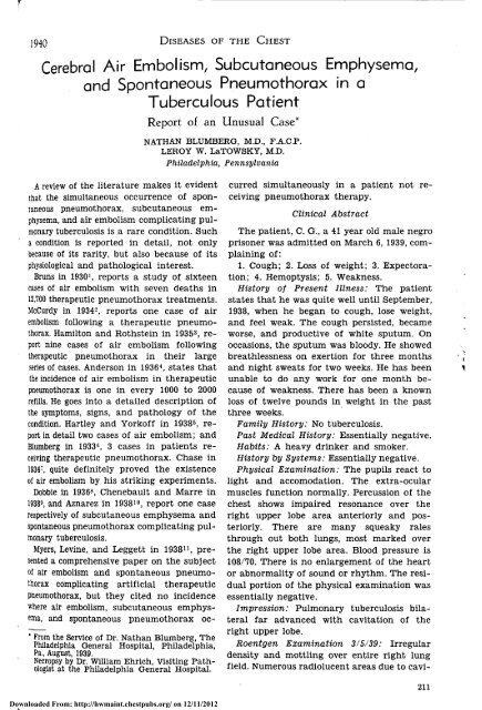
<strong>Cerebral</strong> <strong>Air</strong> <strong>Embolism</strong>, <strong>Subcutaneous</strong> <strong>Emphysema</strong>,<br />
<strong>and</strong> Spontaneous Pneumothorax in a<br />
Tuberculous Patient<br />
Report of an Unusual Case*<br />
A review of the literature makes it evident<br />
that the simultaneous occurrence of spontaneous<br />
pneumothorax, subcutaneous emphysema,<br />
<strong>and</strong> air embolism complicating pulmonary<br />
tuberculosis is a rare condition. Such<br />
a condition is reported in detail, not only<br />
because of its rarity, but also because of its<br />
physiological <strong>and</strong> pathological interest.<br />
Bruns in 1930 1 , reports a study of sixteen<br />
cases of air embolism with seven deaths in<br />
12,700 therapeutic pneumothorax treatments.<br />
McCurdy in 1934 2 , reports one case of air<br />
embolism following a therapeutic pneumothorax.<br />
Hamilton <strong>and</strong> Rothstein in 1935 3 , report<br />
nine cases of air embolism following<br />
therapeutic pneumothorax in their large<br />
series of cases. Anderson in 1936 4 , states that<br />
the incidence of air embolism in therapeutic<br />
pneumothorax is one in every 1000 to 2000<br />
refills. He goes into a detailed description of<br />
the symptoms, signs, <strong>and</strong> pathology of the<br />
condition. Hartley <strong>and</strong> Yorkoff in 1938 5 , report<br />
in detail two cases of air embolism; <strong>and</strong><br />
Blumberg in 1933 6 , 3 cases in patients receiving<br />
therapeutic pneumothorax. Chase in<br />
1934 7 , quite definitely proved the existence<br />
of air embolism by his striking experiments.<br />
Dobbie in 1936 8 , Chenebault <strong>and</strong> Marre in<br />
1938 9 , <strong>and</strong> Aznarez in 1938 10 , report one case<br />
respectively of subcutaneous emphysema <strong>and</strong><br />
spontaneous pneumothorax complicating pulmonary<br />
tuberculosis.<br />
Myers, Levine, <strong>and</strong> Leggett in 1938 11 , presented<br />
a comprehensive paper on the subject<br />
of air embolism <strong>and</strong> spontaneous pneumothorax<br />
complicating artificial therapeutic<br />
pneumothorax, but they cited no incidence<br />
where air embolism, subcutaneous emphysema,<br />
<strong>and</strong> spontaneous pneumothorax oc-<br />
' From the Service of Dr. Nathan Blumberg, The<br />
Philadelphia General Hospital, Philadelphia,<br />
Pa., August, 1939.<br />
Necropsy by Dr. William Ehrich, Visiting Pathologist<br />
at the Philadelphia General Hospital.<br />
Downloaded From: http://hwmaint.chestpubs.org/ on 12/11/2012<br />
NATHAN BLUMBERG, M.D., F.A.C.P.<br />
LEROY W. LaTOWSKY, M.D.<br />
Philadelphia, Pennsylvania<br />
curred simultaneously in a patient not receiving<br />
pneumothorax therapy.<br />
Clinical Abstract<br />
The patient, C. G., a 41 year old male negro<br />
prisoner was admitted on March 6, 1939, complaining<br />
of:<br />
1. Cough; 2. Loss of weight; 3. Expectoration;<br />
4. Hemoptysis; 5. Weakness.<br />
History of Present Illness: The patient<br />
states that he was quite well until September,<br />
1938, when he began to cough, lose weight,<br />
<strong>and</strong> feel weak. The cough persisted, became<br />
worse, <strong>and</strong> productive of white sputum. On<br />
occasions, the sputum was bloody. He showed<br />
breathlessness on exertion for three months<br />
<strong>and</strong> night sweats for two weeks. He has been<br />
unable to do any work for one month because<br />
of weakness. There has been a known<br />
loss of twelve pounds in weight in the past<br />
three weeks.<br />
Family History: No tuberculosis.<br />
Past Medical History: Essentially negative.<br />
Habits: A heavy drinker <strong>and</strong> smoker.<br />
History by Systems: Essentially negative.<br />
Physical Examination: The pupils react to<br />
light <strong>and</strong> accomodation. The extra-ocular<br />
muscles function normally. Percussion of the<br />
chest shows impaired resonance over the<br />
right upper lobe area anteriorly <strong>and</strong> posteriorly.<br />
There are many squeaky rales<br />
through out both lungs, most marked over<br />
the right upper lobe area. Blood pressure is<br />
108/70. There is no enlargement of the heart<br />
or abnormality of sound or rhythm. The residual<br />
portion of the physical examination was<br />
essentially negative.<br />
Impression: Pulmonary tuberculosis bilateral<br />
far advanced with cavitation of the<br />
right upper lobe.<br />
Roentgen Examination 3/5/39: Irregular<br />
density <strong>and</strong> mottling over entire right lung<br />
field. Numerous radiolucent areas due to cavi-<br />
211
tation. Early bronchogenic spread to middle<br />
portion of left lung. Heart not enlarged or<br />
displaced.<br />
Impression: Fibroulcerative tuberculosisright.<br />
Laboratory: Sputum negative for Tubercle<br />
Bacillus. Kahn negative. Blood Sugar 77 mg.%<br />
Blood Urea 10 mg.%<br />
Progress Notes<br />
3/10/39—Therapeutic Conference: Treatment<br />
to be bed rest for the time being.<br />
5/4/39—Therapeutic Conference: Advised<br />
therapeutic pneumothorax on right.<br />
5/18/39—On three separate occasions artificial<br />
pneumothorax attempted, but no pleural<br />
space could be found, <strong>and</strong> therefore was<br />
discontinued. Patient was placed on bed rest.<br />
6/19/39—Patient developed a generalized<br />
edema of the face, back, penis, <strong>and</strong> ankles.<br />
The abdomen was distended with fluid. The<br />
liver was not palpable, there was no venous<br />
engorgement, <strong>and</strong> the cardiac examination<br />
was negative. Blood Pressure 105/70. He also<br />
complained of nocturia 4-5 times.<br />
Laboratory: Urine—albumen 3 plus, hyaline<br />
<strong>and</strong> granular casts.<br />
Blood: RBC 3,980,000; Hemo. 9.5 gm.; WBC<br />
23,700; Polys 60; Lymph 34; Mono 2; Eosin 4.<br />
Total Protein 4.1; Album. 2.3; Glob. 1.8; A/G<br />
1.2. Congo Red test showed disappearance of<br />
the dye to be 70 per cent.<br />
Impression: 1. Nephrotic syndrome; 2.<br />
Amyloid disease of the kidneys; 3. General<br />
amyloidosis; 4. Chronic far advanced pulmonary<br />
tuberculosis.<br />
Therapy: Restrict fluids, no salt, high protein<br />
diet, mercupurin.<br />
7/14/39—Edema has entirely disappeared.<br />
Patient's general condition seems improved.<br />
Mercupurin stopped, but continued on low<br />
fluids <strong>and</strong> salt restriction.<br />
7/24/39—At 6:30 p. m. the patient was found<br />
leaning against the wall in the bath room.<br />
He was in a confused state. He was placed<br />
in bed by the nurse. Examination at 6:45 p. m.<br />
revealed a stuporous patient lying quietly in<br />
bed experiencing some difficulty on inspiration.<br />
The respiratory rate was not increased.<br />
He responded slowly to questions <strong>and</strong> did not<br />
complain of pain. Physical examination<br />
showed extensive subcutaneous emphysema<br />
over the right anterior chest, neck, face,<br />
right arm, <strong>and</strong> right leg. Crepitation was felt<br />
212<br />
Downloaded From: http://hwmaint.chestpubs.org/ on 12/11/2012<br />
DISEASES OF THE CHEST Jill:<br />
in all these areas. There was marked halloo:.ing<br />
of the scrotum. It measured 20 cm. ::<br />
diameter <strong>and</strong> was tympanitic on percussicr.<br />
The chest was hyper-resonant througho 1 ;<br />
even over the precordial area. The left pup.<br />
was relatively enlarged, neither pupil reacted<br />
to light or accomodation. There was;<br />
horizontal nystagmus. The right arm ar.:<br />
right leg showed a complete motor paralys;<br />
of all muscles. The tendon reflexes were absent<br />
<strong>and</strong> there was a positive Babinski c:<br />
the right.<br />
Impression: 1. Spontaneous pneumothorax<br />
2. <strong>Subcutaneous</strong> emphysema; 3. Right heir.:plegia<br />
due to air embolism to left cerebra<br />
area.<br />
7/25/39—Neurological Consultation: Patier.<br />
shows right hemiplegia, partial aphasia, ar.;<br />
no hemianesthesia.<br />
Impression: The sudden appearance c:<br />
right hemiplegia with subcutaneous emptysema<br />
<strong>and</strong> evidence of massive collapse poin:<br />
to air embolism as a causative factor of tii:<br />
patient's hemiplegia. <strong>Cerebral</strong> thrombos;<br />
must be ruled out however.<br />
7/25/39—Roentgen Examination: There:<br />
extensive subcutaneous emphysema throughout<br />
the tissues of the neck <strong>and</strong> thorax. Considerable<br />
irregular density over the entirright<br />
lung <strong>and</strong> probably a pneumothorax or.<br />
this side. No definite involvement on the lei'<br />
side at this time. Heart is not displaced.<br />
Impression: Extensive subcutaneous eir.physema<br />
with spontaneous pneumothorax.<br />
7/26/39—Spread of emphysema over the le:<br />
thorax <strong>and</strong> left leg. Blood Pressure 80/60.<br />
7/28/39—Patient gradually becoming mor<br />
stuporous. Death.<br />
Clinical Diagnosis: 1. Pulmonary tuberci<br />
losis bilateral far advanced with cavitation c<br />
the entire right lung; 2. Amyloid disease^<br />
the kidney; 3. General amyloidosis; 4. Spontaneous<br />
pneumothorax right; 5. Subcutanec<br />
emphysema; 6. <strong>Air</strong> embolism (cerebral):"<br />
Right hemiplegia.<br />
Necropsy Report<br />
External Examination: The body is that-'<br />
a moderately well nourished, 41 year old, bla^<br />
male, of large stature. The skin is emphysrmatous<br />
throughout, with the exception of tt<br />
of the left arm. The scrotum <strong>and</strong> penis s:<br />
tremendously emphysematous. The superb<br />
ial lymph nodes are not palpable.
1940 DISEASES OF THE CHEST<br />
Internal Examination: The right pleural<br />
cavity is partially obliterated by dense fibrous<br />
adhesions, the free part contains air <strong>and</strong><br />
purulent material; the left pleural cavity is<br />
free of adhesions. It contains about 200 cc.<br />
of a slightly purulent fluid <strong>and</strong> air. The pericardial<br />
sac contains a great deal of a slightly<br />
purulent fluid <strong>and</strong> the abdominal cavity contains<br />
600 cc. of a markedly purulent fluid.<br />
Heart: Weighs 210 gms. The pericardial<br />
surface is smooth. The myocardium is moderately<br />
firm. The ventricles are not dilated<br />
<strong>and</strong> contain no air. Ostia <strong>and</strong> valves are not<br />
changed. The myocardium is of normal color<br />
<strong>and</strong> is free of scars. The coronary arteries<br />
are not altered.<br />
Aorta: Apparently not changed.<br />
Lungs: Left weighs 920 gms., right 610<br />
gms. The right lung is partly destroyed by<br />
cavities which are found throughout all lobes.<br />
The remaining tissue of the right lung is<br />
indurated <strong>and</strong> shows many patches of mottled<br />
grayish white tubercles throughout both<br />
lobes. The left lung contains air in most<br />
places, <strong>and</strong> many small patches of gray tubercles<br />
throughout both lobes. The bronchial<br />
lymph nodes are enlarged <strong>and</strong> caseous. The<br />
bronchi contain some purulent material. The<br />
pulmonary vessels are not altered.<br />
Spleen: Weighs 250 gms. The surface is<br />
covered with str<strong>and</strong>s of fibrin. The consistency<br />
is rather firm <strong>and</strong> the mahogony brown<br />
cut surface shows several whitish-yellow<br />
nodules.<br />
Kidneys: Weigh 210 gms., each. The capsules<br />
strip easily leaving cortical surfaces<br />
which show a few yellowish gray nodules.<br />
The cortex is much enlarged, yellowish in<br />
color <strong>and</strong> well demarcated from medulla. The<br />
pelves <strong>and</strong> ureters are essentially normal.<br />
Urinary Bladder: Contains very little urine.<br />
The mucosa is not altered.<br />
Genitalia: The right testis shows several<br />
scarred areas.<br />
G. /. Tract: This is in an advanced state<br />
of autolysis. On the lesser curvature of the<br />
stomach there is a round ulcer, the largest<br />
diameter of which measures 0.6 cm. There<br />
are several irregular ulcers with yellowish<br />
nodules in their base in the ileum.<br />
Large veins: Contain little blood.<br />
Downloaded From: http://hwmaint.chestpubs.org/ on 12/11/2012<br />
Liver: Weighs 1280 gms. The capsular surface<br />
<strong>and</strong> cut surface show a number of small<br />
yellowish gray discrete nodules. Otherwise,<br />
the liver is in an advanced state of autolysis.<br />
The gall bladder contains a small amount of<br />
brown bile. The bile ducts are patent.<br />
Pancreas: This is in an advanced state<br />
of autolysis.<br />
Adrenals: These are of usual size. The<br />
lipoid in the cortices is rather well preserved.<br />
Brain: Weighs 1160 gms. It shows encephalomalacia<br />
in the distribution of the left<br />
anterior recurrent artery of Huebner. There<br />
are anomalies of the circle of Willis.<br />
Pathological Diagnosis<br />
1. Marked subcutaneous emphysema over<br />
entire body with exception of left arm;<br />
pyopneumothorax bilateral, purulent pericarditis<br />
<strong>and</strong> peritonitis; extensive fibrous<br />
pleural adhesions.<br />
2. Heart: Moderate atrophy.<br />
3. Aorta: Apparently normal.<br />
4. Lungs: Chronic pulmonary tuberculosis<br />
with widespread cavitation in right lung <strong>and</strong><br />
widespread, chiefly bronchogenic, extension<br />
in both lungs; caseous tuberculosis of bronchial<br />
lymph nodes.<br />
5. Spleen: Tuberculosis; amyloidosis.<br />
6. Kidneys: Amyloidosis; lipoid nephrosis;<br />
tuberculosis.<br />
7. Bladder: Apparently normal.<br />
8. Genitalia: Scars in right testis.<br />
9. G. I. Tract: Autolysis; small peptic ulcer<br />
of stomach; multiple tuberculous ulcers in<br />
ileum.<br />
10. Liver: Autolysis; tuberculosis.<br />
11. Pancreas: Autolysis.<br />
12. Adrenals: Apparently normal.<br />
13. Brain: Encephalomalacia—distribution<br />
of left anterior recurrent artery of Huebner.<br />
Anomalies of circle of Willis.<br />
Summary<br />
An unusual case of spontaneous pneumothorax,<br />
subcutaneous emphysema, <strong>and</strong> cerebral<br />
air embolism is presented. These conditions<br />
were not due to any mechanical interference<br />
or induced by artificial pneumothorax<br />
therapy.<br />
1922 Spruce Street.<br />
213
References<br />
1 Bruns, E. H.: "<strong>Air</strong> <strong>Embolism</strong> as a Complication<br />
of Artificial Pneumothorax Therapy," Colo.<br />
Med., 27, 237, 1930.<br />
2 McCurdy, T.: "<strong>Air</strong> <strong>Embolism</strong> in Artificial<br />
Pneumothorax," Amer. Rev. of Tuber., 30, 88,<br />
1934.<br />
3 Hamilton, C. E., <strong>and</strong> Rothstein, E.: "<strong>Air</strong> <strong>Embolism</strong>,"<br />
J. A. M. A., 104, 2226, 1935.<br />
4 Anderson, D. L.: "<strong>Air</strong> <strong>Embolism</strong> <strong>and</strong> Pleural<br />
Shock," Virg. Med. Monthly, 63, 371, 1936.<br />
5 Hartley, G. S., <strong>and</strong> Yorkoff, F. H.: "<strong>Air</strong> <strong>Embolism</strong><br />
or Pleural Shock—Report of Two<br />
Cases," Virg. Med. Monthly, 65, 234, 1938.<br />
6 Blumberg, N.: "Artificial Pneumothorax in<br />
The Treatment of Pulmonary Tuberculosis<br />
with Report of Three Cases of <strong>Air</strong> <strong>Embolism</strong>,"<br />
Robert Koch was probably one of the first<br />
to interest the profession in this problem of<br />
tuberculosis among nurses. Upon his suggestion,<br />
a survey was made in Germany of the<br />
hospitals, clinics, <strong>and</strong> sanatoria from 1906-<br />
1910. It was learned that, "in the general<br />
hospital . . infections of physicians <strong>and</strong> nurses<br />
were about four times as frequent in the<br />
special tuberculosis wards as in the dispensary<br />
departments" 1 .<br />
The problem was more forcibly brought to<br />
the attention of the profession by Heimbeck<br />
in Oslo, Norway, when in 1927 he published<br />
figures showing that only about one-half of<br />
the nurses entering training in the Oslo<br />
General Hospital were tuberculin-positive;<br />
<strong>and</strong> further, that the negative reactors contracted<br />
the active disease with far greater<br />
frequency than did the initially positive<br />
reactors 2 .<br />
In 1928 Shipman <strong>and</strong> Davis at the University<br />
of California Training School for nurses<br />
reported on a three year study, after observing<br />
a sharp increase in the incidence of the<br />
disease during the preceding years. A laudable<br />
health program was instituted for the nurses<br />
with frequent physical <strong>and</strong> roentgenological<br />
examinations. There was a corresponding<br />
sharp decrease in the incidence of the disease<br />
3 .<br />
There was a very able <strong>and</strong> practical discussion<br />
of the problem by J. A. Myers in 1930,<br />
when changing opinions were expressed <strong>and</strong><br />
new concepts in the light of recent works<br />
214<br />
Downloaded From: http://hwmaint.chestpubs.org/ on 12/11/2012<br />
DISEASES OF THE CHEST JULY<br />
Med. Jour, <strong>and</strong> Record, October 18,1933.<br />
7 Chase, W. H.: "Anatomical <strong>and</strong> Experimental<br />
Observation on <strong>Air</strong> <strong>Embolism</strong>," Surg. Gyn. anc<br />
Obst., 59, 569, 1934.<br />
8 Dobbie, D. N.: "Non-Traumatic Surgical <strong>Emphysema</strong><br />
in Association with Active Phthisis.<br />
Lancet, 1, 365, 1936.<br />
9 Chenebault, J., <strong>and</strong> Marre, P.: "Un Cas D<br />
Emphyseme Sous-Cutane' et Mediastina;<br />
Spontane' chez un Tuberculeux Pulmonaire.<br />
Rev. de la tuberc., 4, 477, 1938.<br />
10 Aznarex, E.: "Neumotorax bilateral simultaneo<br />
y enfisema mediastinal," La Prem<br />
Med. Argentina, 25, 1037, 1938.<br />
11 Myers, J. A., Levine, I., <strong>and</strong> Leggett, E. A<br />
"<strong>Air</strong> <strong>Embolism</strong> <strong>and</strong> Spontaneous Pneumothorax<br />
Complicating Artificial Pneumothorax,"<br />
Brit. Jour, of Tuber., 31, 77, 1937.<br />
Tuberculosis Among Nurses<br />
PAUL VINCENT DAVIS, M.D.<br />
Warrenton, Virginia<br />
were pronounced 4 . He cited Braeuning's study<br />
in Germany on eighteen. nurses whose past<br />
health had been perfect, but who had pulmonary<br />
tuberculosis at the time of the study.<br />
Fifteen had nursed tuberculous patients<br />
Myers very aptly said: "The greatest of all<br />
causes for the high incidence of tuberculosis<br />
among nurses is the exposure they suffer<br />
to the disease while in hospital or school<br />
residence."<br />
That the disease exists among nurses to a<br />
far greater extent than one would anticipate,<br />
was made clear in a report by the Relief<br />
Fund Committee of the American Nurses<br />
Association in 1930. This report, covering<br />
twenty years (from 1911 to 1930), showed<br />
that of the 543 nurses receiving aid froir.<br />
the Association for illnesses, 48 per cent, or<br />
nearly one-half had tuberculosis 5 .<br />
Investigating the incidence of tuberculosis<br />
among nurses at the Ancker Hospital in St.<br />
Paul, Geer 6 , in 1932, observed that the previous<br />
nine year period showed three times the<br />
normal expected incidence among youn?<br />
women of the same age group. Intracutaneou. ;<br />
testing was begun, with positive reactors having<br />
roentgenograms of the chest taken. I:<br />
was found that about 30 per cent of the<br />
nurses were positive reactors on entering<br />
training while nearly 100 per cent were positive<br />
on graduation. A rigid program of aseptic<br />
nursing technique was instituted. From September<br />
1930 to September 1934 the incidence<br />
of the disease dropped sharply. The three


