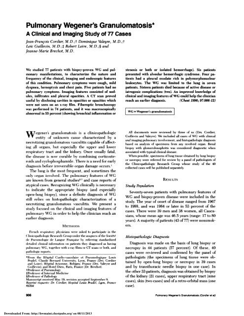
Pulmonary Wegener's Granulomatosis*
Pulmonary Wegener's Granulomatosis*
Pulmonary Wegener's Granulomatosis*

You also want an ePaper? Increase the reach of your titles
YUMPU automatically turns print PDFs into web optimized ePapers that Google loves.
<strong>Pulmonary</strong> Wegener’s <strong>Granulomatosis*</strong><br />
A Clinical and Imaging Study of 77 Cases<br />
Jean-Fran#{231}ois Cordier, M.D.;t Dominique Valeyre, M.D.;t<br />
LoIc Guillevin, M.D.; Robert Loire, M.D.; and<br />
Jeanne-Marie Brechot, Al. D.<br />
We studied 77 patients with biopsy-proven WG and pul-<br />
monary manifestations, to characterize the nature and<br />
frequency of the clinical, imaging and endoseopic features<br />
of this condition. <strong>Pulmonary</strong> symptoms were cough, mild<br />
dyspnea, hemoptysis and chest pain. Five patients had no<br />
pulmonary symptoms. Imaging features consisted of nodules,<br />
infiltrates and pleural opacities. A CT scan proved<br />
useful by disclosing cavities in opacities or opacities which<br />
were not seen on an x-ray film. Fiberoptic bronchoscopy<br />
was performed in 74 patients, and it was macroscopicaily<br />
abnormal in 55 percent (showing bronchial inflammation or<br />
W egeners granuio)matosis is a clinicopathologic<br />
entity (Of tlflknOWfl cause characterized by a<br />
necrotizing granulomatoous vasculitis capable of affect-<br />
ing all organs, hut especially the upper and lower<br />
respirato)ry tract and the kidney. Once usually fatal,<br />
the disease is flow curable by combining corticoste-<br />
roids and cycloophosphamide. There is a need for early<br />
diagnoosis before irreversible organ damage oc’<br />
The lung is the moost frequent, and sometimes the<br />
oonly organ invoolved. The pulmonary features (of WC<br />
are known froom general studies27 and case reports of<br />
atypical cases. Recognizing WG clinically is necessary<br />
to) indicate the appropriate biopsy (and especially<br />
o)pen-lung biopsy), since a definite diagnosis of WG<br />
still relies on histopathologic characterization of a<br />
necnotizing granulomatous vasculitis. We present a<br />
study focused on the clinical and imaging features (of<br />
pulmoonary WG in (order to) help the clinician reach an<br />
earlier diagnoosis.<br />
NI ETHODS<br />
Fremich respirator physicians were asked too participate in the<br />
Clinicoopathoohogic Research Crooump imnder the auspices oofthe Soci#{233}t#{233}<br />
de Pneummologie de Langume Fran#{231}aise by referring standardized<br />
detailed clinical imiformatioon oon patients they diagnosed as having<br />
outlmoonary \VC, toogether with x-ray films our CT scans our both, and<br />
oathologic repourts.<br />
* From the H#{244}pital Cardio-vasculaire et Pneumoologique Luuis<br />
Pradel, Clauode Bermiard Universit,; Lyon, France (Drs. Cordier<br />
an(l Loire); HApital Avicenne, Bohigro France (Drs. Valeyre and<br />
(;uoillevin); and Hootel Dieto, Paris, France (Dr. Brechoot).<br />
tProfessoor oof Pneuomoohugy<br />
Prufessor ouf Internal Medicine<br />
§Professoor oof Patholoogv<br />
Manuscript received May 19; revisioumi accepted Septeniber 8.<br />
Reprint requests: Dr Cordier. Hopital louis Pradel, Ltpn, France<br />
69394<br />
stenosis or both or isolated hemorrhage). Six patients<br />
presented with alveolar hemorrhagic syndrome. Four pa-<br />
tients had a pleural exudate rich in polymorphonuclear<br />
leukocytes. The WG was limited to the lung in seven<br />
patients. Sixteen patients died because of active disease or<br />
iatrogenic complications (two). An improved knowledge of<br />
clinical and imaging features ofWG could help the clinician<br />
reach an earlier diagnosis. (Chest 1990; 97:906-12)<br />
[ WG = Wegener’s granulomatosis I<br />
All documents were reviewed by three of uos (Drs. Cordier,<br />
Cuillevin and Valeyre). We included all cases ouf WG with clinical<br />
and imaging pulmonary involvement, and histopathologic diagnosis<br />
based on analysis of specimens from any involved organ. Renal<br />
biopsy with glomerulonephritis was considered diagnostic when<br />
assouciated with typical clinical disease.<br />
When possible, specimens ofliong tissue obtained by lung biopsy<br />
or miecropsy were referred for review by a panel of pathologists of<br />
the Clinicopathologic Research Group whose study of the 40<br />
collected cases will be published separately.<br />
Study Population<br />
906 <strong>Pulmonary</strong> Wegeners Granulomatosis (Cordier et a!)<br />
Downloaded From: http://hwmaint.chestpubs.org/ on 08/11/2013<br />
RESULTS<br />
Seventy-seven patients with pulmonary features of<br />
WG and biopsy-proven disease were included in the<br />
study. The year of onset of disease ranged from 1967<br />
to 1988, and was 1984 or later in 53 percent of the<br />
cases. There were 39 men and 38 women, all Cauca-<br />
sians, whose mean age was 46.5 years (range: 17 to 80<br />
years). A majo)nty ofpatients (43 of77) were nonsmok-<br />
ers.<br />
Histopathologic Diagnosis<br />
Diagnosis was made on the basis of lung biopsy or<br />
necropsy in 44 patients (57 percent). Of these, 40<br />
cases were reviewed and confirmed by the panel of<br />
pathologists (the specimens of lung tissue were oh-<br />
tamed by open-lung biopsy or necropsy in 39 cases<br />
and by transthoracic needle biopsy in one case). In<br />
the other 33 patients, diagnosis was obtained by biopsy<br />
of the kidney (21 cases), upper respiratory tract (nine<br />
cases), skin (two cases) and ofa retro-orbital mass (one<br />
case).
Table 1 -Clinical Manifestations in 77 Patients with<br />
<strong>Pulmonary</strong> Wegener’s Granulomatosis<br />
IoIamiifestatiouim* Nou . (%)<br />
Ltommg + kidney + (JET + other 42 (55)<br />
Lmong + kidney + outher 8 (10)<br />
Lung + URT + oother 7 (9)<br />
Lung ounl 7 (9)<br />
Lung + URT 5 (7)<br />
Lumig + kidney 4 (5)<br />
Lung + URT + kidney 3 (4)<br />
Lung + oother 1 (1)<br />
*URT: topper respiratoumy tract; (other: any clinical mamiifestations imi<br />
amm orgamm other timan time lumig, kidne; oor URT<br />
General Presentation of Patients<br />
Sixty-four patients (83 percent) presented with fever<br />
and 50 (65 percent) with weight boss. The mean weight<br />
loss (known in 48 cases) was 6.4 kg. As a result of the<br />
inclusion criteria, all patients had pulmoonary features.<br />
Upper respiratory tract and ear features were present<br />
in 57 patients (74 percent), renal in 57 (74 percent),<br />
amid others in 58 (75 percent). Tile combinations of<br />
clinical features related to WG are listed in Table 1.<br />
The onset oof disease was generally progressive and<br />
multifocal, and no clear-cut chronologic sequence of<br />
manifestations could be clearly identified. Four pa-<br />
tients had upper respiratory tract and three had renal<br />
involvement more than two) years before the pulmo-<br />
nary manifestations l)ecame evident.<br />
<strong>Pulmonary</strong> Clinical Presentation<br />
Cough was present in 60 patients (78 percent),<br />
dyspnea in 43 (56 percent), hemoptysis in 30 (39<br />
percent) and chest pain in 25 (32 percent). Cough<br />
usually was unproductive, but in seven patients pu-<br />
rulent hronchoorrhea -as present (in two patients it<br />
was massive, due to) necrosis of a voluminous pulmo-<br />
nary mass). Dyspnea was mild, except in some patients<br />
with diffuse pulmonary involvement in whom it was<br />
ooccasio)nally severe. The physical signs depended on<br />
the type o)fpulmonary involvement and had noo specific<br />
features. Five patients (6 percent) had no pulmonary<br />
symptoollls, pulmonary involvement being foound on<br />
systematic chest x-ray film.<br />
Superinfectioon of pulmoonary lesio)ns was doocu-<br />
mented in only one patient who had acute fever and<br />
enlargement ofa thin wall cavity with an air-fluid level<br />
leading to lobar resection. Subglottic stenosis was<br />
encountered in two patients.<br />
Pleuro-pulmonary Imaging<br />
The iinaglllg features identified by chest x-ray films,<br />
tomograms and CT scans are listed in Table 2. Both<br />
sides of the chest were involved in 63 cases and one<br />
side was involved in 14. There was no obvious pattern<br />
Downloaded From: http://hwmaint.chestpubs.org/ on 08/11/2013<br />
Table 2-Features ofPleuro-pulmonary lmaging in 77<br />
Patients with <strong>Pulmonary</strong> Wegener’s Granulomatosis5<br />
Type ofOpacity No. of Cases (%) Cavitary Opacities<br />
Nodules 53 (69) 49% of nodules (26/53)<br />
Unilateral 14<br />
Bilateral 39<br />
Infiltrates 41 (53) 17% ofinfiltrates (7/41)<br />
Unilateral 15<br />
Bilateral 26<br />
Pletoral opacity 9 (12)<br />
Atelectasis 3 (4)<br />
*Miscellaneoosis: calcification: 2; mediastinal adenopathy: 1.<br />
of lung zone involvement but the apices usually were<br />
spared. Nodules and infiltrates were the most commoon<br />
lesions and (often coexisted.<br />
Nodules<br />
Nodules consisted of rounded lesions with well-<br />
defined margins. Their size varied from about 5 to 100<br />
mm, the largest being usually excavated. Excavated<br />
nodules had generally medium-sized walls and fluid<br />
levels were uncommon. Some nodules which were no)t<br />
excavated on the chest x-ray film had a typical central<br />
necrotic content and/or small cavities as seen on the<br />
CT scan. The number of nodules varied but was<br />
generally fewer than ten. In the course ofthe disease,<br />
nodules tended to increase, particularly in size but<br />
also in number. Furthermore, most nodules became<br />
excavated in the course of untreated disease.<br />
Infiltrates<br />
Infiltrates consisted ofalveolar opacities with a wide<br />
range (Of appearance. We distinguished three categoo-<br />
ries of infiltrates, all of which could be found in the<br />
same patient, together or at different stages of the<br />
disease.<br />
In the first category, the infiltrates were bilateral,<br />
diffuse and of low density as shown by the x-ray film.<br />
The six patients with alveolar hemorrhage, as dis-<br />
cussed later, had opacities which fell into this category<br />
(Fig 1). In the second category, the infiltrates were<br />
more patchy and oflow density (Fig 2). When foollowed<br />
by sequential imaging, some spontaneously cleared,<br />
some increased in density (with or without a reductiooml<br />
in size) and others progressed, with coalescence oof<br />
adjacent opacities.<br />
In the third category, the infiltrates were dense and<br />
localized (consolidation) (Fig 3 and 4) with ill-defined<br />
margins. In some, an air bronchogram or foci oof<br />
excavation, or both, were present.<br />
Atelectasis<br />
Lobar or segmental atelectasis was present in tilree<br />
cases. Limited atelectatic zones were more frequently<br />
CHEST I 97 I 4 I APRIL 1990 907
Fm;tii: 1 . Infiltrat-s oof tlit first category Bilateral diffmose opacities<br />
of how density in a patient scith alveoolar hemoorrhage.<br />
encooumltered and were assoociated with oother abnor-<br />
malities.<br />
Pleural Opacities<br />
Pleural oopacities were not doocumented by CT scan.<br />
Frank 1)lellral effusion was documented in foour cases,<br />
as discussed in the miext sectio)n.<br />
fiscclla UCOU5<br />
Calcifications were present in two cases (in a con-<br />
Fmounm: 2. Infiltrates (of the seco)mid categ(or\: Patchy bilateral<br />
opacities (Of lO)W demisit:<br />
FmCuRE 3. Infiltrates of the third categom-\: Dense and localized<br />
c-onsolidatuun. Samne patient as in Figure 2. The right infiltrate has<br />
spomitamoeoottsly cleared, whereas the demosity amid size ouf the left<br />
infiltrate increased.<br />
so)lidation and iii the area of a previoously resected<br />
noodule, respectively). Hilar and mediastinal lymph<br />
Ilo)(les ere found 0)11 the CT scan in tone patient with<br />
parenchymal consolidation.<br />
Fiberoptie Bronchoscopy<br />
Seventy-four l)atients Elllderiveilt fiberoptic i)ron-<br />
choscopv ‘hich as Illacroscopically aI)n(ormal in 41<br />
(55 percellt). Abnoormal endobronchial aspects coonsisted<br />
oof bronchial steticisis in 13, ulceratioons or<br />
pseudo-tumor in seven, 11 Ham nlatorv lesions without<br />
stenoosis ill tell, isO)late(1 hemorrhage in ten and isolated<br />
purulent secretioons in ooiie.<br />
FmcuooE 4. Coexistence of excavated thin-walled nodule and localized<br />
consolidation on CT scan in the same patient.<br />
908 <strong>Pulmonary</strong> Wegenes Granutomatosis (Cordier et a!)<br />
Downloaded From: http://hwmaint.chestpubs.org/ on 08/11/2013
Biopsy of bronchial lesions showed inflammatory<br />
cells (lymphocytes, plasma cells, polymorphonuclear<br />
leukocytes) and giant cells, but definite vasculitis was<br />
not seen.<br />
Bronchoalveolar Lavage<br />
Bronchoalveolar lavage done in 20 cases showed the<br />
following mean (range) percentages of leukocytes:<br />
lymphocytes, 12.6 percent (0-53 percent); neutrophils,<br />
22.0 percent (0-93 percent); eosinophils, 2.6 percent<br />
(0-25 percent).<br />
Pleural Fluid<br />
Pleural fluid analysis was done in four cases and in<br />
each revealed an exudate (with 38, 42, 47 and 57 g/L<br />
of protein, respectively) with a predominance of poly-<br />
morphonuclear leukocytes.<br />
Lung Function Tests<br />
These tests were done only occasionally, showing<br />
principally a restrictive defect related to space-occu-<br />
pyinglesions in the chest. No characteristic obstructive<br />
defect was found.<br />
Alveolar Hemorrhage<br />
Six patients (8 percent) presented with the alveolar<br />
hemorrhagic syndrome, characterized by dyspnea,<br />
hemoptysis (with blood originating from distal airways<br />
as seen by fiberoptic bronchoscopy), severe anemia<br />
and diffuse bilateral alveolar infiltrates on chest im-<br />
aging. All six patients had further renal and upper<br />
respiratory tract involvement, and five had involve-<br />
ment of other (organs. One of the six patients died<br />
postciperatively after diagnostic open-lung biopsy.<br />
WG Limited to the Lung<br />
Seven patients (four women, three men) had only<br />
pulmonary involvement. Five of the seven presented<br />
at the time of the chest x-ray film with nodules<br />
(excavated in two), and two patients had pulmonary<br />
infiltrates (excavated in one). When compared with<br />
classic WG, only two of the seven cases were in any<br />
way remarkable: one patient refused treatment and<br />
remained well with persistent multiple lung nodules<br />
for four years offollow-up and the other had two lobar<br />
resections (in 1972 and 1974) before diagnosis was<br />
made by reviewing the pathologic findings of the<br />
resected lobes when she developed more lung nodules<br />
in 1982.<br />
Laboratory Data<br />
The erythrocyte sedimentation rate was greater<br />
than 40 mm in 62 patients (81 percent) and greater<br />
than 80 mm in 43 (56 percent). Anemia (hemoglobin,<br />
used , Vitil surgical excisioon of pulmonary lesions<br />
sometimes i)eing done four diagncmstic purposes. On the<br />
V1loole, treatment led rapidly too the regresson of the<br />
bug lesiomos witllouut significamit residual pulnlonary<br />
impairlllellt. The chest x-ray film generally returned<br />
to) oloorillal, with ounly tumor fibrotic sequelae in s(Omne<br />
patielits. When relapse of the disease occttried after<br />
reducing or sto))ping treatment, it did noot involve the<br />
lung in all cases.<br />
Sixteen l)atients (20.8 percent) in this series died<br />
i)ecause oof active disease (14) o)r because of iatrogenic<br />
complications (two). Of the 14 patients who died with<br />
active disease, 13 had renal invo)lvement. Diagnosis of<br />
WC had llO)t i)eeII obtained before necropsy in four<br />
patiellts (5 percent) who died ofactive disease. Three<br />
patients died postoperatively after diagnostic open-<br />
lung biopsy (two) oor pneum(onect(omy (one) with<br />
uncontrolled disease, and in all three the surgical<br />
procedure undoubtedly contributed to death. Four<br />
patients with severe widespread disease died within<br />
twoo Iflooiltils cof diagmlosis despite treatment. One pa-<br />
tient died o)fadute respiratory failure of undetermined<br />
cause, six years after diagnoosis; he had chronic obstruc-<br />
tive pulmonary disease before the onset of WG, and<br />
active widespread WG was present at the time of<br />
death despite treatment. One patient died seven years<br />
after diagnoosis (of WG because of widespread active<br />
disease and associated metastatic colonic carcinoma,<br />
both diagnoses being confirmed at the time of nec-<br />
ropsy. One patient died with extrathoracic WG without<br />
pulmo)nary relapse after previous excision of a pul-<br />
moonary no)dule. Two patients died of iatrogenic com-<br />
plications, tune because of brain toxoplasmosis in the<br />
course ofAIDS acquired through a blood transfusion,<br />
and the other because of Pneunzocystis carinii pneu-<br />
monia while undergoing immunoosuppressive treat-<br />
inent.<br />
DIstTSs1oN<br />
The aim ofthe present study was to precisely define<br />
the nature and frequency (ofthe features of pulmonary<br />
WC . Cases were collected at primary specialist cen-<br />
ters but stll)jected too unifoorm systematic analysis. This<br />
avooided the po)ssible selection bias inherent in case<br />
ccollectioons froom referral centers which may for ex-<br />
ample exclude patients who die rapidly or refuse to<br />
participate ill treatment protocols. On the (other hand,<br />
sour study has tile disadvantages of all retrospective<br />
studies, and in particular yields no useful information<br />
on treatfllent.<br />
The mean age of our patients (46.5 years) is similar<br />
to that ofootiler series.30’ The sex ratio was about equal.<br />
The Inajo)rity were noonsmokers. Smoking habits have<br />
tRot l)eIl characterized in previous studies, and this<br />
needs further evaluation. Most patients in this series<br />
presented with fever, weight loss and extrapulmonary<br />
clinical features. Twenty patients (26 percent) had no<br />
renal involvement, thus falling into the category of<br />
“limited WG,”M but on1y seven patients (9 percent)<br />
had WC strictly limited to) the lung.<br />
The frequency of pulmoonary symptoonis in tour<br />
patients is higher than in other studies.3#{176}’ This is not<br />
surprising since our case definition was based on<br />
pulmonary involvement. Only 6 percent had no) pul-<br />
monary symptoms, and their pulmonary involvement<br />
was found by systematic chest x-ray films.<br />
The most classic imaging appearance of WG is<br />
bilateral nodules, which are highly suggestive of this<br />
condition when excavated. Nevertheless, pulmonary<br />
infiltrates are common, and we could distinguish on<br />
the chest x-ray film and CT scan three broad categ(ories<br />
of infiltrates which easily can be recognized in the<br />
data of previously published studies.3’4’#{176}#{176}#{176}3 Some pa-<br />
tients present with diffuse bilateral infiltrates of low<br />
density, characteristically associated with alveolar<br />
hemorrhagic syndrome. In others, the infiltrates are<br />
less diffuse and more patchy In our experience, the<br />
classic spontaneous clearing of pulmonary o)pacities in<br />
WG4 involves this sec(ond category of infiltrates. In<br />
the third group, the infiltrates are dense and localized<br />
(consolidation). The CT scan was most useful for<br />
further characterizing the imaging features. First it<br />
revealed lesions which were not seen on the chest x-<br />
ray film. Furthermore, the CT scan often showed<br />
previously unsuspected necrotic cavities or necrotic<br />
content in nodules or infiltrates. In the latter, an air<br />
bronchogram was sometimes present. The varied<br />
appearance of infiltrates most probably reflects the<br />
wide spectrum of lesions seen in pathologic studies,<br />
which range from alveolar hemorrhage to pneumonialike<br />
fibrinous exudation in addition to the classic<br />
vascular necrotizing granulomatous lesions. oos Calci-<br />
fication and mediastinal adenopathy were rare in this<br />
study as in others.<br />
Fiberoptic bronchoscopy was done in almost all<br />
patients in this series, and was abnormal in 55 percent.<br />
In most cases, endobronchial involvement consisted<br />
of nonspecific inflammatory lesions with or without<br />
stenosis. The bronchial lesions seemed to be more<br />
associated with the surrounding parenchymal involve-<br />
ment than isolated primary bronchial disease, and<br />
atelectasis of healthy pulmonary parenchyma as a<br />
consequence of isolated bronchial stenosis was most<br />
uncommon. Bronchial biopsies showed inflammatory<br />
and giant cells, but were not diagnostic since no frank<br />
vasculitis was seen. Nevertheless, they are suggestive<br />
ofactive disease in a typical clinical context. Fiberoptic<br />
bronchoscopy also helped in differentiating the origin<br />
of blood in patients with hemoptysis, is, blood origi-<br />
nating from focal bronchial lesions vs diffuse bleeding<br />
from distal airways suggesting alveolar hemorrhage.<br />
The only relevant finding at bronchoalveolar lavage<br />
910 <strong>Pulmonary</strong> Wegeners Granulomatosis (Cordier et a!)<br />
Downloaded From: http://hwmaint.chestpubs.org/ on 08/11/2013
was the increase in polymorphonuclear leukocytes at<br />
the time of the differential cell count, but this is not a<br />
feature peculiar to WG, and its diagnostic value is<br />
therefore limited.<br />
Pleural involvement in WG rarely results in much<br />
effusion, and no published information on pleural fluid<br />
is available.’ In our four cases with pleural fluid<br />
analysis, there was an exudate with a predominance<br />
(of polymorphonuclear leukocytes.<br />
Lung functioon tests have been used in the staging<br />
and the follow up o)f patients with pulmonary WG,<br />
and the most common abnormality was airflow (ob-<br />
struction.#{176} Detailed lung function tests were rarely<br />
available in the present study, thus allowing rio com-<br />
ment (on this point.<br />
The alveolar hemorrhagic syndrome has become a<br />
more frequently recognized pulmonary manifestation<br />
ofWC,’7”5’21 and it occurred in 8 percent of patients<br />
in this series. Patients present with hemoptysis, and<br />
blood originating from distal airways is seen at bron-<br />
choscopy. The chest x-ray film shows diffuse bilateral<br />
alveolar infiltrates. Alveolar hemorrhage is generally<br />
acute and even fulminant in soome cases. Death fre-<br />
quently occurs in untreated patients or if treatment is<br />
delayed.’8 On the oother hand, resolution is generally<br />
complete with early treatment and may sometimes<br />
occur spontaneously.2#{176} Alveolar hemorrhagic WG may<br />
be confused with Gooodpasture syndrome especially<br />
since the latter also is usually assoociated with renal<br />
disease. However, patients with WG have in addition<br />
extrapulmonary and extrarenal disease. They also have<br />
antineutrophil antibodies, whereas they lack the anti-<br />
basement membrane antibodies found in Gooodpas-<br />
ture’s syndrome. The pathologic diagnosis of WC at<br />
the time of the lung biopsy often is difficult when<br />
typical lesions are absent and capillaritis is overshad-<br />
owed by alveolar 0708<br />
We fcound no striking differences between the pill-<br />
monary manifestations of “limited” WG and those oof<br />
“classic” WC (ie, with renal invoolvement). The cooncept<br />
O)f tile limited form o)f WG has been proposed princi-<br />
pally to differentiate patients with and without renal<br />
invoolvemellt, the former having the poorer proogno-<br />
sis.#{176}27Iii this series, only OOC oof the 14 patients whoo<br />
died with active disease had “limited” WC.<br />
Usual laboratory tests are oof little help in the<br />
diagnosis<br />
matoorytation<br />
of WG,<br />
syndrome<br />
rate , anenlia,<br />
since<br />
witil<br />
they only point<br />
a raised erythroocyte<br />
hyperleukoocytosis<br />
to) an inflamsedimen-<br />
and hyperthroombocytosis.)6<br />
Circulating immune co)mplexes<br />
were Present iii at least 16 percent oofour patients and<br />
at least 19 percent of Fauci’s,#{176} and they (occasionally<br />
have been reported by o)thers. The roole of immune<br />
complexes in WG is uncertain but may he similar to<br />
that in other vasculitic syndromes. Several recent<br />
studies have shown that anti-neutroophil antibodies are<br />
Downloaded From: http://hwmaint.chestpubs.org/ on 08/11/2013<br />
present in the serum oof patients with active WG and<br />
could be invoolved in the pathogenesis of the dis-<br />
ease.’#{176} In the present series, antineutro)phil antihod-<br />
ies were present in ten out ofthe 12 patients tested.<br />
We shall not discuss the clinical manifestations<br />
outside the respiratory tract o)ther than to make the<br />
point that the multi-organ involvement can guide the<br />
diagnosis of the pulmonary disease. The prognoosis of<br />
WG has been transformed by therapy with corticooste-<br />
rooids and cycloophosphamide.3 The case fatality rate of<br />
7 percent reported by Fauci3 was especially low hut<br />
related to) patients enrolled in a treatment protocoi at<br />
a referral center, thus excluding patients dying before<br />
or just after diagnosis in other institutions. In the<br />
present and two other series,6’ the case fatality rate<br />
ranged from 20.8 to) 28 percent. In the present series,<br />
seven of 77 (9 percent) patients died without treat-<br />
ment. The fact that three patients died shortly after a<br />
diagncstic thoracotomy underlines the risk (of this<br />
procedure in patients with severe pulmonary WC. On<br />
the other hand, treated patients generally improve<br />
without significant sequelae, although the hazards of<br />
immunoosuppressive drugs are a major limiting factor<br />
foir long-term survivai.’ Cotrimoxazole has been<br />
advocated as an alternative treatment, especially when<br />
WG is limited to the lung.’#{176}”#{176}’#{176}<br />
Our study shows that patients die from WG mainly<br />
as a result of delays in diagnoosis. It is therefore<br />
necessary to recognize the protean clinical and imag-<br />
ing presentation cof WG so that the approopriate diag-<br />
noostic investigations are done and the patient is treated<br />
earl)<br />
ACKNOWLEDGMENT: Ve thank L. D. Cnoer for reviewing the<br />
translation o)f this paper and NI. C. Thevenet four secretarial<br />
assistamice.<br />
APPENDIX<br />
The foollowing French clinicians participated in the Clinicoopathoulogic<br />
Research<br />
contributing one<br />
Crotop’s<br />
our moore<br />
study<br />
patients:<br />
on Wegener’s<br />
C. Akotmn,<br />
granuloumnatosis<br />
Paris; J. P Bernard,<br />
b)V<br />
Lvun; J. Bignon, Cr#{233}teil; F. Blanc-Jouvan, Grenoble; F. Bo)nns010d,<br />
Limoges;<br />
Caries,<br />
P A. Boudes, Paris;<br />
Tooulouuse; J. Cerrina,<br />
J. Bran,<br />
Paris; J. C.<br />
Rouemm;<br />
Dalphin,<br />
P Camus, Dijon;<br />
Besan#{231}’omi; M .<br />
P<br />
Dc<br />
Lajartre,<br />
A. Dhers,<br />
Nantes;<br />
Macon;<br />
P. Delaval,<br />
P 1)ugioe,<br />
Rennes;<br />
Crasse;<br />
P Deteix, Clermont-Ferramod;<br />
J. NI. Durand, Marseille; A.<br />
Emnoomiot, Saint-Etienne; P Godard, Moontpo’llier; J. C. Cto#{233}rimi,<br />
Lyon; A. llaloumomi,<br />
Kerhrouimc’h, Brest;<br />
Nantes;<br />
A.<br />
B. Ilerer,<br />
Krivitzk;<br />
Paris;<br />
Paris:<br />
P llyvernat,<br />
J. Lacromiiojmme,<br />
Lyon;<br />
Paris:<br />
J. F.<br />
0.<br />
L5UO(110e, TO)(Ol000Se; NI. Laville, Lyon; F. X. Lehas, Le Mans; NI. C.<br />
Level, Verolumi; C. Mayaud, Paris; B. Milleron, Paris; F. Natali,<br />
Paris; C. Nootovet, Rouuemo; R. Panente, Paris; F. Patte, Poitiers; J. N.<br />
Prevost. Rooiocn; 0. Rigatid, Crenooble: D. Roohert, Lyon; E. Rowgel.<br />
Straslouomrg; B. Saoovezie, Clermoont-Ferrand; C . Tenipelhouff,<br />
Rooanmme; J. M. V#{235}rgnoomm, Saint-Etiemone.<br />
REFERENCES<br />
1 Leavitt R1 Fatoci AS. Pimlmummary vasculitis. Am Rev Ito-spir Dis<br />
1986; 134:149-66<br />
2 Dc Remee BA, McDoonakl TJ, liarrisomi Jr EC, Cooles DT<br />
Wegener’s granuhumatosis: anatoomic correlates, a proposed clas-<br />
sificatioun. Mayoo Clin Proc 1975; 51:777-8 1<br />
3 Fauci AS, Haynes BF, Katz P, Wolff SM. Vegener granioloomso-<br />
tosis: prospective clinical and therapeutic experiemice with 85<br />
patients for 21 years. Ann lntern Med 1983; 98:76-85<br />
CHEST I 97 I 4 I APRIL 1 990 911
4 Famoci AS, Woolf SM. Wegener’s granulomnatosis: studies in<br />
eigimteemm patiemots amid a review ofthe literature. Medicine 1973;<br />
52:535-61<br />
5 Israel ilL, Patchefskv AS. Wegener’s gramoulooniatosis ouf lung:<br />
distgmooosis stood treatmmoemot. Experiemmc-e with 12 cases. Amimi Interim<br />
Me(l 1971; 74:881-91<br />
6 I.e Thi Ilimomog l)U, Wechsler B, Cabane J, Piette JC, Herreman<br />
(;. c;muillt’simm L, et al. Cranuloumatoose<br />
c-limmiqomes, probl#{232}momes noosolougiqtoes: revue<br />
de Vegener: aspects<br />
de Ia litt#{233}rature a<br />
do’ 30 ouioservatiomos. Amomo Med Imotern 1988; 139: 169-82<br />
7 Valtoon E\V. ciamit cell granulomna (of the respirat(ury tract<br />
(Wegemier’s gramiuloommiatcosis). Br Med J 1958; 2:265-70<br />
8 Carringtomi CB, Lieb)O)w AA. Limited formns oof angiitis and<br />
granuloumnatosis ouf \Vegemiers type. Am J Med 1966; 41:497-527<br />
9 Lamo(lman 5, Burgemmer F. <strong>Pulmonary</strong> manifestatioomis in Wegener’s<br />
grantoloonoatousis. AJR 1974; 122:750-57<br />
10 Bamloery P. Katariya S, Sakhuja V. Kaur U, Behera 0, Malik<br />
SK, et al. \Vegener’s graniokumatoosis in Nourth India: radicokogic<br />
manifestations in eleven patients. Acta Radiol 1988; 29: 1 1-13<br />
1 1 Farrelly CA. Wegener’s granulomatousis: a radioukugical review ouf<br />
the pulmomonary manifestatiouns at initial presentation and during<br />
relapse. Clin R.adiool 1982; 33:545-51<br />
12 (;oomolez L, Van Ordstramiol uS. Wegener’s granuloomnatousis:<br />
review of 11 cases. Radioologv 1973; 107:295-300<br />
13 Maguire R, Fauci AS, Douppman JL, Wolff SM. Untossoal<br />
radiographic features ouf Vegener’s granuloumatousis. AJR 1978;<br />
130:233-38<br />
14 Katzemisteimi AL, Askimm FB . Surgical pathology ouf non-moeouplastic<br />
hommg (lisesose. Philadelphia: WB Samomiders, 1982:166-202<br />
15 Lieiooow AA. Pulmoummarv angiitis and grammlomatusis. Am Rev<br />
Respir Dis 1973; 108:1-18<br />
16 Mark EJ, Msotsuloara 0, Tan-Liti NS, Fiemoberg R. The pimlmwunary<br />
b)iOOI)5V iii the early diagmiosis ouf Wegemoer’s (pathergic) granuloo-<br />
mmosotosis: a study based oomm35 o)pemo lung biopsies. hum Pathol<br />
1988; 19:1(16.5-71<br />
17 Myers JL, Katzemisteimm AL. Wegemmer’s granomloumatosis presenting<br />
witim mnassive iotolmmomisiry Imefli(OmThage and cal)illaritis. Ama J Stmrg<br />
Patinol 1987; 11:895-98<br />
18 ‘l’ravis \VI). Carpenter hA, Lii’ JT. Diffuse poolmoummary henioor-<br />
rhage: ammitmoeooninmoommmmmammifestatioommouf\Vegemmers granulooniatousis.<br />
Atto J Smorg Patlooul 1987; 11:702-08<br />
19 Sahn SA. The pleura: state (ufthe art. Am Rev Respir Dis 1988;<br />
138:184-234<br />
20 Rosemmberg 1)M, Weinberger SE, Fulmer JO, Flye MV, Fauci<br />
AS, Crystal RC. Functional correlates (of lung invoulvement in<br />
\Vegemiers grantmloomnatoosis: use oof puimnoummary functiomm tests in<br />
stagimig amid foollouw oil). Amn J Med 1980; 69:387-94<br />
21 llawurth SJ, Savage COS, Carr D, hughes JBM, Rees AJ.<br />
Pulmnoonary haemn(orrhage com)licating \Vegemier’.s gm-ammmoloumnato-<br />
sis amid micr(osco)I)ic po)lyarteritis. Br Med J 198.5; 290:1775-78<br />
22 Leathermami JW, Davies SF, Iloidal JR. Alveolar hemorrhage<br />
svmidromes: diffuse momicroovascular Imomog hemourrhage in immumme<br />
and idioopathic disoorders. Medicimie 1984; 63:343-61<br />
23 Stookes TC, Mc Canmo BC, Rees RT, Sims Eli, Harrisoun BDW.<br />
Acute fulminating intrapulmonary haemourrhage in Vegemier’s<br />
granuloumat(usis. Thorax 1982; 37:315-16<br />
24 Yoshikawa Y, Watanabe T. Pulmo)narv lesioumis in Wegemier’s<br />
granuhumatosis: a climmicou-patholoogic study of 22 autopsy cases.<br />
I-bum Pathol 1986; 17:401-10<br />
25 Coolbv ‘IV. Diffuse pulmoummary hemnourrhage in \Vegeners grammim-<br />
lomnatusis. Semin Respir Med 1989; 10:136-40<br />
26 Braudwein 5, Esdaile J, Dammoff D, Tannenbaimni H. \Vegemoers<br />
grammimloomimatousis: clinic-al features amid ouutcoome iii 13 patiemmts.<br />
Arch Imitern Med 1983; 143:476-79<br />
27 Cassamm SM , Coules DT, Ilarrisomi Jr EC. The comicept ouf limited<br />
forms ouf Wegener’s grantolomnatosis. Am J Med 1970; 49:366-79<br />
28 howell SB, Epstein WV. Circulating imnmnionogloobulimi coumplexes<br />
in \Vegeneris granulomatousis. Amn J Med 1976; 60:259-68<br />
29 Cross CE, Lillington GA. Serodiagnosis o)f \Vegener’s grantolo-<br />
matousis: pathobiouloogic amid clinical implicatioons. Mayo Clin Proc<br />
1989; 64:119-22<br />
30 Falk RJ, Jennette JC. Anti-neutrouphil cyto)plasmnic autouantihoud-<br />
ies with specificity for myeloperoxydase in patients with systemic<br />
vasciolitis and idiopathic necrotizing and crescentic gloumenolo-<br />
nephritis. N Emigl J Med 1988; 318: 1651-57<br />
31 Harrisomi DJ, Simpson R, Neary C, Wathen CC. Renal bbopsy<br />
and antineutrophil antiiooudies in the diagnosis and assessment<br />
ouf Wegener’s granulouma. Br J Dis Chest 1988; 82:398-404<br />
32 Lockwood CM, Bakes D, Jones S, Whitaker KB, Mouss DW,<br />
Savage COS . Association ouf alkaline phoosphatase with an auto-<br />
antigems recogmmized by circulating anti-neutrophil antibodies in<br />
systemic vasciolitis. Lancet 1987; 1:716-20<br />
33 Savage COS, Wimmearls CG, Jones S. Marshall PD, Lockwood<br />
CM . Prcnpective study oof radiouimmoonoassay four antibodies<br />
against neutrouphil cytoplasm in the diagmnosis of systemic vas-<br />
colitis. Lancet 1987; i:1389-93<br />
34 Specks U, Wheatley CL, McDonald TJ, Rohrbach MS. De<br />
Remee RA. Amiticytoplasmnic autoamtilxxlies in the diagnousis and<br />
follow-top oof Wegener’s granuloniatosis. Mayoo Clin Proc 1989;<br />
912 <strong>Pulmonary</strong> Wegenes Granulomatosis (Cordier et a!)<br />
Downloaded From: http://hwmaint.chestpubs.org/ on 08/11/2013<br />
64:28-36<br />
35 Van Der%Vouole FJ, Rasmussen N, Louloatto 5, Wiik A, Permami 11,<br />
Van Es LA, et al. Atotooantibodies agaimmst neutroopimils and<br />
miooomoox-ytes: toool for diagruosis 5(0(1 marker of disease activity in<br />
Vegemmer’s granuloomatousis. Lamicet 1985; 1:425-29<br />
36 Dc Remnee RA, McDoonald TJ, Weilamod LII. Aspekte ztor<br />
Therapie mind Verlatofsheobachtungen der \Vegenerschemo Cranti-<br />
loumatouse. Medwelt 1987; 38:470-73<br />
37 De Remee BA. Respiratoory vasculitis: a current coummentary.<br />
Semin Respir Med 1989; 10:189-90<br />
38 Dc Remee BA. The treatment ofWegener’s grantolomatousis with<br />
trimethoprim/sulfamethouxazole: illusiumi or visioon? Arthritis<br />
Rhetomn 1988; 31:1068-72<br />
39 Dc Remnee BA, McDounald TJ, Weiland LII. Wegemier’s granu-<br />
lomatosis: observations omi treatment with antimicrooioial agents.<br />
Mayo Clin Prox: 1985; 60:27-32<br />
40 West BC, TOdd JR. King JW. Wegener granuloumatosis and<br />
trimethoprim-sulfaniethouxazoole: complete remissiom after a<br />
twenty-year course. Amimo Intern Med 1987; 106:840-42