Global Initiative for Chronic Obstructive Lung Disease - GOLD
Global Initiative for Chronic Obstructive Lung Disease - GOLD
Global Initiative for Chronic Obstructive Lung Disease - GOLD

Create successful ePaper yourself
Turn your PDF publications into a flip-book with our unique Google optimized e-Paper software.
<strong>GOLD</strong>_WR_05 8/18/05 12:56 PM Page 37<br />
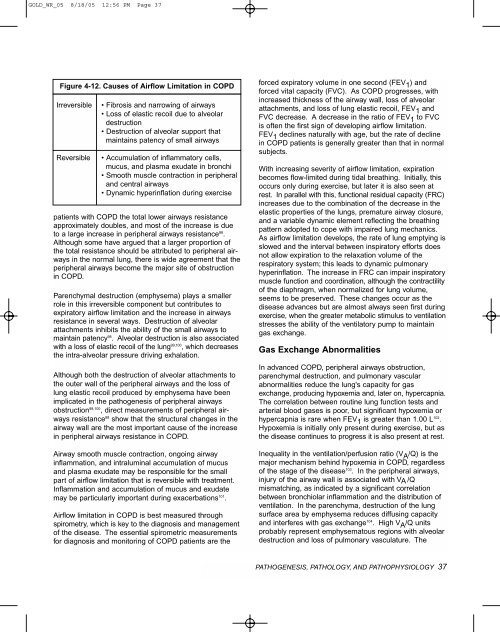
Figure 4-12. Causes of Airflow Limitation in COPD<br />
Irreversible<br />
Reversible<br />
• Fibrosis and narrowing of airways<br />
• Loss of elastic recoil due to alveolar<br />
destruction<br />
• Destruction of alveolar support that<br />
maintains patency of small airways<br />
• Accumulation of inflammatory cells,<br />
mucus, and plasma exudate in bronchi<br />
• Smooth muscle contraction in peripheral<br />
and central airways<br />
• Dynamic hyperinflation during exercise<br />
patients with COPD the total lower airways resistance<br />
approximately doubles, and most of the increase is due<br />
to a large increase in peripheral airways resistance 88 .<br />
Although some have argued that a larger proportion of<br />
the total resistance should be attributed to peripheral airways<br />
in the normal lung, there is wide agreement that the<br />
peripheral airways become the major site of obstruction<br />
in COPD.<br />
Parenchymal destruction (emphysema) plays a smaller<br />
role in this irreversible component but contributes to<br />
expiratory airflow limitation and the increase in airways<br />
resistance in several ways. Destruction of alveolar<br />
attachments inhibits the ability of the small airways to<br />
maintain patency 98 . Alveolar destruction is also associated<br />
with a loss of elastic recoil of the lung 99,100 , which decreases<br />
the intra-alveolar pressure driving exhalation.<br />
Although both the destruction of alveolar attachments to<br />
the outer wall of the peripheral airways and the loss of<br />
lung elastic recoil produced by emphysema have been<br />
implicated in the pathogenesis of peripheral airways<br />
obstruction 98,100 , direct measurements of peripheral airways<br />
resistance 88 show that the structural changes in the<br />
airway wall are the most important cause of the increase<br />
in peripheral airways resistance in COPD.<br />
Airway smooth muscle contraction, ongoing airway<br />
inflammation, and intraluminal accumulation of mucus<br />
and plasma exudate may be responsible <strong>for</strong> the small<br />
part of airflow limitation that is reversible with treatment.<br />
Inflammation and accumulation of mucus and exudate<br />
may be particularly important during exacerbations 101 .<br />
Airflow limitation in COPD is best measured through<br />
spirometry, which is key to the diagnosis and management<br />
of the disease. The essential spirometric measurements<br />
<strong>for</strong> diagnosis and monitoring of COPD patients are the<br />
<strong>for</strong>ced expiratory volume in one second (FEV 1 ) and<br />
<strong>for</strong>ced vital capacity (FVC). As COPD progresses, with<br />
increased thickness of the airway wall, loss of alveolar<br />
attachments, and loss of lung elastic recoil, FEV 1 and<br />
FVC decrease. A decrease in the ratio of FEV 1 to FVC<br />
is often the first sign of developing airflow limitation.<br />
FEV 1 declines naturally with age, but the rate of decline<br />
in COPD patients is generally greater than that in normal<br />
subjects.<br />
With increasing severity of airflow limitation, expiration<br />
becomes flow-limited during tidal breathing. Initially, this<br />
occurs only during exercise, but later it is also seen at<br />
rest. In parallel with this, functional residual capacity (FRC)<br />
increases due to the combination of the decrease in the<br />
elastic properties of the lungs, premature airway closure,<br />
and a variable dynamic element reflecting the breathing<br />
pattern adopted to cope with impaired lung mechanics.<br />
As airflow limitation develops, the rate of lung emptying is<br />
slowed and the interval between inspiratory ef<strong>for</strong>ts does<br />
not allow expiration to the relaxation volume of the<br />
respiratory system; this leads to dynamic pulmonary<br />
hyperinflation. The increase in FRC can impair inspiratory<br />
muscle function and coordination, although the contractility<br />
of the diaphragm, when normalized <strong>for</strong> lung volume,<br />
seems to be preserved. These changes occur as the<br />
disease advances but are almost always seen first during<br />
exercise, when the greater metabolic stimulus to ventilation<br />
stresses the ability of the ventilatory pump to maintain<br />
gas exchange.<br />
Gas Exchange Abnormalities<br />
In advanced COPD, peripheral airways obstruction,<br />
parenchymal destruction, and pulmonary vascular<br />
abnormalities reduce the lung's capacity <strong>for</strong> gas<br />
exchange, producing hypoxemia and, later on, hypercapnia.<br />
The correlation between routine lung function tests and<br />
arterial blood gases is poor, but significant hypoxemia or<br />
hypercapnia is rare when FEV 1 is greater than 1.00 L 102 .<br />
Hypoxemia is initially only present during exercise, but as<br />
the disease continues to progress it is also present at rest.<br />
Inequality in the ventilation/perfusion ratio (V A /Q) is the<br />
major mechanism behind hypoxemia in COPD, regardless<br />
of the stage of the disease 103 . In the peripheral airways,<br />
injury of the airway wall is associated with VA /Q<br />
mismatching, as indicated by a significant correlation<br />
between bronchiolar inflammation and the distribution of<br />
ventilation. In the parenchyma, destruction of the lung<br />
surface area by emphysema reduces diffusing capacity<br />
and interferes with gas exchange 104 . High V A /Q units<br />
probably represent emphysematous regions with alveolar<br />
destruction and loss of pulmonary vasculature. The<br />
PATHOGENESIS, PATHOLOGY, AND PATHOPHYSIOLOGY 37
![Di Bari [NO].pdf - GOLD](https://img.yumpu.com/21544924/1/190x143/di-bari-nopdf-gold.jpg?quality=85)