Amino acid transmitters in the mammalian central nervous system
Amino acid transmitters in the mammalian central nervous system
Amino acid transmitters in the mammalian central nervous system
You also want an ePaper? Increase the reach of your titles
YUMPU automatically turns print PDFs into web optimized ePapers that Google loves.
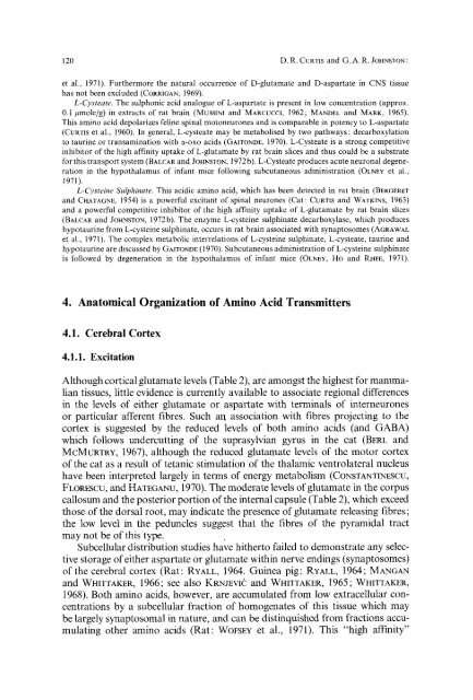
120 D.R. CURTlS and G. A. R. JOHNSTON :<br />
et al., 1971). Fur<strong>the</strong>rmore <strong>the</strong> natural occurrence of D-glutamate and D-aspartate <strong>in</strong> CNS tissue<br />
has not been excluded (CORRIGAN, 1969).<br />
L-Cysteate. The sulphonic <strong>acid</strong> analogue of L:aspartate is present <strong>in</strong> low concentration (approx.<br />
0.1 /~mole/g) <strong>in</strong> extracts of rat bra<strong>in</strong> (MussINI and MARCU¢¢k 1962; MANDEL and MARK, 1965).<br />
This am<strong>in</strong>o <strong>acid</strong> depolarizes fel<strong>in</strong>e sp<strong>in</strong>al motoneurones and is comparable <strong>in</strong> potency to L-aspartate<br />
(CURTIS et al., 1960). In general, L-cysteate may be metabolised by two pathways: decarboxylation<br />
to taur<strong>in</strong>e or transam<strong>in</strong>ation with a-oxo <strong>acid</strong>s (GAITONDE, 1970). L-Cysteate is a strong competitive<br />
<strong>in</strong>hibitor of <strong>the</strong> high aff<strong>in</strong>ity uptake of L-glutamate by rat bra<strong>in</strong> slices and thus could be a substrate<br />
for this transport <strong>system</strong> (BALCAR and JOHNSTON, 1972 b). L-Cysteate produces acute neuronal degeneration<br />
<strong>in</strong> <strong>the</strong> hypothalamus of <strong>in</strong>fant mice follow<strong>in</strong>g subcutaneous adm<strong>in</strong>istration (OLNEY et al.,<br />
1971).<br />
L-Cyste<strong>in</strong>e Sulph<strong>in</strong>ate. This <strong>acid</strong>ic am<strong>in</strong>o <strong>acid</strong>, which has been detected <strong>in</strong> rat bra<strong>in</strong> (BERGERET<br />
and CHAXAGNE, 1954)is a powerful excitant of sp<strong>in</strong>al neurones (Cat: CURTIS and WATKINS, 1963)<br />
and a powerful competitive <strong>in</strong>hibitor of <strong>the</strong> high aff<strong>in</strong>ity uptake of L-glutamate by rat bra<strong>in</strong> slices<br />
(BALeAR and JOHNSTON, 1972b). The enzyme L-cyste<strong>in</strong>e sulph<strong>in</strong>ate decarboxylase, which produces<br />
hypotaur<strong>in</strong>e from L-cyste<strong>in</strong>e sulph<strong>in</strong>ate, occurs <strong>in</strong> rat bra<strong>in</strong> associated with synaptosomes (AGRAWAL<br />
et al., 1971). The complex metabolic <strong>in</strong>terrelations of L-cyste<strong>in</strong>e sulph<strong>in</strong>ate, L-cysteate, taur<strong>in</strong>e and<br />
hypotaur<strong>in</strong>e are discussed by GAITONDE (1970). Subcutaneous adm<strong>in</strong>istration of L-cyste<strong>in</strong>e sulph<strong>in</strong>ate<br />
is followed by degeneration <strong>in</strong> <strong>the</strong> hypothalamus of <strong>in</strong>fant mice (OLN~Y, HO and RHEE, 1971).<br />
4. Anatomical Organization of <strong>Am<strong>in</strong>o</strong> Acid Transmitters<br />
4.1. Cerebral Cortex<br />
4.1.1. Excitation<br />
Although cortical glutamate levels (Table 2), are amongst <strong>the</strong> highest for <strong>mammalian</strong><br />
tissues, little evidence is currently available to associate regional differences<br />
<strong>in</strong> <strong>the</strong> levels of ei<strong>the</strong>r glutamate or aspartate with term<strong>in</strong>als of <strong>in</strong>terneurones<br />
or particular afferent fibres. Such arl association with fibres project<strong>in</strong>g to <strong>the</strong><br />
cortex is suggested bY <strong>the</strong> reduced levels of both am<strong>in</strong>o <strong>acid</strong>s (and GABA)<br />
which follows undercutt<strong>in</strong>g of <strong>the</strong> suprasylvian gyrus <strong>in</strong> <strong>the</strong> cat (BERL and<br />
MCMVRTRY, 1967), although <strong>the</strong> reduced glutamate levels of <strong>the</strong> motor cortex<br />
of <strong>the</strong> cat as a result of tetanic stimulation of <strong>the</strong> thalamic ventrolateral nucleus<br />
have been <strong>in</strong>terpreted largely <strong>in</strong> terms of energy metabolism (CONSTANTINESCU,<br />
FLOR~SCU, and HATEGANU, 1970). The moderate levels of glutamate <strong>in</strong> <strong>the</strong> corpus<br />
callosum and <strong>the</strong> posterior portion of <strong>the</strong> <strong>in</strong>ternal capsule (Table 2), which exceed<br />
those of <strong>the</strong> dorsal root, may <strong>in</strong>dicate <strong>the</strong> presence of glutamate releas<strong>in</strong>g fibres;<br />
<strong>the</strong> low level <strong>in</strong> <strong>the</strong> peduncles suggest that <strong>the</strong> fibres of <strong>the</strong> pyramidal tract<br />
may not be of this type.<br />
Subcellular distribution studies have hi<strong>the</strong>rto failed to demonstrate any selective<br />
storage of ei<strong>the</strong>r aspartate or glutamate with<strong>in</strong> nerve end<strong>in</strong>gs (synaptosomes)<br />
of <strong>the</strong> cerebral cortex (Rat: RYALL, 1964. Gu<strong>in</strong>ea pig: RYALL, 1964 ; MANGAN<br />
and WHITTAKER, 1966; see also KRNJEVIC and WHITTAKER, 1965; WHITTAKER,<br />
1968). Both am<strong>in</strong>o <strong>acid</strong>s, however, are accumulated from low extracellular concentrations<br />
by a subcellular fraction of homogenates of this tissue which may<br />
be largely synaptosomal <strong>in</strong> nature, and can be dist<strong>in</strong>quished from fractions accumulat<strong>in</strong>g<br />
o<strong>the</strong>r am<strong>in</strong>o <strong>acid</strong>s (Rat: WOFSEY et al., 1971). This "high aff<strong>in</strong>ity"


