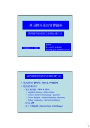
Components of Central Nervous System (CNS)
Components of Central Nervous System (CNS)
Components of Central Nervous System (CNS)

Create successful ePaper yourself
Turn your PDF publications into a flip-book with our unique Google optimized e-Paper software.
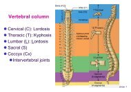
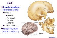
<strong>Components</strong> <strong>of</strong> <strong>Central</strong> <strong>Nervous</strong> <strong>System</strong> (<strong>CNS</strong>)<br />
Neurons, Glia: gray matter, white matter<br />
Neuropil (nerve fibers + glia processes)<br />
Synapse<br />
Glia (Neuroglia)<br />
Astrocyte (Astroglia)<br />
Microglia<br />
Oligodendrocyte (Oligodendroglia)<br />
Ependyma (choroid plexus epithelium)<br />
Histo: <strong>CNS</strong> - 1
<strong>Components</strong>: Neurons, Glia and Neuropil<br />
Gray matter: Neurons (+ processes), Glia<br />
White matter: Nerve fibers (processes), Glia<br />
Neuropil: nerve processes + glia processes<br />
Neuropil<br />
Histo: <strong>CNS</strong> - 2
Synapses<br />
Functional classifications<br />
Chemical synapse<br />
Neurotransmitters,<br />
Neuropeptides<br />
Electronic synapse: gap<br />
junction (nexus), connexin<br />
Morphological classification (EM)<br />
Histo: <strong>CNS</strong> - 3
Chemical Synapses<br />
Ultrastructures<br />
Presynaptic element: synaptic<br />
vesicles<br />
Synaptic cleft<br />
Postsynaptic element: clustering <strong>of</strong><br />
receptors/channels<br />
Neurotransmitters<br />
acetylcholine (ACh), noradrenaline<br />
(NE), gamma-aminobutyric acid<br />
(GABA), serotonin (5-HT),<br />
dopamine (DA), glycine, glutamate,<br />
aspartate et al.<br />
Neuropeptides (peptidergic):<br />
calcitonin gene-related peptide<br />
(CGRP), et al.<br />
Histo: <strong>CNS</strong> - 4
Structural classification <strong>of</strong><br />
Synapses<br />
axosomatic synapse<br />
axodendritic synapse<br />
axoaxonic synapse<br />
Axosomatic Axodendritic Axoaxonic<br />
Histo: <strong>CNS</strong> - 5
Neuroglia<br />
Microglia (macrophage)<br />
Histo: <strong>CNS</strong> - 6
Neuroglia<br />
Non-nervous elements;<br />
more abundant than<br />
neurons<br />
protective, supportive,<br />
nutritive<br />
Exist in the extraneuronal<br />
space<br />
as ionic sink<br />
little true tissue space, no<br />
lymphatics<br />
Proliferate in <strong>CNS</strong> injury<br />
Histo: <strong>CNS</strong> - 7
Types <strong>of</strong> neuroglia<br />
Astrocytes (Astroglia)<br />
Protoplasmic, Fibrous<br />
Oligodendrocytes<br />
(Oligodendroglia)<br />
Microglia (Mesoglia)<br />
Ependyma cells, Choroid<br />
epithelium<br />
Histo: <strong>CNS</strong> - 8
Astrocytes<br />
Perivascular feet (Foot<br />
plates)<br />
“glia limitans” (LM) on<br />
brain surface<br />
subpia foot <strong>of</strong><br />
astrocytes<br />
basal lamina <strong>of</strong> pia<br />
matter (EM)<br />
cytoplasm: glial fibrillary<br />
acidic protein (GFAP)<br />
?Functions: barriers,<br />
metabolism<br />
Histo: <strong>CNS</strong> - 9
Protoplasmic astrocytes<br />
in gray matter<br />
Stellate form with<br />
multiple radiating<br />
processes<br />
A larger and paler<br />
nucleus than other glial<br />
cells<br />
Processes ending as<br />
vascular feet/pedicles<br />
(perivascular feet) and<br />
perineural feet<br />
Histo: <strong>CNS</strong> - 10
Protoplasmic astrocyte<br />
Blood vessel<br />
Cajal method Silver<br />
impregnation<br />
GFAP<br />
Histo: <strong>CNS</strong> - 11
Fibrous astrocytes<br />
in white matter, and<br />
periventricular gray<br />
matter<br />
an ovoid euchromatic<br />
nucleus with<br />
pale-staining<br />
cytoplasm and long,<br />
thin processes<br />
Histo: <strong>CNS</strong> - 12
Fibrous astrocyte<br />
Immunocytochemistry GFAP<br />
Silver-gold impregnation<br />
Histo: <strong>CNS</strong> - 13
Microglia<br />
Mononuclear<br />
phagocytic cells<br />
A dense oval or<br />
elongate nucleus<br />
with scant<br />
cytoplasm<br />
Tortuous processes<br />
with spines<br />
Upon injury:<br />
proliferate, loose<br />
spines, become<br />
swollen (“amoeboid”)<br />
Histo: <strong>CNS</strong> - 14
Microglia<br />
Microgliosis<br />
Silver impregnation<br />
Immunohistochemistry: MHCII (major<br />
histocompatibility complex II) for<br />
activated microglia<br />
Histo: <strong>CNS</strong> - 15
Oligodendrocytes<br />
Form myelin sheath in <strong>CNS</strong><br />
Smaller size than astrocytes<br />
Small irregularly shaped<br />
deeply stained nucleus<br />
Various classifications and<br />
Types<br />
Perineural<br />
oligodendrocytes<br />
Perifascicular<br />
oligodendrocytes<br />
Histo: <strong>CNS</strong> - 16
Perineuronal<br />
oligodendrocytes<br />
oligo<br />
Mainly in gray matter<br />
Closely associated with<br />
soma <strong>of</strong> neurons<br />
? Functions: metabolic<br />
interdependency<br />
Histo: <strong>CNS</strong> - 17
Perifascicular<br />
oligodendrocytes<br />
Mainly in white<br />
matter<br />
arranged in rows<br />
between bundles <strong>of</strong><br />
axons<br />
Analogous to<br />
Schwann cells in<br />
PNS<br />
forming myelin<br />
sheaths <strong>of</strong> several<br />
parallel axons, up to<br />
50 or more<br />
Histo: <strong>CNS</strong> - 18
Oligodendrocytes<br />
Perineuronal<br />
oligodendrocytes<br />
Perifascicular<br />
oligodendrocytes<br />
19<br />
Histo: <strong>CNS</strong> - 19
Different patterns <strong>of</strong> myelination between <strong>CNS</strong> and PNS: 1/3<br />
1. Surface <strong>of</strong> the node: contacted by astrocyte processes in<br />
<strong>CNS</strong>; covered by Schwann cell processes in PNS.<br />
2. Cytoplasm <strong>of</strong> myelinating cells near the node <strong>of</strong> Ranvier: (-)<br />
for Oligo; (+) for Schwann cells.<br />
Histo: <strong>CNS</strong> - 20
Different patterns <strong>of</strong> myelination between <strong>CNS</strong> and PNS: 2/3<br />
3. One Oligo for several axons; One Schwann for a single axon<br />
4. Distance between cell body and myelin sheath: Oligo ><br />
Schwann<br />
Histo: <strong>CNS</strong> - 21
Different patterns <strong>of</strong> myelination between <strong>CNS</strong> and PNS: 3/3<br />
5. Basal lamina associated with myelin sheath: (-) in <strong>CNS</strong>,<br />
(+) in PNS<br />
6. Supporting connective tissue for myelinated axons: (-) in<br />
<strong>CNS</strong>, (+) in PNS<br />
Histo: <strong>CNS</strong> - 22
Demyelinating diseases<br />
Myelin proteins<br />
only in PNS: peripheral myelin protein 22 (PMP22) et al<br />
only in <strong>CNS</strong>: proteolipid protein (PLP) et al<br />
in PNS and <strong>CNS</strong>: myelin-associated glycoprotein (MAG)<br />
et al<br />
Demyelinating diseases<br />
PNS: Guillain-Barré syndrome (acute inflammatory<br />
demyelinating polyradiculopathy) 急 性 神 經 根 炎<br />
<strong>CNS</strong>: multiple sclerosis 多 發 性 硬 化 症<br />
Histo: <strong>CNS</strong> - 23
Guillain-Barré syndrome (GBS)<br />
Progressive<br />
weakness<br />
Day 1<br />
Lower limbs<br />
Day 3<br />
Upper limbs<br />
Day 7<br />
Respiratory failure<br />
Histo: <strong>CNS</strong> - 24
Ependymal cells<br />
Lining cells <strong>of</strong> ventricular system<br />
Ventricles<br />
Aqueduct<br />
<strong>Central</strong> canal<br />
Choroid plexus: continuous with<br />
ependyma cells <strong>of</strong> ventricles<br />
Histo: <strong>CNS</strong> - 25
Ependymal cells<br />
Histo: <strong>CNS</strong> - 26
Ependymal cells<br />
C: cilia<br />
M:microvilli<br />
JC: junctional complex<br />
BB: basal body<br />
G: Golgi apparatus<br />
Histo: <strong>CNS</strong> - 27
Choroid plexus<br />
Ependyma layer + vascular core<br />
(vessels + loose connective tissues)<br />
Histo: <strong>CNS</strong> - 28
Blood-Brain Barrier (BBB)<br />
Nonfenestrated endothelial<br />
cells (tight junctions)<br />
thick basal lamina with<br />
outer surfaces enclosed by<br />
Astrocytes’ foot processes<br />
(perivascular feet, end feet)<br />
Histo: <strong>CNS</strong> - 29
Blood-Brain Barrier<br />
Capillary <strong>of</strong> continuous type; Thick<br />
basal lamina; Perivascular feet<br />
Functions: impermeable to foreign<br />
substances for preventing<br />
Autoimmune damage<br />
Blood vessel<br />
Histo: <strong>CNS</strong> - 30
Meninges<br />
Dural mater<br />
(pachymeninx)<br />
periosteum <strong>of</strong><br />
skull<br />
subdural space<br />
Pia-arachnoid<br />
(leptomenginges)<br />
Arachnoid mater<br />
subarachnoid<br />
space / cistern<br />
Intact vessels<br />
Pia mater<br />
Histo: <strong>CNS</strong> - 31
Meninges<br />
Histo: <strong>CNS</strong> - 32
Pia-Arachnoid<br />
Histo: <strong>CNS</strong> - 33
Organization <strong>of</strong> <strong>CNS</strong><br />
Cerebellum: 3 layers<br />
Cerebral cortex (Neocortex): 6<br />
layers (cytoarchitecture:<br />
Broadmann area)<br />
Histo: <strong>CNS</strong> - 34
Mol: molecular layer<br />
Pkj: Purkinje cell layer<br />
Gr: granular layer<br />
Cerebellum<br />
Cerebellar cortex<br />
Histo: <strong>CNS</strong> - 35
Pyramidal cells (neurons)
Cerebral cortex<br />
(Neocortex): 6 layers<br />
1. Plexiform (molecular) layer<br />
2. Outer granular layer<br />
3. Pyramidal cell layer<br />
4. Inner granular layer<br />
5. Ganglionic layer<br />
6. Multiform cell layer<br />
White matter<br />
Histo: <strong>CNS</strong> - 37
Response <strong>of</strong> a nerve to injury<br />
Histo: <strong>CNS</strong> - 38
Nerve Degeneration and Regeneration<br />
Histo: <strong>CNS</strong> - 39
Nerve injury: PNS vs. <strong>CNS</strong><br />
Histo: <strong>CNS</strong> - 40
Nerve Degeneration and Regeneration<br />
Wallerian degeneration: disintegration <strong>of</strong> axonal<br />
cytoskeleton distal to the site <strong>of</strong> injury<br />
Cell body: chromatolysis (loss <strong>of</strong> Nissl substance)<br />
Responses<br />
PNS: proliferation <strong>of</strong> Schwann cells; migration <strong>of</strong><br />
macrophage<br />
<strong>CNS</strong>: proliferation <strong>of</strong> astrocytes<br />
Regeneration<br />
PNS: as a rule<br />
<strong>CNS</strong>: negligible<br />
inhibitory molecules, glial scar et al.<br />
Histo: <strong>CNS</strong> - 41
Review <strong>of</strong> <strong>CNS</strong> Histology<br />
<strong>Components</strong> <strong>of</strong> <strong>CNS</strong>: neurons, glia<br />
Neurons, Glia: gray matter, white matter<br />
Neuropil (nerve fibers + glia processes)<br />
Synapse<br />
Glia (Neuroglia)<br />
Meninges (Menix)<br />
Blood-Brain Barrier<br />
Organization <strong>of</strong> the <strong>CNS</strong>: layers<br />
Responses to nerve injury<br />
Histo: <strong>CNS</strong> - 42