Skull Cranial skeleton (Neurocranium) Calarvia Frontal, Temporal ...
Skull Cranial skeleton (Neurocranium) Calarvia Frontal, Temporal ...
Skull Cranial skeleton (Neurocranium) Calarvia Frontal, Temporal ...

Create successful ePaper yourself
Turn your PDF publications into a flip-book with our unique Google optimized e-Paper software.
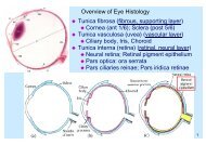
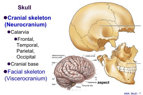
<strong>Skull</strong><br />
•<strong>Cranial</strong> <strong>skeleton</strong><br />
(<strong>Neurocranium</strong>)<br />
•<strong>Calarvia</strong><br />
•<strong>Frontal</strong>,<br />
<strong>Temporal</strong>,<br />
Parietal,<br />
Occipital<br />
•<strong>Cranial</strong> base<br />
•Facial <strong>skeleton</strong><br />
(Viscerocranium)<br />
ANA: <strong>Skull</strong> - 1
<strong>Neurocranium</strong>: cranial vault<br />
• <strong>Frontal</strong>, Parietal, <strong>Temporal</strong><br />
• Mainly membranous bone formation<br />
ANA: <strong>Skull</strong> - 2
<strong>Neurocranium</strong>:<br />
cranial base<br />
•Midline<br />
•Ethmoid<br />
•Sphenoid<br />
•Occipital<br />
•Bilateral<br />
•<strong>Temporal</strong><br />
ANA: <strong>Skull</strong> - 3
ANA: <strong>Skull</strong> - 4<br />
Viscerocranium:<br />
anterior view<br />
•Viscerocranium<br />
•Ethmoid,<br />
Vomer,<br />
Mandible<br />
•Maxilla, Zygoma,<br />
Nasal, Lacrimal,<br />
Inferior nasal<br />
chonae, Palatine
Viscerocranium: inferior view<br />
• Palatine<br />
• Maxilla<br />
• Zygoma<br />
ANA: <strong>Skull</strong> - 5
Sutures and Fontanelles<br />
• Coronal suture<br />
• Sagittal suture<br />
• Lambdoid suture<br />
• Metopic suture<br />
coronal<br />
ANA: <strong>Skull</strong> - 6
<strong>Skull</strong>: posterior<br />
view<br />
• external occipital<br />
protuberance<br />
(inion)<br />
• external occipital<br />
crest<br />
• superior nuchal<br />
line<br />
• inferior nuchal line<br />
ANA: <strong>Skull</strong> - 7
Superior nuchal line<br />
• Attachment of back muscles; e.g. Splenius capitis from spinous<br />
process of C7/T1-3 to superior nuchal line; draw head backwards<br />
ANA: <strong>Skull</strong> - 8<br />
Superior nuchal line<br />
Splenius capitis
ANA: <strong>Skull</strong> - 9<br />
<strong>Skull</strong>: lateral view<br />
• Frankfurt plane<br />
(anatomical position,<br />
OrbitoMeatal line):<br />
upper margin of ext.<br />
acoustic meatus - orbit<br />
floor horizontal<br />
• superior temporal line;<br />
inferior temporal line<br />
• external acoustic<br />
meatus; mastoid<br />
process<br />
• level of ant., mid., post.<br />
cranial fossae
ANA: <strong>Skull</strong> - 10<br />
OrbitoMeatal line (OM line) in<br />
radiology<br />
• from lateral canthus to<br />
external acoustic meatus
ANA: <strong>Skull</strong> - 11<br />
Pterion
ANA: <strong>Skull</strong> - 12<br />
Superior temporal line and temporalis<br />
muscle
<strong>Skull</strong> from front<br />
• Supercillary arch<br />
• Zygomatic proc.<br />
of frontal bone<br />
• Glabella<br />
• Zygomatic bone<br />
• <strong>Frontal</strong> proc. of<br />
maxilla<br />
• <strong>Frontal</strong> proc. of<br />
zygoma<br />
ANA: <strong>Skull</strong> - 13
Internal surface of the skull: The roof (vault)<br />
• sagittal fissure<br />
• coronal fissure<br />
• lamboid fissure<br />
• grooves for<br />
middle<br />
meningeal vein,<br />
artery<br />
ANA: <strong>Skull</strong> - 14
ANA: <strong>Skull</strong> - 15<br />
Grooves for middle mengigeal arteries<br />
Foramen<br />
spinosum
Sagittal fissure: superior sagittal sinus<br />
• sup. sagittal sinus (SSS)<br />
• depressions for arachnoid granulation: along SSS<br />
ANA: <strong>Skull</strong> - 16
ANA: <strong>Skull</strong> - 17<br />
Sagittal fissure: falx cerebri<br />
• Dura extending from skull
ANA: <strong>Skull</strong> - 18<br />
Dural sinuses in Posterior cranial fossa<br />
• groove for transverse sinus; confluence of the sinus - internal<br />
occipital protuberance<br />
• tentorium cerebelli: separating occipital lobe from cerebellum<br />
• internal occipital crest [- falx cerebelli]
Inner surface of Anterior cranial fossa<br />
• <strong>Frontal</strong> bone: Orbital plate: thin except near superciliary arch; frontal<br />
air sinus<br />
• Lesser wing of sphenoid: ant. clinoid processes<br />
• Ethmoid bone: crista galli (attachment of falx cerebri)<br />
• foramen caecum; cribriform plate<br />
ANA: <strong>Skull</strong> - 19
ANA: <strong>Skull</strong> - 20<br />
Inner surface of Middle cranial fossa (1/2)<br />
• <strong>Temporal</strong> bone: petrous part: thick, contains inner ear<br />
• hypophyseal fossa = sella turcica (Turk‘s saddle); tuberculum sellae,<br />
dorsum sellae<br />
• ant. clinoid process (clinoid in Latin: bed-side); post. clinoid<br />
process; diaphragma sellae<br />
Anterior clinoid process
ANA: <strong>Skull</strong> - 21<br />
Sella turcica<br />
• Tuberculum sella<br />
• Hypohyseal fossa:<br />
pituitary gland<br />
• Dorsum sella
ANA: <strong>Skull</strong> - 22<br />
Sella turcica in<br />
sphenoid bone
Inner surface of Middle cranial fossa (2/2)<br />
Superior orbital fissure (V1), foramen Rotudum (V2), foramen Ovale (V3)<br />
foramen spinosum (mid menigeal art)<br />
ANA: <strong>Skull</strong> - 23
Inner surface of Posterior fossa<br />
• petrous portion of temporal (inner ear)<br />
• int. acoustic meatus<br />
• to and from neck: foramen magnum; jugular foramen; hypoglossal canal<br />
ANA: <strong>Skull</strong> - 24
ANA: <strong>Skull</strong> - 25<br />
Inner surface of Occipital bone, <strong>Temporal</strong> bone<br />
• Occipital bone: Basilar part, [clivus]; Lateral parts;<br />
Squamous part (squama occipitalis)<br />
• <strong>Temporal</strong> bone: Squamous part; Petrous part; (Mastoid<br />
part; Sytloid process)
ANA: <strong>Skull</strong> - 26<br />
Floor (Outer surface) of middle cranial fossa-1<br />
• foramen ovale (V3); foramen spinosum: (spine of<br />
sphenoid bone close to foramen) for middle meningeal a.<br />
• foramen lacerum: cartilage (internal carotid artery)
ANA: <strong>Skull</strong> - 27<br />
Floor (Outer surface) of middle cranial fossa-2<br />
• mandibular fossa, articular tubercle
Outer surface of<br />
Post. cranial fossa<br />
• sphenoid and<br />
occipital fused in the<br />
midline<br />
• opening of carotid<br />
canal (ICA)<br />
• stylomastoid<br />
foramen (CN VII)<br />
• pharyngeal tubercle<br />
ANA: <strong>Skull</strong> - 28
carotid canal for<br />
internal carotid artery<br />
http://dc311.4shared.com/doc/WfGJhex5/preview.html<br />
ANA: <strong>Skull</strong> - 29
internal carotid artery (ICA)<br />
http://dermatologic.com.ar/7.htm<br />
ANA: <strong>Skull</strong> - 30
ANA: <strong>Skull</strong> - 31<br />
Outer surface of<br />
Post. cranial fossa<br />
• jugular foramen<br />
• occipital condyle<br />
• hypoglossal canal<br />
(medial opening hidden<br />
under condyle)<br />
• styloid process:<br />
stylohyoid lig.<br />
• mastoid process: air<br />
cell middle ear<br />
•sternocleidomastoid<br />
muscle attaches
ANA: <strong>Skull</strong> - 32<br />
Floor of posterior cranial fossa-1<br />
• inferior nuchal line
Floor of posterior cranial fossa-2<br />
• mastoid notch = groove medial to mastoid process: origin<br />
of digastric muscle (digastric groove)<br />
• sulcus for occipital a.: medial to digastric groove<br />
ANA: <strong>Skull</strong> - 33<br />
Mastoid notch
Mastoid notch and digastric muscle<br />
ANA: <strong>Skull</strong> - 34<br />
• mastoid notch and digastric groove<br />
(temporal bone)<br />
• greater horn (hypoid bone)<br />
• symphysis menti (mandible bone)<br />
• elevates hypoid bone
<strong>Skull</strong> (cranial <strong>skeleton</strong>): review<br />
• <strong>Neurocranium</strong><br />
•External surface<br />
•Interior (cranial fossa)<br />
•Anterior<br />
•Middle<br />
•Posterior<br />
• Openings and contents<br />
through them<br />
ANA: <strong>Skull</strong> - 35
ANA: <strong>Skull</strong> - 36<br />
Facial <strong>skeleton</strong><br />
•<strong>Cranial</strong> <strong>skeleton</strong> (<strong>Neurocranium</strong>)<br />
•Facial <strong>skeleton</strong><br />
(Viscerocranium)<br />
•Ear<br />
•Orbit<br />
•Nasal cavity / Nasopharynx<br />
•Oral cavity / Palate and Jaw<br />
•Mandible<br />
•Bones<br />
•<strong>Temporal</strong> bone<br />
•Sphenoid bone
ANA: <strong>Skull</strong> - 37<br />
Ear • External ear<br />
•External auditory meatus<br />
• Middle ear<br />
•Mastoid antrum<br />
•Pharyngotympanic tube<br />
•Auditroy ossicles<br />
• Inner ear (petrous part)
external auditory meatus<br />
ANA: <strong>Skull</strong> - 38
3 ossicles<br />
• malleus (handle<br />
on tympanic<br />
membrane)<br />
• Incus<br />
• stapes (rest on<br />
fenestra vestibuli)<br />
ANA: <strong>Skull</strong> - 39
Inner ear: petrous part of temporal bone<br />
• bony labyrinth<br />
•Cochlea<br />
•Vestibula<br />
• internal accoustic meatus:<br />
vestibulocochlear nerve (8 th ) and facial<br />
nerve (7 th )<br />
ANA: <strong>Skull</strong> - 40
ANA: <strong>Skull</strong> - 41<br />
Facial canal: internal acoustic meatus;<br />
stylomastoid foramen
<strong>Temporal</strong> bone<br />
• External surface<br />
• Squamous part<br />
• Zygomatic process<br />
• Mastoid part<br />
• Styloid process<br />
• Internal surface<br />
• Petrous part<br />
ANA: <strong>Skull</strong> - 42
ANA: <strong>Skull</strong> - 43<br />
Bony orbits: roof<br />
• superciliary arch of frontal bone: thickened<br />
• supraorbital notch (foramen): supraorbital n. (V 1<br />
), vessels<br />
• floor of ant. cranial fossa: thin<br />
• lesser wing of sphenoid<br />
• Lacrimal gland<br />
Lesser wing,<br />
Sphenoid
ANA: <strong>Skull</strong> - 44<br />
Sphenoid bone<br />
• Greater wing<br />
• Lesser wing<br />
• Sphenoid body<br />
• Medial pterygoid plate<br />
• Lateral pterygoid plate<br />
• Superior orbital fissure
Bony orbits: lateral wall-1<br />
• frontal proc. of<br />
zygomatic bone<br />
• zygomatic proc. of<br />
frontal bone<br />
ANA: <strong>Skull</strong> - 45
Zygomatic bone<br />
• <strong>Frontal</strong> process<br />
• Maxillary process<br />
• <strong>Temporal</strong> process<br />
F<br />
T<br />
M<br />
ANA: <strong>Skull</strong> - 46
Bony orbits: lateral<br />
wall-2<br />
• Greater wing of<br />
sphenoid<br />
• superior orbital<br />
fissure (where roof<br />
and lateral wall meet)<br />
• inferior orbital<br />
fissure (where lateral<br />
wall and floor meet)<br />
ANA: <strong>Skull</strong> - 47
Bony orbits: floor-1<br />
• thin orbital floor<br />
• maxillary process of zygomatic bone<br />
•zygomaticofacial foramen on malar surface<br />
ANA: <strong>Skull</strong> - 48<br />
Maxillary proc.,<br />
Zygoma
Bony orbits: floor-2<br />
• Maxilla<br />
•maxillary sinus<br />
ANA: <strong>Skull</strong> - 49
ANA: <strong>Skull</strong> - 50<br />
Bony orbits: floor-3<br />
• infraorbital n. : enters<br />
from pterygopalatine<br />
fossa through inf.<br />
orbital fissure <br />
infraorbital groove <br />
infraorbital canal <br />
infraorbital foramen<br />
Infraorbital<br />
foramen
Bony orbits:<br />
medial wall-1<br />
• frontal process of maxilla<br />
• frontal bone<br />
• lacrimal bone<br />
• fossa for lacrimal sac <br />
nasolacrimal canal <br />
inferior nasal meatus<br />
• orbital plate of ethmoid:<br />
(ethmoidal air cells medial<br />
to this)<br />
lateral<br />
<strong>Frontal</strong> bone<br />
<strong>Frontal</strong><br />
process<br />
of<br />
maxilla<br />
medial<br />
ANA: <strong>Skull</strong> - 51
Bony orbits:<br />
medial wall-2<br />
• orbital plate of<br />
palatine bone<br />
• body of sphenoid:<br />
completes the lower<br />
part of optic canal<br />
• optic canal (where<br />
roof and medial wall<br />
meet)<br />
Body of<br />
sphenoid<br />
ANA: <strong>Skull</strong> - 52
Bony orbits:<br />
medial wall-3<br />
•ant. & post. ethmoidal<br />
foramina<br />
•transmitting<br />
corresponding vessels<br />
(br. of ophthalmic a.) to<br />
supply nasal cavity<br />
and ethmoidal air cells<br />
<strong>Frontal</strong> bone<br />
<strong>Frontal</strong><br />
process<br />
of<br />
maxilla<br />
ANA: <strong>Skull</strong> - 53
Openings of orbit<br />
• 1. optic foramen (canal)<br />
• 2. sup. orbital fissure<br />
• 3. inf. orbital fissure<br />
• 4. nasolacrimal canal<br />
• 5. ant. & post.<br />
ethmoidal foramina<br />
<strong>Frontal</strong> bone<br />
<strong>Frontal</strong><br />
process<br />
of<br />
maxilla<br />
ANA: <strong>Skull</strong> - 54
ANA: <strong>Skull</strong> - 55<br />
Contents of orbital openings<br />
III, IV, V 1 , VI<br />
V 2
ANA: <strong>Skull</strong> - 56<br />
Nasal cavity-1<br />
• piriform aperture; cartilage<br />
plates; maxilla; nasal bone
ANA: <strong>Skull</strong> - 57<br />
nasal cavities and nasopharynx-2<br />
• nasal septum: perpendicular plate of ethmoid + vomer
ANA: <strong>Skull</strong> - 58<br />
Roof of nasal<br />
cavity<br />
• cribriform plate<br />
of ethmoid;<br />
frontal & nasal<br />
bone anteriorly;<br />
sphenoid<br />
posteriorly
Floor of nasal cavity<br />
• ant. 2/3: palatine<br />
process of maxilla<br />
• post. 1/3: horizontal<br />
plate of palatine<br />
ANA: <strong>Skull</strong> - 59
Lateral wall of nasal cavity-1<br />
• Superior: ethmoid (ethmoidal air cells); inferior: maxilla<br />
(maxillary sinus); posterior: perpendicular plate of palatine<br />
• perpendicular plate separates the nasal cavity from<br />
pterygopalatine fossa<br />
ANA: <strong>Skull</strong> - 60<br />
Ethmoid bone
Lateral wall of nasal cavity-2<br />
• sphenopalatine foramen<br />
opens from nasal cavity to<br />
pterygopalatine fossa.<br />
• further posteriorly, the<br />
medial surface of medial<br />
pterygoid plate completes<br />
the lateral wall<br />
Medial pterygoid<br />
plate, palatine bone<br />
ANA: <strong>Skull</strong> - 61
Extension of bones into Nasal cavity-1<br />
• Shells of bones<br />
extending into the<br />
nasal cavities:<br />
• 1) superior concha:<br />
part of ethmoid<br />
• 2) middle concha: part<br />
of ethmoid<br />
• 3) inferior concha: a<br />
separate bone<br />
ANA: <strong>Skull</strong> - 62
Extension of bones into Nasal cavity-2<br />
• 4 channels in nasal cavity:<br />
• 1) sphenoethmoidal recess: above superior concha, 2)<br />
sup. 3) mid. and 4) inf. nasal meatus<br />
ANA: <strong>Skull</strong> - 63
Paranasal sinuses<br />
•communicate with nasal cavities and nasopharynx<br />
•frontal sinus, ethomidal air cells, maxillary & sphenoidal sinus<br />
ANA: <strong>Skull</strong> - 64
Nasopharynx<br />
ANA: <strong>Skull</strong> - 65
Bony walls of nasopharynx<br />
• pharynx (raphe) attaches to pharyngeal tubercle; medial<br />
pterygoid plate; auditory tube (cartilaginous part)<br />
ANA: <strong>Skull</strong> - 66
Oral cavity: upper jaw<br />
• maxilla; hard palate; alveolar process<br />
ANA: <strong>Skull</strong> - 67
ANA: <strong>Skull</strong> - 68<br />
Bony palate-1<br />
• incisor fossa/foramen: septal br. of sphenopalatine a. +<br />
nasopalatine n. (connection between oral and nasal cavity)
ANA: <strong>Skull</strong> - 69<br />
Bony palate-2<br />
• midline suture; transverse suture: palatine/ maxilla<br />
• greater palatine foramen: pterygopalatine fossa<br />
• pterygoid hamulus of medial pterygoid plate<br />
ransverse suture
Pterygoid of sphenoid bone<br />
• Lateral pterygoid plate (lamina)<br />
• Medial pterygoid plate (lamina)<br />
• Pterygoid hamulus<br />
ANA: <strong>Skull</strong> - 70
ANA: <strong>Skull</strong> - 71<br />
lower jaw (mandible)<br />
• body; alveolar process; angle; ramus: head of mandible<br />
(condylar proc.); coronoid proc.<br />
Coronoid process
ANA: <strong>Skull</strong> - 72<br />
Medial surface of mandible-1<br />
•mandibular foramen<br />
•inf. alveolar n. (branch<br />
of V 3<br />
), vessels<br />
•Entrance into<br />
mandibular canal<br />
•Lingula: triangular, for<br />
sphenomandibular lig.<br />
•mylohyoid groove:<br />
mylohyoid n., vessels<br />
•mylohyoid line:<br />
attachment of mylohyoid<br />
muscle<br />
Mylohyoid groove
ANA: <strong>Skull</strong> - 73<br />
Medial surface of mandible-2<br />
• rough area at angle and post. margin of ramus:<br />
med. pterygoid muscle attachment
ANA: <strong>Skull</strong> - 74<br />
Lateral surface of mandible<br />
• masseter muscle attachment;<br />
mental foramen
ANA: <strong>Skull</strong> - 75<br />
Mandibular foramen; Mental foramen
ANA: <strong>Skull</strong> - 76<br />
Temporomandibular<br />
joint (TM joint, TMJ)<br />
• head of mandible <br />
mandibular fossa +<br />
articular tubercle
Hyoid bone<br />
• Body<br />
• greater cornu<br />
• lesser cornu<br />
(stylohyoid lig.)<br />
ANA: <strong>Skull</strong> - 77
<strong>Skull</strong>: review<br />
• <strong>Neurocranium</strong><br />
•External surface<br />
•Interior: Anterior, Middle,<br />
Posterior cranial fossa<br />
• Facial <strong>skeleton</strong><br />
•Ear, Orbit<br />
•Nasal and Oral cavities<br />
•Mandible<br />
• Openings and contents<br />
through them<br />
ANA: <strong>Skull</strong> - 78