here - Health Promotion Agency
here - Health Promotion Agency
here - Health Promotion Agency

You also want an ePaper? Increase the reach of your titles
YUMPU automatically turns print PDFs into web optimized ePapers that Google loves.
Antenatal care and antenatal classes<br />
56<br />
L ATER VISITS<br />
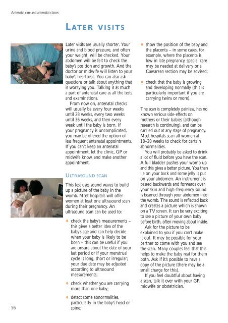
Later visits are usually shorter. Your<br />
urine and blood pressure, and often<br />
your weight, will be checked. Your<br />
abdomen will be felt to check the<br />
baby’s position and growth. And the<br />
doctor or midwife will listen to your<br />
baby’s heartbeat. You can also ask<br />
questions or talk about anything that<br />
is worrying you. Talking is as much<br />
a part of antenatal care as all the tests<br />
and examinations.<br />
From now on, antenatal checks<br />
will usually be every four weeks<br />
until 28 weeks, every two weeks<br />
until 36 weeks, and then every<br />
week until the baby is born. If<br />
your pregnancy is uncomplicated,<br />
you may be offered the option of<br />
less frequent antenatal appointments.<br />
If you can’t keep an antenatal<br />
appointment, let the clinic, GP or<br />
midwife know, and make another<br />
appointment.<br />
ULTRASOUND SCAN<br />
This test uses sound waves to build<br />
up a picture of the baby in the<br />
womb. Most hospitals will offer<br />
women at least one ultrasound scan<br />
during their pregnancy. An<br />
ultrasound scan can be used to:<br />
•<br />
check the baby’s measurements –<br />
this gives a better idea of the<br />
baby’s age and can help decide<br />
when your baby is likely to be<br />
born – this can be useful if you<br />
are unsure about the date of your<br />
last period or if your menstrual<br />
cycle is long, short or irregular;<br />
your due date may be adjusted<br />
according to ultrasound<br />
measurements;<br />
•<br />
check whether you are carrying<br />
more than one baby;<br />
•<br />
detect some abnormalities,<br />
particularly in the baby’s head or<br />
spine;<br />
• show the position of the baby and<br />
the placenta – in some cases, for<br />
example, w<strong>here</strong> the placenta is<br />
low in late pregnancy, special care<br />
may be needed at delivery or a<br />
Caesarean section may be advised;<br />
•<br />
check that the baby is growing<br />
and developing normally (this is<br />
particularly important if you are<br />
carrying twins or more).<br />
The scan is completely painless, has no<br />
known serious side-effects on<br />
mothers or their babies (although<br />
research is continuing), and can be<br />
carried out at any stage of pregnancy.<br />
Most hospitals scan all women at<br />
18–20 weeks to check for certain<br />
abnormalities.<br />
You will probably be asked to drink<br />
a lot of fluid before you have the scan.<br />
A full bladder pushes your womb up<br />
and this gives a better picture. You then<br />
lie on your back and some jelly is put<br />
on your abdomen. An instrument is<br />
passed backwards and forwards over<br />
your skin and high-frequency sound<br />
is beamed through your abdomen into<br />
the womb. The sound is reflected back<br />
and creates a picture which is shown<br />
on a TV screen. It can be very exciting<br />
to see a picture of your own baby<br />
before birth, often moving about inside.<br />
Ask for the picture to be<br />
explained to you if you can’t make<br />
it out. It may be possible for your<br />
partner to come with you and see<br />
the scan. Many couples feel that this<br />
helps to make the baby real for them<br />
both. Ask if it’s possible to have a<br />
copy of the picture (t<strong>here</strong> may be a<br />
small charge for this).<br />
If you feel doubtful about having<br />
a scan, talk it over with your GP,<br />
midwife or obstetrician.