15th Annual Scientific Meeting - Heart Failure Society of America
15th Annual Scientific Meeting - Heart Failure Society of America
15th Annual Scientific Meeting - Heart Failure Society of America
Create successful ePaper yourself
Turn your PDF publications into a flip-book with our unique Google optimized e-Paper software.
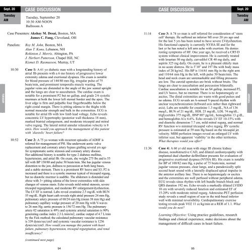
Sept. 20<br />
Tuesday<br />
AM<br />
CASE DISCUSSION<br />
Tuesday, September 20<br />
10:30 AM-NOON<br />
Ballroom A<br />
CASE DISCUSSION<br />
Sept. 20<br />
Tuesday<br />
AM<br />
TuesdAY<br />
Case Presenters: Akshay M. Desai, Boston, MA<br />
James C. Fang, Cleveland, OH<br />
Panelists:<br />
Roy M. John, Boston, MA<br />
Alan T. Kono, Lebanon, NH<br />
Rohinton J. Morris, Abington, PA<br />
J. Herbert Patterson, Chapel Hill, NC<br />
Kismet D. Rasmusson, Murray, UT<br />
10:30 Case 1: A 61 yo diabetic man with a longstanding history <strong>of</strong><br />
atrial fib presents with a 6 mo history <strong>of</strong> progressive lower<br />
extremity edema and exertional dyspnea. His exam is notable<br />
for blood pressure <strong>of</strong> 144/80 mm Hg, irregular pulse <strong>of</strong> 75<br />
beats/min, and prominent temporalis muscle wasting. The<br />
jugular veins are distended to the angle <strong>of</strong> the jaw seated upright<br />
and the lungs are clear to auscultation. The cardiac exam is<br />
notable for a prominent P2 but no gallop, and grade 2/6 systolic<br />
murmurs at both the lower left sternal border and the apex. The<br />
liver edge is firm and palpable four fingerbreadths below the<br />
right costal margin. There is pitting edema to the thighs with<br />
normal muscular strength and bulk in the extremities. ECG is<br />
notable for atrial fib with low limb lead voltage. Echo reveals<br />
concentric LV hypertrophy (posterior wall thickness 18 mm),<br />
marked biatrial enlargement, and moderate tricuspid and mitral<br />
valve regurg. The lateral mitral annular relaxation velocity is 8<br />
cm/s. How would you approach the management <strong>of</strong> this patient<br />
with ‘diastolic’ heart failure?<br />
10:52 Case 2: A 75 yo woman with recurrent episodes <strong>of</strong> ADHF is<br />
referred for management <strong>of</strong> PH. She underwent aortic valve<br />
replacement and coronary artery bypass grafting several yrs ago<br />
for symptomatic aortic stenosis and coronary artery disease.<br />
Her additional history is notable for type 2 diabetes mellitus,<br />
hypertension, and atrial fib. On exam, she weighs 275 lbs and is 55<br />
tall with BP 150/80 and pulse 50 beats/min. She has jugular venous<br />
distention to the jaw, dullness to percussion at both lung bases,<br />
and a stable sternum. There is a prominent RV heave and S3; P2 is<br />
increased and there is a systolic murmur typical <strong>of</strong> tricuspid regurg,<br />
but no diastolic murmur is audible. The abdomen is distended and<br />
obese with 3+ pitting edema <strong>of</strong> the lower extremities with skin<br />
changes <strong>of</strong> venous stasis. Echo reveals mild mitral stenosis, severe<br />
tricuspid regurgitation, and moderate RV enlargement/dysfunction.<br />
The LV EF is normal. Labs reveal creatinine 2.5 mg/dL with BUN<br />
55 mg/dL. Right heart cath reveals right atrial pressure 24 mm Hg,<br />
pulmonary artery pressure <strong>of</strong> 68/24 mm Hg (mean 38 mm Hg) and<br />
pulmonary capillary wedge pressure <strong>of</strong> 20 mm Hg with V-waves<br />
to 26 mm Hg; aortic pressure is 156/72 mm Hg. The pulmonary<br />
artery 02 saturation is 49% and systemic arterial saturation is 90%<br />
generating cardiac index 2.3 L/min/m2, cardiac output <strong>of</strong> 4.7 L/min<br />
by the Fick method; the calculated pulmonary vascular resistance<br />
is 339 dynes/sec/cm5 and systemic vascular resistance is 1256<br />
dynes/sec/cm5. How would you manage this patient with heart<br />
failure, pulmonary hypertension, tricuspid regurgitation, and renal<br />
insufficiency?<br />
11:14 Case 3: A 74 yo man is self referred for consideration <strong>of</strong> ‘stem<br />
cell’ therapy. He suffered an inferior MI over 20 yrs ago and<br />
for the last 5 yrs has been noted to have severe LVdysfunction.<br />
His functional capacity is currently NYHA III and for the<br />
last yr he has noted a left arm ache with exertion. He denies<br />
resting symptoms <strong>of</strong> HF. One year ago, he received a CRT-D<br />
system without clinical improvement. He is currently treated<br />
with losartan 50 mg daily, carvedilol CR 40 mg daily, and<br />
aspirin 325 mg daily. On exam, he is a pleasant elderly man<br />
in no acute distress. He is 5’10” and 197 lbs with body mass<br />
index <strong>of</strong> 28 kg/m2. His BP is 116/61 mm Hg in the right arm<br />
and 110/64 mm Hg in the left, with pulse 50 beats/min. The<br />
head and neck exam are unremarkable and filling pressures<br />
are low. The carotid upstrokes are brisk without bruits. The<br />
lungs are clear to auscultation and percussion bilaterally.<br />
Cardiac auscultation is notable for an S4 gallop, increased P2,<br />
and LV heave, but no murmur. There is no hepatomegaly or<br />
ascites. The distal extremities are warm with good pulses and<br />
no edema. ECG reveals an A-sensed V-paced rhythm with<br />
unclear resynchronization (leftward axis rather than rightward<br />
axis). Labs are notable for creatinine 1.3 mg/dL, NA <strong>of</strong> 136<br />
meq/L, BUN <strong>of</strong> 21 mg/dL, HDL 23 mg/dL, LDL 74 mg/dL,<br />
triglycerides 375 mg/dL, BNP 887 pg/mL, hemoglobin 13 g/dL,<br />
and hemoglobin A1c 6.6%. Echo reveals LV EF 10-15% with<br />
end diastolic dimension 7.7 cm, mild mitral regurg, and normal<br />
RV function w/o minimal tricuspid valve regurg. RV systolic<br />
pressure is estimated at 59 mm Hg based on the tricuspid jet<br />
velocity. MIBI perfusion images reveal an enlarged LV with<br />
inferior scar, but apparent ‘viability’ in the other territories.<br />
What therapies would you <strong>of</strong>fer?<br />
11:36 Case 4: A 60 yr old man with stage III chronic kidney<br />
disease, nonobstructive CAD, and dilated cardiomyopathy with<br />
implanted dual chamber defibrillator presents for evaluation <strong>of</strong><br />
progressive exertional dyspnea (NYHA III). His exam is notable<br />
for BP <strong>of</strong> 100/82 mm Hg, a pulse <strong>of</strong> 75 beats/min, normal<br />
jugular venous pressure, clear lungs, and a paradoxically split<br />
second heart sound with a laterally displaced apical impulse in<br />
the anterior axillary line. There is no hepatomegaly or ascites<br />
and the extremities are well perfused without peripheral edema.<br />
ECG reveals sinus rhythm with left bundle branch block and<br />
QRS duration 192 ms. Echo reveals a markedly dilated LVIDD<br />
10 cm with severely reduced function and estimated EF <strong>of</strong><br />
15-20% with moderate mitral regurg. Adenosine-PET perfusion<br />
study reveals a small region <strong>of</strong> scar in the mid-basal inferior<br />
wall with minimal reversibility. Cardiopulmonary exercise<br />
testing reveals peak VO2 11 cc/kg/min at a RER <strong>of</strong> 1.1. What<br />
would you do next?<br />
Learning Objective: Using practice guidelines, research<br />
findings and clinical experience, make decisions about the<br />
management <strong>of</strong> difficult cases in heart failure.<br />
TuesdAY<br />
(continued next page)<br />
94<br />
95


