

Volume 1 · No. 2 · December 2010 V o lu m e 1 · N o ... - IMA Fungus
Volume 1 · No. 2 · December 2010 V o lu m e 1 · N o ... - IMA Fungus
Volume 1 · No. 2 · December 2010 V o lu m e 1 · N o ... - IMA Fungus

Create successful ePaper yourself
Turn your PDF publications into a flip-book with our unique Google optimized e-Paper software.
Physiological differences between wet- and drydistributed<br />
conidia<br />
The distinction between fungi which<br />
produce conidia in slimy heads<br />
(“wet-spored” and waterborne) and<br />
those which form them in powdery masses<br />
or chains (“dry-spored” and airborne) has<br />
been recognized as of fundamental importance<br />
in the characterization of genera since<br />
the early 20 th century. <strong>No</strong>w, some c<strong>lu</strong>es as<br />
to the basis of this distinction have been generated<br />
by van Leeuwen et al. (<strong>2010</strong>). The<br />
freshly harvested condia of two wet-spored<br />
(Fusarium oxysporum and Lecanicillium<br />
fungicola 1 ) and two dry-spored species (Aspergil<strong>lu</strong>s<br />
niger and Penicillium discolor) were<br />
compared physiologically using electron<br />
spin resonance (ESR) to test for cytoplasmic<br />
viscosity and staining with the f<strong>lu</strong>orescent<br />
dye filipin to indicate the presence of ergosterol.<br />
Striking differences were found; the<br />
wet-spored species had lower cytoplasmic<br />
viscosity than the dry-spored pair, and the<br />
staining showed ergosterol was present in<br />
the plasma membrane only of the wetspored<br />
ones. Whether this correlation can<br />
be used as a generalization must, however,<br />
await studies on a much more diverse range<br />
of fungi.<br />
RESEARCH NEWS<br />
Leeuwen MR van, Doorn TH van, Golovina EA, Stark J, Dijksterhuis J (<strong>2010</strong>) Water- and<br />
air-distributed conidia differ in sterol content and cytoplasmic microviscosity. Applied and<br />
Environmental Microbiology 76: 366–369.<br />
1<br />
The authors used the name Verticillium fungicola, but that species has been transferred to<br />
Lecanicillium (Zare R, Gams W, Mycological Research 112: 818, 2008).<br />
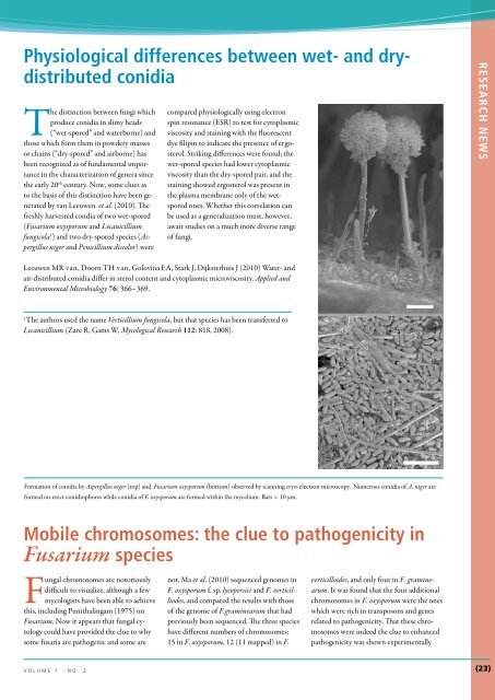
Formation of conidia by Aspergil<strong>lu</strong>s niger (top) and Fusarium oxysporum (bottom) observed by scanning cryo-electron microscopy. Numerous conidia of A. niger are<br />
formed on erect conidiophores while conidia of F. oxysporum are formed within the mycelium. Bars = 10 µm.<br />
Mobile chromosomes: the c<strong>lu</strong>e to pathogenicity in<br />
Fusarium species<br />
Fungal chromosomes are notoriously<br />
difficult to visualize, although a few<br />
mycologists have been able to achieve<br />
this, inc<strong>lu</strong>ding Punithalingam (1975) on<br />
Fusarium. <strong>No</strong>w it appears that fungal cytology<br />
could have provided the c<strong>lu</strong>e to why<br />
some fusaria are pathogenic and some are<br />
not. Ma et al. (<strong>2010</strong>) sequenced genomes in<br />
F. oxysporum f. sp. lycopersici and F. verticilliodes,<br />
and compared the results with those<br />
of the genome of F.graminearum that had<br />
previously been sequenced. The three species<br />
have different numbers of chromosomes:<br />
15 in F. oxysporum, 12 (11 mapped) in F.<br />
verticilloides, and only four in F. graminearum.<br />
It was found that the four additional<br />
chromosomes in F. oxysporum were the ones<br />
which were rich in transposons and genes<br />
related to pathogenicity. That these chromosomes<br />
were indeed the c<strong>lu</strong>e to enhanced<br />
pathogenicity was shown experimentally<br />
v o l u m e 1 · n o . 2 <br />
(23)


