

Volume 1 · No. 2 · December 2010 V o lu m e 1 · N o ... - IMA Fungus
Volume 1 · No. 2 · December 2010 V o lu m e 1 · N o ... - IMA Fungus
Volume 1 · No. 2 · December 2010 V o lu m e 1 · N o ... - IMA Fungus

You also want an ePaper? Increase the reach of your titles
YUMPU automatically turns print PDFs into web optimized ePapers that Google loves.
Modelling fungal colonies and communities: challenges and opportunities<br />
rapidly increasing computing power, and faster algorithms<br />
for reconstruction and data processing make large vo<strong>lu</strong>me<br />
scanning at high reso<strong>lu</strong>tions feasible. The state-of-the-art<br />
equipment at the UGCT (Centre for X-ray Tomography at<br />
Ghent University) is highly flexible, with in-house developed<br />
software for scanner control, sample reconstruction,<br />
analysis, and visualisation. This set-up allows scanning<br />
with a reso<strong>lu</strong>tion of 0.2 mm for samples of 37 cm in<br />
diameter down to approximately 400 nm for objects about<br />
the size of a splinter. As such, apart from visualisation, 3D<br />
quantitative information can be retrieved from objects with<br />
a broad range of sizes. Sub-micron reso<strong>lu</strong>tion scanning<br />
should enable the visualisation of fungal hyphae and by<br />
using time-lapse tomography the growth of these tubular<br />
structures could be monitored (van den Bulcke et al. 2009).<br />
The latter procedure however has associated challenges.<br />
First, fungal growth can interfere with scanning during<br />
moderately long scan times. Second, with lab-based X-ray<br />
sources, polychromatic X-rays, scattering, f<strong>lu</strong>orescence and<br />
noise disturb the ideal acquisition (Vidal et al. 2005). Third,<br />
at sub-micron reso<strong>lu</strong>tion phase contrast emerges especially<br />
at sharp edges, complicating thresholding and segmentation.<br />
Fourth, tube shift during long scans at sub-micron reso<strong>lu</strong>tion<br />
can reduce image quality. Fifth, hyphal tubes are hollow<br />
thin-walled structures, as such having a very low X-ray<br />
attenuation. A drastic so<strong>lu</strong>tion to some of the problems is the<br />
use of synchrotron radiation, having a monochromatic X-ray<br />
bundle, allowing faster scanning with less heating of the<br />
samples, but access to such facilities is a major bottleneck.<br />
Especially the available beam time is limited and as such<br />
this is not an option for long-running experiments, of the<br />
order of days to weeks, and for repeated experiments. Many<br />
of the aforementioned problems are handled at the UGCT<br />
facility. Post-processing can contribute to the enhancement<br />
of image quality; the phase contrast phenomenon can<br />
be solved using dedicated filtering (Boone et al. 2009, De<br />
Witte et al. 2009); and tube shift can be counteracted with<br />
correction software. Proper scanning and processing can<br />
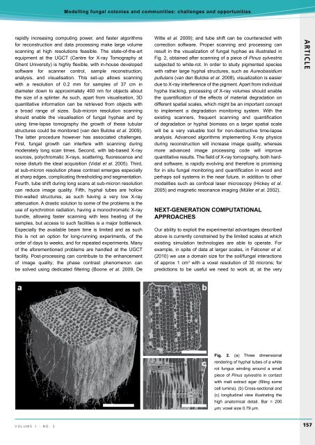
result in the visualization of fungal hyphae as il<strong>lu</strong>strated in<br />
Fig. 2, obtained after scanning of a piece of Pinus sylvestris<br />
subjected to white-rot. In order to study pigmented species<br />
with rather large hyphal structures, such as Aureobasidium<br />
pul<strong>lu</strong>lans (van den Bulcke et al. 2008), visualization is easier<br />
due to X-ray interference of the pigment. Apart from individual<br />
hypha tracking, processing of X-ray vo<strong>lu</strong>mes should enable<br />
the quantification of the effects of material degradation on<br />
different spatial scales, which might be an important concept<br />
to implement a degradation monitoring system. With the<br />
existing scanners, frequent scanning and quantification<br />
of degradation or hyphal biomass on a larger spatial scale<br />
will be a very va<strong>lu</strong>able tool for non-destructive time-lapse<br />
analysis. Advanced algorithms implementing X-ray physics<br />
during reconstruction will increase image quality, whereas<br />
more advanced image processing code will improve<br />
quantitative results. The field of X-ray tomography, both hardand<br />
software, is rapidly evolving and therefore is promising<br />
for in situ fungal monitoring and quantification in wood and<br />
perhaps soil systems in the near future, in addition to other<br />
modalities such as confocal laser microscopy (Hickey et al.<br />
2005) and magnetic resonance imaging (Müller et al. 2002).<br />
Next-generation computational<br />
approaches<br />
Our ability to exploit the experimental advantages described<br />
above is currently constrained by the limited scales at which<br />
existing simulation technologies are able to operate. For<br />
example, in spite of data at larger scales, in Falconer et al.<br />
(<strong>2010</strong>) we use a domain size for the soil/fungal interactions<br />
of approx 1 cm 3 with a voxel reso<strong>lu</strong>tion of 30 microns; for<br />
predictions to be useful we need to work at, at the very<br />
ARTICLE<br />
Fig. 2. (a) Three dimensional<br />
rendering of hyphal tubes of a white<br />
rot fungus winding around a small<br />
piece of Pinus sylvestris in contact<br />
with malt extract agar (filling some<br />
cell <strong>lu</strong>mina). (b) Cross-sectional and<br />
(c) longitudinal view il<strong>lu</strong>strating the<br />
high anatomical detail. Bar = 200<br />
µm; voxel size 0.79 µm.<br />
v o l u m e 1 · n o . 2 <br />
157


