

Acquisition, optimization and interpretation of X-ray computed ...
Acquisition, optimization and interpretation of X-ray computed ...
Acquisition, optimization and interpretation of X-ray computed ...

You also want an ePaper? Increase the reach of your titles
YUMPU automatically turns print PDFs into web optimized ePapers that Google loves.
Computers & Geosciences 27 (2001) 381–400<br />
<strong>Acquisition</strong>, <strong>optimization</strong> <strong>and</strong> <strong>interpretation</strong> <strong>of</strong> X-<strong>ray</strong><br />
<strong>computed</strong> tomographic imagery: applications<br />
to the geosciences<br />
Richard A. Ketcham*, William D. Carlson<br />
Department <strong>of</strong> Geological Sciences, University <strong>of</strong> Texas at Austin, Austin, TX 78712, USA<br />
Abstract<br />
High-resolution X-<strong>ray</strong> <strong>computed</strong> tomography (CT) is a novel technology ideally suited to a wide range <strong>of</strong> geological<br />
investigations. It is a quick <strong>and</strong> nondestructive method to produce images that correspond closely to serial sections<br />
through an object. Sequential contiguous images are compiled to create three-dimensional representations that can be<br />
manipulated digitally to perform efficiently a large ar<strong>ray</strong> <strong>of</strong> measurement <strong>and</strong> visualization tasks. Optimal data<br />
acquisition <strong>and</strong> <strong>interpretation</strong> require proper selection <strong>of</strong> scanning configuration, use <strong>of</strong> suitable X-<strong>ray</strong> sources <strong>and</strong><br />
detectors, careful calibration, <strong>and</strong> attention to origins <strong>and</strong> modes <strong>of</strong> artifact suppression. Visualization <strong>of</strong> CT data<br />
typically pr<strong>of</strong>its from the ability to view arbitrarily oriented sections through the three-dimensional volume represented<br />
by the data, <strong>and</strong> from the capability to extract features <strong>of</strong> interest selectively <strong>and</strong> display perspective views <strong>of</strong> them using<br />
methods <strong>of</strong> isocontouring or volume rendering. Geological applications include interior examination <strong>of</strong> one-<strong>of</strong>-a-kind<br />
fossils or meteorites; textural analysis <strong>of</strong> igneous <strong>and</strong> metamorphic rocks; geometric description <strong>and</strong> quantification <strong>of</strong><br />
porosity <strong>and</strong> permeability in rocks <strong>and</strong> soils; <strong>and</strong> any other application dem<strong>and</strong>ing three-dimensional data that<br />
formerly required physical serial sectioning. # 2001 Elsevier Science Ltd. All rights reserved.<br />
Keywords: Visualization; Iunage analysis<br />
1. Introduction<br />
X-<strong>ray</strong> <strong>computed</strong> tomography (CT) is an established<br />
<strong>and</strong> rapidly evolving technology <strong>of</strong> proven value for<br />
geological investigations. Because <strong>of</strong> its origin outside <strong>of</strong><br />
the earth sciences, the potential for application <strong>of</strong> CT<br />
imagery to geological problems is only beginning to be<br />
explored. This article provides the interested geoscientist<br />
with an underst<strong>and</strong>ing <strong>of</strong> the rudiments <strong>of</strong> X-<strong>ray</strong> CT,<br />
how scanning instrumentation <strong>and</strong> methods can be<br />
optimized for particular imaging tasks, <strong>and</strong> some <strong>of</strong> the<br />
issues that influence proper utilization <strong>of</strong> CT data,<br />
*Corresponding author. Tel.: 1-512-471-0260; fax: 1-512-471-<br />
9425.<br />
E-mail address: richk@maestro.geo.utexas.edu (R.A. Ketcham).<br />
together with a description <strong>of</strong> several past <strong>and</strong> current<br />
geologic applications <strong>of</strong> X-<strong>ray</strong> CT.<br />
CT provides nondestructive three-dimensional visualization<br />
<strong>and</strong> characterization, creating images that map<br />
the variation <strong>of</strong> X-<strong>ray</strong> attenuation within objects, which<br />
relates closely to density. Because density transitions<br />
usually correspond to boundaries between materials or<br />
phases, these data are <strong>of</strong>ten straightforward <strong>and</strong><br />
intuitive for the geologist to interpret. The imagery is<br />
commonly analogous to data that would be obtained<br />
more tediously <strong>and</strong> laboriously with serial sectioning.<br />
Furthermore, because the data are digital, the method<br />
lends itself more easily to both quantitative analysis <strong>and</strong><br />
widespread dissemination.<br />
Two vivid examples <strong>of</strong> these advantages are found in<br />
the fields <strong>of</strong> paleontology <strong>and</strong> metamorphic petrology.<br />
A serial-sectioning study <strong>of</strong> a skull <strong>of</strong> the mammalian<br />
ancestor Thrinaxodon was undertaken by Fourie (1974).<br />
0098-3004/01/$ - see front matter # 2001 Elsevier Science Ltd. All rights reserved.<br />
PII: S 0098-3004(00)00116-3
382<br />
R.A. Ketcham, W.D. Carlson / Computers & Geosciences 27 (2001) 381–400<br />
The sample was ground down in 200-mm increments,<br />
<strong>and</strong> at each stage drawings were made <strong>and</strong> acetate peels<br />
were taken, a process that required 2 years to complete.<br />
When the results were published (Fourie, 1974), only a<br />
small subset <strong>of</strong> the images that were obtained could be<br />
presented, <strong>and</strong> the specimen had been destroyed. In<br />
contrast, a high-resolution CT scan <strong>of</strong> another Thrinaxodon<br />
skull was completed by Rowe et al. (1993) in only<br />
6 h, roughly the time it took to complete <strong>and</strong> fully<br />
document a single serial section in the original study,<br />
<strong>and</strong> the sample remains available for further research.<br />
Interpretation <strong>of</strong> the digital data was greatly aided by<br />
image-processing s<strong>of</strong>tware, <strong>and</strong> all <strong>of</strong> the collected data<br />
were published on a single compact disk (Rowe et al.,<br />
1993).<br />
A similar example is provided by studies focused on<br />
quantitative analysis <strong>of</strong> metamorphic textures. Since the<br />
1960s it has been recognized that the sizes <strong>and</strong> threedimensional<br />
spatial disposition <strong>of</strong> porphyroblasts in<br />
metamorphic rocks contain information about the<br />
atomic-scale processes that control crystal nucleation<br />
<strong>and</strong> growth (Kretz, 1966, 1969). Kretz (1993) acquired<br />
the requisite data on crystal sizes <strong>and</strong> locations <strong>of</strong> garnet<br />
by mechanically dissecting a rock using a steel chisel <strong>and</strong><br />
brush while recording the positions <strong>of</strong> each <strong>of</strong> the<br />
porphyroblasts found. A total <strong>of</strong> 91 crystals were<br />
located, measured <strong>and</strong> subsequently analyzed. In contrast,<br />
high-resolution X-<strong>ray</strong> <strong>computed</strong> tomography has<br />
also been used to reveal the locations <strong>of</strong> garnet<br />
porphyroblasts in several rocks (Carlson <strong>and</strong> Denison,<br />
1992; Denison et al., 1997). Each was scanned in a few<br />
hours, <strong>and</strong> analysis <strong>of</strong> the data for each rock required<br />
only a few weeks, once the computational process was<br />
streamlined. Numbers <strong>of</strong> crystals measured in a single<br />
sample ranged up to 12,000; such large numbers are<br />
important, because analyses <strong>of</strong> the statistical tests<br />
applied to the data indicate that at least 1000 crystals<br />
should be measured to ensure accurate results (Denison<br />
et al., 1997, Appendix 1). Furthermore, the rocks<br />
remained available for further study, such as electron<br />
microprobe analysis <strong>of</strong> selected crystals (e.g., Chern<strong>of</strong>f<br />
<strong>and</strong> Carlson, 1997, 1999).<br />
Many similar examples <strong>of</strong> geological application <strong>of</strong><br />
high-resolution CT are accumulating as knowledge <strong>and</strong><br />
utilization <strong>of</strong> this technology becomes more prevalent.<br />
The synopses <strong>of</strong> recent CT investigations that conclude<br />
this article should provide a sense <strong>of</strong> the breadth <strong>and</strong><br />
applicability <strong>of</strong> this technique to geological problems.<br />
1.1. Industrial <strong>computed</strong> tomography<br />
First developed for widespread use in medicine for<br />
the imaging <strong>of</strong> s<strong>of</strong>t tissue <strong>and</strong> bone, X-<strong>ray</strong> CT was<br />
subsequently extended <strong>and</strong> adapted to a wide variety <strong>of</strong><br />
industrial tasks. These latter developments, which<br />
dem<strong>and</strong>ed imagery <strong>of</strong> denser objects across a range <strong>of</strong><br />
size classes <strong>and</strong> resolution requirements, provided key<br />
advances that greatly enhanced the potential for application<br />
<strong>of</strong> this technology to geological investigations.<br />
To maximize their effectiveness in differentiating<br />
tissues while minimizing patient exposure, medical CT<br />
systems need to use a limited dose <strong>of</strong> relatively lowenergy<br />
X-<strong>ray</strong>s ( 125 keV). To obtain as much data as<br />
possible given these requirements, they use relatively<br />
large (mm-scale), high-efficiency detectors. Because<br />
industrial CT systems face no limitations on acceptable<br />
radiation dose, they are able to take advantage <strong>of</strong><br />
several <strong>optimization</strong>s. X-<strong>ray</strong>s for industrial systems can<br />
be <strong>of</strong> higher energy, <strong>and</strong> exposure times have no firm<br />
upper limit. Smaller detectors can be used, which leads<br />
to higher resolution because the decline in signal that<br />
accompanies smaller surface area can be compensated<br />
by higher X-<strong>ray</strong> intensities or exposure times. Finally,<br />
because the objects being scanned are inanimate, more<br />
accurate <strong>and</strong> precise positioning is possible.<br />
CT scanners can be generally grouped into four<br />
categories, based on their spatial resolution <strong>and</strong> the size<br />
<strong>of</strong> objects they are most suitable for scanning. A<br />
proposed classification is summarized in Table 1. Most<br />
medical systems fall into the category <strong>of</strong> conventional<br />
CT, although some specialized systems for scanning<br />
appendages could be termed high resolution. Industrial<br />
scanners can span a wide range <strong>of</strong> scales, from<br />
conventional or larger for scanners designed to image<br />
large objects such as engine blocks, down to ultra-high<br />
resolution. To achieve the resolution requirements for<br />
micro-CT, synchrotron X-<strong>ray</strong> sources are required (e.g.,<br />
Coker et al., 1996; Flannery et al., 1987; Kinney et al.,<br />
1993). The University <strong>of</strong> Texas (UT) high-resolution X-<br />
<strong>ray</strong> CT Facility houses a t<strong>and</strong>em scanner, with both a<br />
high-resolution system <strong>and</strong> an ultra-high-resolution<br />
system. Each is described in detail in a later section.<br />
1.2. Geological applications<br />
The natural application <strong>of</strong> X-<strong>ray</strong> CT to paleontology<br />
was quickly recognized (Conroy <strong>and</strong> Vannier, 1984).<br />
Although medical scanners were able to obtain only<br />
low-resolution data on a limited subset <strong>of</strong> specimens,<br />
the fact that CT could acquire interior information<br />
Table 1<br />
General classification <strong>of</strong> <strong>computed</strong> tomography<br />
Type<br />
Scale <strong>of</strong><br />
observation<br />
Scale <strong>of</strong><br />
resolution<br />
Conventional m mm<br />
High-resolution dm 100 mm<br />
Ultra-high-resolution cm 10 mm<br />
Microtomography mm mm
R.A. Ketcham, W.D. Carlson / Computers & Geosciences 27 (2001) 381–400 383<br />
nondestructively from irreplaceable specimens made it a<br />
valuable technique nonetheless (Haubitz et al., 1988).<br />
Similarly, CT was also applied to meteorites at an early<br />
stage to facilitate study <strong>of</strong> one-<strong>of</strong>-a-kind specimens<br />
(Arnold et al., 1982). Other successful early geological<br />
applications <strong>of</strong> X-<strong>ray</strong> CT using medical scanners include<br />
investigations in fluid flow, soil science, <strong>and</strong> sedimentology.<br />
Petroleum engineers used CT data to study tw<strong>of</strong>luid<br />
coreflood experiments in reservoir lithologies<br />
(Wellington <strong>and</strong> Vinegar, 1987; Withjack, 1988), <strong>and</strong><br />
those techniques were later adapted to imaging air<br />
sparging as used in environmental remediation (Chen<br />
et al., 1996). CT data have been exploited to examine<br />
soil transport properties, through both direct imaging <strong>of</strong><br />
fluid-flow experiments (Anderson et al., 1992; Heijs<br />
et al., 1995) <strong>and</strong> characterization <strong>of</strong> soil <strong>and</strong> pore-space<br />
morphology (Peyton et al., 1992; Zeng et al., 1996).<br />
Medical CT data have also been applied to <strong>of</strong>fshore<br />
sediments for characterization <strong>of</strong> morphology (Orsi <strong>and</strong><br />
Anderson, 1995; Orsi et al., 1994) <strong>and</strong> density distribution<br />
(Amos et al., 1996). Scale models <strong>of</strong> faulting have<br />
also been studied using medical CT systems, which have<br />
become fast enough to allow real-time analysis (Schreurs<br />
<strong>and</strong> Ha¨ nni, 1998).<br />
High-resolution industrial X-<strong>ray</strong> CT allows many <strong>of</strong><br />
these inquiries to be conducted more effectively by<br />
generating much finer-detailed imagery. Paleontological<br />
investigations have moved from imaging gross structures<br />
to imaging such fine details as tooth replacement in<br />
marsupials (Cifelli <strong>and</strong> Muizon, 1998a; Cifelli <strong>and</strong><br />
Muizon, 1998b; Cifelli et al., 1996) <strong>and</strong> inner-ear<br />
structure in early mammals (Rowe, 1996). Meteorite<br />
investigations have progressed from solely finding<br />
inclusions to mapping out the shape <strong>and</strong> size distributions<br />
<strong>of</strong> their mineralogical components, providing<br />
textural clues about their origins (Kuebler et al., 1999).<br />
Scans <strong>of</strong> soil <strong>and</strong> sediment cores are capable <strong>of</strong> imaging<br />
individual pores <strong>and</strong> fractures at much higher resolutions<br />
(better by an order <strong>of</strong> magnitude or more than<br />
achievable with medical scanners), enabling more<br />
detailed research into solute <strong>and</strong> tracer transport<br />
description <strong>and</strong> modeling (Moline et al., 1997). Highresolution<br />
CT has also allowed more detailed analysis <strong>of</strong><br />
scale models depicting faulting <strong>and</strong> salt flow (Le Calvez<br />
<strong>and</strong> Vendeville, 1999).<br />
High-resolution imagery has also exp<strong>and</strong>ed the<br />
breadth <strong>of</strong> X-<strong>ray</strong> CT usage to other geologic subdisciplines.<br />
One <strong>of</strong> the first examples was the quantitative<br />
textural analysis <strong>of</strong> porphyroblast crystallization in<br />
metamorphic rocks to determine atomic-scale processes<br />
controlling nucleation <strong>and</strong> growth (Carlson <strong>and</strong> Denison,<br />
1992; Denison <strong>and</strong> Carlson, 1997; Denison et al.,<br />
1997). More recent <strong>and</strong> ongoing textural investigations<br />
include studies <strong>of</strong> flow pathways for melt in migmatites<br />
(Brown et al., 1999) <strong>and</strong> mingling <strong>of</strong> different melt<br />
types in lamprophyres (Ogasawara et al., 1998). Highresolution<br />
CT is also being used to study spiral inclusion<br />
trails in garnet, where the nondestructive acquisition <strong>of</strong><br />
data for large volumes <strong>of</strong> rock allows these features to be<br />
compared among adjacent garnets <strong>and</strong> linked to<br />
surrounding three-dimensional rock texture <strong>and</strong> garnet<br />
morphology (Bauer et al., 1998). CT analysis to<br />
determine the size distribution <strong>and</strong> morphology <strong>of</strong><br />
vesicles in basalt has provided new clues to the physics<br />
<strong>of</strong> eruption processes (Proussevitch et al., 1998).<br />
Imagery <strong>of</strong> fractures <strong>and</strong> pore spaces in reservoir<br />
limestone has improved underst<strong>and</strong>ing <strong>of</strong> the geometrical<br />
components <strong>of</strong> porosity <strong>and</strong> permeability, <strong>and</strong><br />
their relation to geological <strong>and</strong> biological features (Beall<br />
et al., 1996; Gournay <strong>and</strong> Kirkl<strong>and</strong>, 1998).<br />
2. Essentials <strong>of</strong> <strong>computed</strong> tomography<br />
Because industrial X-<strong>ray</strong> CT scanners are typically<br />
custom-built, it is impossible to provide a detailed<br />
description <strong>of</strong> their principles <strong>and</strong> operation that will<br />
apply in all cases. Instead, we provide here a description<br />
<strong>of</strong> each component <strong>of</strong> the CT-scanning process, both in<br />
general terms <strong>and</strong> as specifically applied at the<br />
University <strong>of</strong> Texas High-Resolution X-<strong>ray</strong> CT Facility.<br />
The material presented in this <strong>and</strong> subsequent sections is<br />
a combination <strong>of</strong> information provided with the UT<br />
system by the manufacturer (Bio-Imaging Research,<br />
Inc., <strong>of</strong> Lincolnshire, Illinois), insights gained from<br />
experience, <strong>and</strong> general principles derived from the<br />
literature. For a more complete technical overview <strong>of</strong><br />
CT, we recommend ASTM publication E1441-92a<br />
(ASTM, 1992) as an excellent starting point. Very<br />
readable, if somewhat dated, synopses <strong>of</strong> medical CT<br />
<strong>and</strong> its components <strong>and</strong> history can be found in Hendee<br />
(1979) <strong>and</strong> Newton <strong>and</strong> Potts (1981), the latter <strong>of</strong> which<br />
provides considerable detail about many fundamentals<br />
<strong>of</strong> tomographic scanning.<br />
2.1. Scanning configuration<br />
The simplest common elements <strong>of</strong> X-<strong>ray</strong> radiography<br />
are an X-<strong>ray</strong> source, an object to be imaged through<br />
which the X-<strong>ray</strong>s pass, <strong>and</strong> a series <strong>of</strong> detectors that<br />
measure the extent to which the X-<strong>ray</strong> signal has been<br />
attenuated by the object (Fig. 1). A single set <strong>of</strong> X-<strong>ray</strong><br />
intensity measurements on all detectors for a given<br />
object position <strong>and</strong> scanner geometry is termed a view.<br />
The fundamental principle behind <strong>computed</strong> tomography<br />
is to acquire multiple sets <strong>of</strong> views <strong>of</strong> an object over<br />
a range <strong>of</strong> angular orientations. By this means,<br />
additional dimensional data are obtained in comparison<br />
to conventional X-radiography, in which there is only<br />
one view. These data are used to create two-dimensional<br />
images that are called slices because they correspond to
384<br />
R.A. Ketcham, W.D. Carlson / Computers & Geosciences 27 (2001) 381–400<br />
Fig. 1. Schematic illustration <strong>of</strong> different generations <strong>of</strong> X-<strong>ray</strong> CT scan geometries. Solid arrows indicate movements during data<br />
collection, dashed arrows indicate movement between sequences <strong>of</strong> data collection. Solid lines passing from sources to detectors are <strong>ray</strong><br />
paths, <strong>and</strong> each set <strong>of</strong> solid lines from a single angular orientation constitutes a view. These illustrations show the source <strong>and</strong> detectors<br />
moving around stationary object, as is the case with medical scanners. Motion is relative, however, <strong>and</strong> in many industrial scanners the<br />
object moves while the source <strong>and</strong> detectors are stationary. In all cases, axis <strong>of</strong> rotation is center <strong>of</strong> circle. (A) first-generation,<br />
translate-rotate pencil beam geometry; (B) second-generation, translate-rotate fan beam geometry; (C) third-generation, rotate-only<br />
geometry; (D) third-generation <strong>of</strong>fset-mode geometry.<br />
what would be seen if the object were sliced along the<br />
scan plane.<br />
First- through fourth-generation <strong>computed</strong> tomography<br />
systems utilize only <strong>ray</strong>s that are in the scan plane.<br />
In first-generation CT (Fig. 1A) this is done by directing<br />
a pencil beam through the object to a single detector,<br />
translating the source–detector pair across the extent <strong>of</strong><br />
the object in the scan plane, then repeating the<br />
procedure from a number <strong>of</strong> angular orientations.<br />
Second-generation CT (Fig. 1B) uses the same scanning<br />
procedure, but a fan beam replaces the pencil beam <strong>and</strong><br />
the single detector is replaced by a linear or arcuate<br />
series <strong>of</strong> detectors, leading to a higher rate <strong>of</strong> data<br />
acquisition. In typical third-generation CT (Fig. 1C), the<br />
fan beam <strong>and</strong> detector series are wide enough to<br />
encompass the entire object, <strong>and</strong> thus only rotation <strong>of</strong><br />
the object or the source–detector combination is<br />
required. One variation <strong>of</strong> third-generation scanning<br />
<strong>of</strong>fsets the sample from the center <strong>of</strong> the fan beam so<br />
that a part <strong>of</strong> it is outside <strong>of</strong> the beam, but the center<br />
<strong>of</strong> rotation is within it (Fig. 1D). As the object rotates,<br />
all <strong>of</strong> it passes through the fan beam, which permits
R.A. Ketcham, W.D. Carlson / Computers & Geosciences 27 (2001) 381–400 385<br />
reconstruction <strong>of</strong> a complete image. This technique<br />
allows larger objects to be scanned <strong>and</strong> permits smaller<br />
objects to be moved closer to the source into a narrower<br />
section <strong>of</strong> the fan beam, leading to increased resolution<br />
through enhanced utilization <strong>of</strong> detectors to image<br />
smaller subsections <strong>of</strong> the object in any one view.<br />
Third-generation scanning tends to be much faster than<br />
second-generation, as X-<strong>ray</strong>s are utilized more efficiently.<br />
Most modern medical scanners are fourthgeneration<br />
devices, consisting <strong>of</strong> a fixed complete ring<br />
<strong>of</strong> detectors <strong>and</strong> a single X-<strong>ray</strong> source that rotates<br />
around the object being scanned. In first- through thirdgeneration<br />
scanners the motion between the object being<br />
scanned <strong>and</strong> the source–detector pair is relative, <strong>and</strong> can<br />
be accomplished either by keeping the object stationary<br />
<strong>and</strong> moving the source–detector pair, as is done in<br />
medical CT systems, or vice versa as is more common in<br />
industrial systems.<br />
In volume CT, a cone beam or highly collimated,<br />
thick, parallel beam is used rather than a fan beam, <strong>and</strong><br />
a planar grid replaces the linear series <strong>of</strong> detectors. This<br />
allows for much faster data acquisition, as the data<br />
required for multiple slices can be acquired in one<br />
rotation. However, it is also computationally more<br />
intensive, prone to distortion, <strong>and</strong> in many cases<br />
provides lower-resolution images. Whereas volume CT<br />
has been largely perfected for some <strong>of</strong> the most<br />
advanced medical systems, <strong>and</strong> is ideally suited for<br />
tomography using synchrotron radiation, for industrial<br />
scanners it does not yet provide the same quality <strong>of</strong><br />
imagery as single-slice arrangements.<br />
The UT facility is capable <strong>of</strong> second- <strong>and</strong> thirdgeneration<br />
scanning on the high-energy system, <strong>and</strong><br />
third generation <strong>and</strong> volume scanning on the ultra-highresolution<br />
system. On each system, however, thirdgeneration<br />
scanning is usually the method <strong>of</strong> choice.<br />
clarity. Higher intensities improve the underlying<br />
counting statistics, but <strong>of</strong>ten require a larger focal spot.<br />
Many conventional X-<strong>ray</strong> tubes have a dual filament<br />
that provides two focal-spot sizes, with the smaller spot<br />
size allowing more detailed imagery at a cost in<br />
intensity. Medical CT systems tend to have X-<strong>ray</strong> spot<br />
sizes that range from 0.5 to 2 mm. The high-resolution<br />
system at the UT CT Facility utilizes a dual-spot 420-kV<br />
X-<strong>ray</strong> source (Pantak HF420), with spot sizes <strong>of</strong> 0.8 <strong>and</strong><br />
1.8 mm. The small spot has a maximum load <strong>of</strong> 800 W<br />
(i.e., 2 mA at 400 kV), whereas the large spot has a<br />
maximum load <strong>of</strong> 2000 W. The 200 kV tube used for<br />
ultra-high resolution work (Feinfocus FXE-200.20) has<br />
an adjustable focal spot with a minimum size <strong>of</strong> 510 mm<br />
at 8 W total load, but at higher loads the spot size is<br />
automatically increased to prevent thermal damage to<br />
the target. In most cases a slightly ‘‘defocused’’ beam<br />
(larger spot size) can be used to improve counting<br />
statistics with little cost in resolution. Both sources have<br />
tungsten targets.<br />
The energy spectrum generated is usually described in<br />
terms <strong>of</strong> the peak X-<strong>ray</strong> energy (keV or MeV), but<br />
actually consists <strong>of</strong> a continuum in which the level with<br />
maximum intensity is typically less than half <strong>of</strong> the peak<br />
(Fig. 2). The total ‘‘effective’’ spectrum is determined by<br />
a number <strong>of</strong> factors in addition to the energy input <strong>of</strong><br />
the X-<strong>ray</strong> source itself, including aut<strong>of</strong>iltering both by<br />
absorption <strong>of</strong> photons generated beneath the surface <strong>of</strong><br />
a thick target (Silver, 1994) <strong>and</strong> by passage through the<br />
2.2. X-<strong>ray</strong> source<br />
The important variables that determine how effective<br />
an X-<strong>ray</strong> source will be for a particular task are the size<br />
<strong>of</strong> the focal spot, the spectrum <strong>of</strong> X-<strong>ray</strong> energies<br />
generated, <strong>and</strong> the X-<strong>ray</strong> intensity. The focal-spot size<br />
partially defines the potential spatial resolution <strong>of</strong> a CT<br />
system by determining the number <strong>of</strong> possible source–<br />
detector paths that can intersect a given point in the<br />
object being scanned. The more such source–detector<br />
paths there are, the more blurring <strong>of</strong> features there will<br />
be. The energy spectrum defines the penetrative ability<br />
<strong>of</strong> the X-<strong>ray</strong>s, as well as their expected relative<br />
attenuation as they pass through materials <strong>of</strong> different<br />
density. Higher-energy X-<strong>ray</strong>s penetrate more effectively<br />
than lower-energy ones, but are less sensitive to changes<br />
in material density <strong>and</strong> composition. The X-<strong>ray</strong> intensity<br />
directly affects the signal-to-noise ratio <strong>and</strong> thus image<br />
Fig. 2. Theoretical energy spectra for 420-kV X-<strong>ray</strong> source with<br />
tungsten target, calculated combining 5-keV intervals. Spectra<br />
consist <strong>of</strong> continuous Bremsstrahlung <strong>and</strong> characteristic K-<br />
series peaks at 57–59 <strong>and</strong> 67–69 keV. Upper spectrum is<br />
modified only by inherent beam filtration by 3 mm <strong>of</strong> aluminum<br />
at tube exit port. Mean X-<strong>ray</strong> energy is 114 keV. Lower curve<br />
represents spectrum that has also passed through 5 cm <strong>of</strong><br />
quartz. Preferential attenuation <strong>of</strong> low-energy X-<strong>ray</strong>s causes<br />
average energy to rise to 178 keV.
386<br />
R.A. Ketcham, W.D. Carlson / Computers & Geosciences 27 (2001) 381–400<br />
tube exit port; other beam filtration introduced to<br />
selectively remove low-energy X-<strong>ray</strong>s; beam hardening<br />
in the object being scanned; <strong>and</strong> the relative efficiency <strong>of</strong><br />
the detectors to different energies. As discussed below,<br />
changes in the X-<strong>ray</strong> spectrum caused by passage<br />
through an object can lead to a variety <strong>of</strong> scanning<br />
artifacts unless efforts are made to compensate for them.<br />
2.3. X-<strong>ray</strong> attenuation<br />
As the X-<strong>ray</strong>s pass through the object being scanned,<br />
the signal is attenuated by scattering <strong>and</strong> absorption.<br />
The basic equation for attenuation <strong>of</strong> a monoenergetic<br />
beam through a homogeneous material is Beer’s Law<br />
I ¼ I 0 exp½ mxŠ; ð1Þ<br />
where I 0 is the initial X-<strong>ray</strong> intensity, m is the linear<br />
attenuation coefficient for the material being scanned,<br />
<strong>and</strong> x is the length <strong>of</strong> the X-<strong>ray</strong> path through the<br />
material. If the scan object is composed <strong>of</strong> a number <strong>of</strong><br />
different materials, the equation becomes:<br />
"<br />
I ¼ I 0 exp X #<br />
ð m i x i Þ ; ð2Þ<br />
i<br />
where each increment i reflects a single material with<br />
attenuation coefficient m i over a linear extent x i . To take<br />
into account the fact that the attenuation coefficient is a<br />
strong function <strong>of</strong> X-<strong>ray</strong> energy, the complete solution<br />
would require solving the equation over the range <strong>of</strong> the<br />
effective X-<strong>ray</strong> spectrum<br />
Z "<br />
I ¼ I 0 ðEÞ exp X #<br />
ð m i ðEÞx i Þ dE;<br />
ð3Þ<br />
i<br />
However, such a calculation is usually problematical for<br />
industrial CT, as the precise form <strong>of</strong> the X-<strong>ray</strong> spectrum,<br />
<strong>and</strong> its variation at <strong>of</strong>f-center angles in a fan or cone<br />
beam, is usually only estimated theoretically rather than<br />
measured. Furthermore, most reconstruction strategies<br />
solve Eq. (2), ins<strong>of</strong>ar as they assign a single value to each<br />
pixel rather than some energy-dependent range.<br />
There are three dominant physical processes responsible<br />
for attenuation <strong>of</strong> an X-<strong>ray</strong> signal: photoelectric<br />
absorption, Compton scattering, <strong>and</strong> pair production.<br />
Photoelectric absorption occurs when the total energy <strong>of</strong><br />
an incoming X-<strong>ray</strong> photon is transferred to an inner<br />
electron, causing the electron to be ejected. In Compton<br />
scattering, the incoming photon interacts with an outer<br />
electron, ejecting the electron <strong>and</strong> losing only a part <strong>of</strong><br />
its own energy, after which it is deflected in a different<br />
direction. In pair production, the photon interacts with<br />
a nucleus <strong>and</strong> is transformed into a positron-electron<br />
pair, with any excess photon energy transferred into<br />
kinetic energy in the particles produced.<br />
In general for geological materials, the photoelectric<br />
effect is the dominant attenuation mechanism at low<br />
X-<strong>ray</strong> energies, up to approximately 50–100 keV. Compton<br />
scatter is dominant at higher energies up to 5–<br />
10 MeV, after which pair production predominates.<br />
Thus, unless higher-energy sources are used, only<br />
photoelectric absorption <strong>and</strong> Compton scattering need<br />
to be considered. The practical importance <strong>of</strong> the<br />
distinction between mechanisms is that photoelectric<br />
absorption is proportional to Z 425 , where Z is the<br />
atomic number <strong>of</strong> an atom in the attenuating material,<br />
whereas Compton scattering is proportional only to Z<br />
(Markowicz, 1993). As a result, low-energy X-<strong>ray</strong>s are<br />
more sensitive to differences in composition than higherenergy<br />
ones.<br />
The best way to gain insight into what one might<br />
expect when scanning a geological sample is to plot the<br />
linear attenuation coefficients <strong>of</strong> the component materials<br />
over the range <strong>of</strong> the available X-<strong>ray</strong> spectrum. These<br />
values can be calculated by combining experimental<br />
results for atomic species (e.g., Markowicz, 1993).<br />
Alternatively, mass attenuation coefficients can be<br />
obtained from the XCOM database managed by NIST<br />
(which can be accessed at http://physics.nist.gov/Phys-<br />
RefData/Xcom/Text/XCOM.html). Mass attenuation<br />
coefficients must be multiplied by mass density to<br />
determine linear attenuation coefficients. To illustrate,<br />
Fig. 3 shows curves for four minerals: quartz, orthoclase,<br />
calcite, <strong>and</strong> alm<strong>and</strong>ine garnet. Quartz <strong>and</strong><br />
orthoclase are very similar in mass density (2.65 g/cm 3<br />
vs. 2.59 g/cm 3 ), but at low energy their attenuation<br />
coefficients are quite different because <strong>of</strong> the presence <strong>of</strong><br />
relatively high-Z potassium in the feldspar. With rising<br />
X-<strong>ray</strong> energy, their attenuation coefficients converge,<br />
Fig. 3. Linear attenuation coefficient as function <strong>of</strong> X-<strong>ray</strong><br />
energy for four rock-forming minerals. Such curves, when<br />
combined with the X-<strong>ray</strong> spectrum utilized for scanning<br />
(Fig. 2), allow prediction <strong>of</strong> ability to differentiate between<br />
minerals in CT images.
R.A. Ketcham, W.D. Carlson / Computers & Geosciences 27 (2001) 381–400 387<br />
Fig. 4. Core <strong>of</strong> graphic granite imaged at various energy conditions. Field <strong>of</strong> view diameter for each image is 22 mm, <strong>and</strong> slice<br />
thickness is 100 mm. Scan (A) was created using X-<strong>ray</strong> energy <strong>of</strong> 100 keV <strong>and</strong> no beam filtration; scan (B) was acquired with X-<strong>ray</strong><br />
energy <strong>of</strong> 200 keV <strong>and</strong> 1/8 00 brass filter. Both scans employed ‘‘self-wedge’’ calibration.<br />
<strong>and</strong> at approximately 125 keV they cross; above<br />
125 keV quartz is slightly (but probably indistinguishably)<br />
more attenuating, owing to its higher density.<br />
Thus, one would expect that these two minerals could be<br />
differentiated in CT imagery if the mean X-<strong>ray</strong> energy<br />
used is low enough, but at higher energies they would be<br />
nearly indistinguishable (Fig. 4). Calcite, though only<br />
slightly denser (2.71 g/cm 3 ) than quartz <strong>and</strong> orthoclase,<br />
is substantially more attenuating, owing to the presence<br />
<strong>of</strong> calcium. Here the divergence with quartz persists to<br />
slightly higher energies, indicating that it should be<br />
possible to distinguish the two even on higher-energy<br />
scans. High-density, high-Z phases such as alm<strong>and</strong>ine<br />
are distinguishable at all energies from the other rockforming<br />
minerals examined here.<br />
2.4. X-<strong>ray</strong> detectors<br />
Detectors for CT scanners make use <strong>of</strong> scintillating<br />
materials in which incoming X-<strong>ray</strong>s produce flashes <strong>of</strong><br />
light that are counted. Detectors influence image quality<br />
through their size <strong>and</strong> quantity, <strong>and</strong> through their<br />
efficiency in detecting the energy spectrum generated by<br />
the source. The size <strong>of</strong> an individual detector determines<br />
the amount <strong>of</strong> an object that is averaged into a single<br />
intensity reading, while the number <strong>of</strong> detectors<br />
determines how much data can be gathered simultaneously.<br />
In third-generation scanning, the number <strong>of</strong><br />
detectors also defines the degree <strong>of</strong> resolution possible in<br />
a single view, <strong>and</strong> thus in an image overall. The film used<br />
in conventional X-<strong>ray</strong> radiography is an excellent<br />
detector in that it consists, in essence, <strong>of</strong> a very large<br />
number <strong>of</strong> small <strong>and</strong> sensitive detectors. Unfortunately,<br />
it is not amenable to quickly producing the digital data<br />
needed for <strong>computed</strong> tomography.<br />
The efficiency <strong>of</strong> scintillation detectors varies with X-<br />
<strong>ray</strong> energy, precisely because higher-energy X-<strong>ray</strong>s are<br />
more penetrative than lower-energy ones, indicating that<br />
they are more capable <strong>of</strong> traveling through materials<br />
without interactions. This factor must be taken into<br />
account when determining the level <strong>of</strong> expected signal<br />
after polychromatic X-<strong>ray</strong>s pass through materials.<br />
The high-resolution system at UT has two separate<br />
detectors. The P250D detector consists <strong>of</strong> a linear ar<strong>ray</strong><br />
<strong>of</strong> 512 discrete cadmium tungstate scintillators with<br />
dimensions 0.25 mm 5.0 mm 5.0 mm, packed in a<br />
comb to prevent crosstalk between channels, <strong>and</strong> each<br />
connected to a Si photodiode. Its channel-to-channel<br />
pitch is 381 mm, <strong>and</strong> its total horizontal extent is<br />
195 mm. The RLS (Radiographic Line Scanner) detector<br />
consists <strong>of</strong> a single 0.25-mm thick gadolinium oxysulfide<br />
scintillator screen connected to a 2048-channel linear<br />
photodiode ar<strong>ray</strong> using a fiber optic taper. The detector<br />
spacing is 50 mm, <strong>and</strong> the total detector extent is<br />
approximately 100 mm. The ultra-high-resolution scanner<br />
uses a 9-in image intensifier (Toshiba AI-5764-HVP)<br />
as a detector. The image intensifier consists <strong>of</strong> a partial<br />
sphere <strong>of</strong> cesium iodide scintillators attached to a<br />
photocathode. The signal from the photocathode is<br />
electronically focused onto a phosphor screen, producing<br />
a real-time X-<strong>ray</strong> image. The image on the<br />
phosphor is converted to digital data using a CCD
388<br />
R.A. Ketcham, W.D. Carlson / Computers & Geosciences 27 (2001) 381–400<br />
video system. The video signal, consisting <strong>of</strong> scan lines<br />
divided into pixels, is used to create a set <strong>of</strong> 512 virtual<br />
detectors by s<strong>of</strong>tware.<br />
3. <strong>Acquisition</strong> <strong>of</strong> CT Data<br />
3.1. Sample preparation<br />
Strictly speaking, the only preparation that is<br />
absolutely necessary for CT scanning is to ensure that<br />
the object fits inside the field <strong>of</strong> view <strong>and</strong> that it does not<br />
move during the scan. Because the full scan field for CT<br />
is a cylinder (i.e., a stack <strong>of</strong> circular fields <strong>of</strong> view), the<br />
most efficient geometry to scan is also a cylinder. Thus,<br />
when possible it is <strong>of</strong>ten advantageous to have the object<br />
take on a cylindrical geometry, either by using a coring<br />
drill or drill press to obtain a cylindrical subset <strong>of</strong> the<br />
material being scanned, or by packing the object in a<br />
cylindrical container with either X-<strong>ray</strong>-transparent filler<br />
or with material <strong>of</strong> similar density. For some applications<br />
the sample can also be treated to enhance the<br />
contrasts that are visible. Examples have included<br />
injecting soils <strong>and</strong> reservoir rocks with NaI-laced fluids<br />
to reveal fluid-flow characteristics (Wellington <strong>and</strong><br />
Vinegar, 1987; Withjack, 1988), injecting s<strong>and</strong>stones<br />
with Woods metal to map out the fine-scale permeability,<br />
<strong>and</strong> soaking samples in water to bring out areas<br />
<strong>of</strong> differing permeability, which can help to reveal fossils<br />
(Zinsmeister <strong>and</strong> De Nooyer, 1996).<br />
3.2. Calibration<br />
Calibrations are necessary to establish the characteristics<br />
<strong>of</strong> the X-<strong>ray</strong> signal as read by the detectors under<br />
scanning conditions, <strong>and</strong> to reduce geometrical uncertainties.<br />
The latter calibrations vary widely among<br />
scanners; as a rule flexible-geometry scanners such as<br />
the one at the University <strong>of</strong> Texas require them, whereas<br />
fixed-geometry scanners geared towards scanning a<br />
single object type may not.<br />
The two principal signal calibrations are <strong>of</strong>fset <strong>and</strong><br />
gain, which determine the detector readings with X-<strong>ray</strong>s<br />
<strong>of</strong>f, <strong>and</strong> with X-<strong>ray</strong>s on at scanning conditions,<br />
respectively. An additional signal calibration, called a<br />
wedge, used on some third-generation systems (including<br />
the UT facility) consists <strong>of</strong> acquiring X-<strong>ray</strong>s as they pass<br />
through a calibration material over a 3608 rotation. The<br />
<strong>of</strong>fset-corrected average detector reading is then used as<br />
the baseline from which all data are subtracted. If the<br />
calibration material is air, the wedge is equivalent to a<br />
gain calibration. A typical non-air wedge is a cylinder <strong>of</strong><br />
material with attenuation properties similar to those <strong>of</strong><br />
the scan object. Such a wedge can provide automatic<br />
corrections for both beam hardening <strong>and</strong> ring artifacts<br />
(described later), <strong>and</strong> can allow utilization <strong>of</strong> high X-<strong>ray</strong><br />
intensities that would saturate the detectors during a<br />
typical gain calibration. Although widely employed in<br />
medical systems, which use phantoms <strong>of</strong> water or waterequivalent<br />
plastic to approximate the attenuating<br />
properties <strong>of</strong> tissue, the wedge calibration is relatively<br />
uncommon in industrial systems.<br />
3.3. Collection<br />
The principal variables in collection <strong>of</strong> third-generation<br />
CT data are the number <strong>of</strong> views <strong>and</strong> the signalacquisition<br />
time per view. In most cases, rotation is<br />
continuous during collection. At the UT CT Facility, the<br />
number <strong>of</strong> views used ranges from 600 to 3600 or more.<br />
Each view represents a rotational interval equal to 3608<br />
divided by the total number <strong>of</strong> views. The raw data are<br />
displayed such that each line contains a single set <strong>of</strong><br />
detector readings for a view, <strong>and</strong> time progresses from<br />
top to bottom. This image is called a sinogram, as any<br />
single point in the scanned object corresponds to a<br />
sinusoidal curve. Second-generation CT data are collected<br />
at a small number <strong>of</strong> distinct angular positions<br />
(such as 15 or 30), but the progression <strong>of</strong> relative object<br />
<strong>and</strong> source–detector position combinations allows these<br />
data to complete a fairly continuous sinogram.<br />
3.4. Reconstruction<br />
Reconstruction is the mathematical process <strong>of</strong> converting<br />
sinograms into two-dimensional slice images. The<br />
most widespread reconstruction technique is called<br />
filtered backprojection, in which the data are first<br />
convolved with a filter <strong>and</strong> each view is successively<br />
superimposed over a square grid at an angle corresponding<br />
to its acquisition angle. The primary convolution<br />
filter used at UT is the Laks filter (Ramach<strong>and</strong>ran <strong>and</strong><br />
Lakshmina<strong>ray</strong>anan, 1970), which is preferred when highresolution<br />
images are desired; also available is the Shepp-<br />
Logan filter (Shepp <strong>and</strong> Logan, 1974), which is used<br />
more frequently in medical systems <strong>and</strong> reduces noise at<br />
some expense in spatial resolution (ASTM, 1992).<br />
During reconstruction, the raw intensity data in the<br />
sinogram are converted to CT numbers or CT values that<br />
have a range determined by the computer system. The<br />
most common scale used to date has been 12-bit, in<br />
which 4096 values are possible. On most industrial<br />
scanners, these values correspond to the g<strong>ray</strong>scale in the<br />
image files created or exported by the systems. Although<br />
CT values should map linearly to the effective attenuation<br />
coefficient <strong>of</strong> the material in each voxel, the<br />
absolute correspondence is arbitrary. Medical systems<br />
generally use the Hounsfield Unit (HU), in which air is<br />
given a CT number <strong>of</strong> 1000 <strong>and</strong> water is given a value<br />
<strong>of</strong> 0, causing most s<strong>of</strong>t tissues to have values ranging<br />
from 100 to 100 <strong>and</strong> bone to range from 600 to over<br />
2000 (Zatz, 1981). Industrial CT systems are sometimes
R.A. Ketcham, W.D. Carlson / Computers & Geosciences 27 (2001) 381–400 389<br />
calibrated so that air has a value <strong>of</strong> 0, water <strong>of</strong> 1000, <strong>and</strong><br />
aluminum <strong>of</strong> 2700, so the CT number corresponds<br />
roughly with density (Johns et al., 1993). The calibration<br />
<strong>of</strong> CT values is straightforward for fixed-geometry,<br />
single-use systems, but far less so for systems with<br />
flexible geometry <strong>and</strong> scanning modes, <strong>and</strong> multiple uses<br />
each requiring different <strong>optimization</strong> techniques.<br />
Although a link to a reference scale can be useful in<br />
some circumstances, the chemical variability <strong>of</strong> geological<br />
materials <strong>and</strong> the wide range <strong>of</strong> scanning conditions<br />
used precludes any close correspondence to density in<br />
most cases. Furthermore, because material components<br />
can range from air to native metals, a rigid scale would<br />
be counterproductive. Given the finite range <strong>of</strong> CT<br />
values, a single scale may be insufficiently broad if there<br />
are large attenuation contrasts, or needlessly desensitize<br />
the system if subtle variations are being imaged. For<br />
geological purposes, it is commonly more desirable to<br />
select the reconstruction parameters to maximize the<br />
CT-value contrast for each scanned object. This can be<br />
done by assigning arbitrary low <strong>and</strong> high values near the<br />
limits <strong>of</strong> the available range to the least <strong>and</strong> most<br />
attenuating features in the scan field. In general we try to<br />
ensure that no CT value is generated beyond either end<br />
<strong>of</strong> the 12-bit range, lest some dimensional data be lost.<br />
For example, the boundary <strong>of</strong> an object being scanned<br />
in air is usually taken to correspond to the CT-value<br />
average between the object <strong>and</strong> air. If air is assigned to a<br />
CT value below zero, the apparent boundary <strong>of</strong> the<br />
object may shift inward.<br />
Two parameters can be used to define a linear<br />
conversion between scanner output <strong>and</strong> the CT value<br />
range, corresponding to a slope m <strong>and</strong> an intercept b.<br />
The intercept defines the CT value that the wedge<br />
calibration material will have in the final image. Given<br />
an initial image with reconstruction parameters m 1 <strong>and</strong><br />
b 1 , minimum <strong>and</strong> maximum CT values <strong>of</strong> g min 1 <strong>and</strong><br />
g max 1 , <strong>and</strong> desired minimum <strong>and</strong> maximum CT values<br />
<strong>of</strong> g min 2 <strong>and</strong> g max 2 , the appropriate parameters m 2 <strong>and</strong><br />
b 2 are:<br />
m 2 ¼ m 1 ðg max 2 g min 2 Þ= ðg max 1 g min 1 Þ; ð4Þ<br />
b 2 ¼ g min 2 m 2 ðg min 1 b 1 Þ=m 1 : ð5Þ<br />
Care should also be taken to establish CT values for<br />
features <strong>of</strong> interest that may not be directly represented<br />
in the scan images. For example, in pore <strong>and</strong> fracture<br />
analysis the CT value <strong>of</strong> air or water commonly must be<br />
known, even if there is no void space large enough to<br />
allow this CT value to be achieved in an image.<br />
4. Artifacts <strong>and</strong> partial volume effects<br />
Although the output <strong>of</strong> <strong>computed</strong> tomography is<br />
visual in nature <strong>and</strong> thus lends itself to straightforward<br />
<strong>interpretation</strong>, subtle complications can render the data<br />
more problematic for quantitative use. Scanning artifacts<br />
can obscure details <strong>of</strong> interest, or cause the CT<br />
value <strong>of</strong> a single material to change in different parts <strong>of</strong><br />
an image. Partial-volume effects, if not properly<br />
accounted for, can lead to erroneous determinations <strong>of</strong><br />
feature dimensions <strong>and</strong> component volume fractions. In<br />
this section we discuss commonly encountered problems,<br />
<strong>and</strong> some approaches for solving them.<br />
4.1. Beam hardening<br />
The most frequently encountered artifact in CT<br />
scanning is beam hardening, which causes the edges <strong>of</strong><br />
an object to appear brighter than the center, even if the<br />
material is the same throughout (Fig. 5a). The artifact<br />
derives its name from its underlying cause } the<br />
increase in mean X-<strong>ray</strong> energy, or ‘‘hardening’’ <strong>of</strong> the<br />
X-<strong>ray</strong> beam as it passes through the scanned object.<br />
Because lower-energy X-<strong>ray</strong>s are attenuated more readily<br />
than higher-energy X-<strong>ray</strong>s, a polychromatic beam<br />
passing through an object preferentially loses the lowerenergy<br />
parts <strong>of</strong> its spectrum. The end result is a beam<br />
that, though diminished in overall intensity, has a higher<br />
average energy than the incident beam (Fig. 2). This also<br />
means that, as the beam passes through an object, the<br />
effective attenuation coefficient <strong>of</strong> any material diminishes,<br />
thus making short <strong>ray</strong> paths proportionally<br />
more attenuating than long <strong>ray</strong> paths. In X-<strong>ray</strong> CT<br />
images <strong>of</strong> sufficiently attenuating material, this process<br />
generally manifests itself as an artificial darkening at the<br />
center <strong>of</strong> long <strong>ray</strong> paths, <strong>and</strong> a corresponding brightening<br />
near the edges. In objects with roughly circular<br />
cross sections this process can cause the edge to appear<br />
brighter than the interior, but in irregular objects it is<br />
commonly difficult to differentiate between beam hardening<br />
artifacts <strong>and</strong> actual material variations.<br />
Beam hardening can be a pernicious artifact because it<br />
changes the CT value <strong>of</strong> a material (or void) depending<br />
upon its location in an image. Thus, the attempt to<br />
utilize a single CT number range to identify <strong>and</strong> quantify<br />
the extent <strong>of</strong> a particular material can become problematic.<br />
One measure that is sometimes taken is to remove<br />
the outer edges <strong>of</strong> the image <strong>and</strong> analyze only the center.<br />
Although this technique removes the worst part <strong>of</strong> the<br />
problem, the artifact is continuous <strong>and</strong> thus even subsets<br />
<strong>of</strong> the image are affected. Furthermore, if the crosssectional<br />
area <strong>of</strong> the object changes from slice to slice,<br />
the extent <strong>of</strong> the beam-hardening artifact also changes,<br />
making such a strategy prone to error.<br />
There are a number <strong>of</strong> possible remedies for beam<br />
hardening, ranging from sample <strong>and</strong> scanning preparation<br />
to data processing. The simplest approach is to use<br />
an X-<strong>ray</strong> beam that is energetic enough to ensure that<br />
beam hardening is negligible, <strong>and</strong> can thus be ignored.<br />
Unfortunately, most materials <strong>of</strong> geological interest are
390<br />
R.A. Ketcham, W.D. Carlson / Computers & Geosciences 27 (2001) 381–400<br />
Fig. 5. Scans through 6-in diameter column <strong>of</strong> saprolite encased in PVC pipe, showing scanning artifacts <strong>and</strong> results <strong>of</strong> various<br />
strategies for remedying them. Scans all represent 1-mm thick slices collected with X-<strong>ray</strong> source at 420 kV <strong>and</strong> acquisition times <strong>of</strong><br />
3 min. Scan (A) shows both ring- <strong>and</strong> beam-hardening artifacts. Latter is visible most obviously as bright ring around outer part <strong>of</strong><br />
PVC. Image (B) is result <strong>of</strong> s<strong>of</strong>tware correction <strong>of</strong> ring artifacts in (A). If g<strong>ray</strong>-scale fluctuations caused by rings are smaller than for<br />
features <strong>of</strong> interest, this approach can be very successful. However, in this case some fractures close to center have been obscured or<br />
altered. Image (C) shows the result <strong>of</strong> pre-filtering the X-<strong>ray</strong> beam by passing it through 6.35 mm <strong>of</strong> brass. Beam-hardening <strong>and</strong> ring<br />
artifacts have been reduced markedly but not totally, <strong>and</strong> image noise has increased considerably. Scan shown in (D) was done using<br />
self-wedge calibration through relatively homogeneous portion <strong>of</strong> column. Bright rim on left was caused by imperfect centering <strong>of</strong><br />
column; image <strong>of</strong> saprolite itself, however, has only very minor ring artifacts <strong>and</strong> no beam hardening. Note that although centers <strong>of</strong><br />
images (B) <strong>and</strong> (D) are similar, edges <strong>of</strong> the saprolite are brighter in image (B). Thus it is evident that beam-hardening artifact in image<br />
(B) was not confined strictly to edge <strong>of</strong> the PVC casing, but was a continuous feature within the saprolite as well. Also, y-intersection <strong>of</strong><br />
fractures just to upper-left <strong>of</strong> center (indicated by arrows) appears discontinuous in s<strong>of</strong>tware-corrected image (B). Sample courtesy <strong>of</strong><br />
Dr. Gerilynn Moline, Oak Ridge National Laboratory.<br />
attenuating enough that beam hardening is noticeable<br />
unless the sample is quite small. Furthermore, higherenergy<br />
beams are less sensitive to attenuation contrasts<br />
in materials, <strong>and</strong> thus may not provide sufficient<br />
differentiation between features <strong>of</strong> interest. Another<br />
possible strategy is to pre-harden (or post-harden) the<br />
X-<strong>ray</strong> beam by passing it through an attenuating filter<br />
before or after it passes through the scanned object<br />
(Fig. 5C). Filters are normally flat or shaped pieces <strong>of</strong><br />
metal such as copper, brass or aluminum. The drawback
R.A. Ketcham, W.D. Carlson / Computers & Geosciences 27 (2001) 381–400 391<br />
to beam filtration is that it typically degrades the X-<strong>ray</strong><br />
signal at all energies to some degree, thus leading to<br />
greater image noise unless longer acquisition times are<br />
used. It is also characteristically only partially effective.<br />
Another method is to employ a wedge calibration using<br />
a material <strong>of</strong> similar attenuation properties to the object<br />
(Fig. 5D), as discussed above. To be effective, the wedge<br />
material should be cylindrical, <strong>and</strong> the scanned object<br />
should either be cylindrical or packed in an attenuating<br />
material (ideally the wedge material) to achieve an<br />
overall cylindrical form. If the latter is necessary, images<br />
may be noisier because <strong>of</strong> the additional X-<strong>ray</strong> attenuation<br />
caused by the packing material. The wedge material<br />
in the images also commonly interferes with 3-D analysis<br />
<strong>of</strong> the object <strong>of</strong> interest, in which case it must be<br />
eliminated during image processing.<br />
Beam hardening is characteristically more difficult to<br />
alleviate at the data-processing stage, <strong>and</strong> such measures<br />
are usually available only in special circumstances. If the<br />
scanned object is materially uniform, a correction can be<br />
applied to the raw scan data that converts each reading<br />
to a non-beam-hardened equivalent before reconstruction<br />
takes place; unfortunately, the requirement <strong>of</strong><br />
uniformity is more <strong>of</strong>ten met in industrial applications<br />
than geological ones. If the object is cylindrical <strong>and</strong><br />
fairly uniform (i.e., a rock core), it may be possible to<br />
construct an after-the-fact wedge correction by compiling<br />
a radial average <strong>of</strong> CT values for a stack <strong>of</strong> slices. A<br />
Fourier filter that removes long-wavelength variations in<br />
CT value has also been effective in some circumstances.<br />
4.2. Ring artifacts<br />
Ring artifacts occur in third-generation scanning,<br />
appearing as full or partial circles centered on the<br />
rotational axis (Fig. 5A). They are caused by shifts in<br />
output from individual detectors or sets <strong>of</strong> detectors,<br />
which cause the corresponding <strong>ray</strong> or <strong>ray</strong>s in each view<br />
to have anomalous values; the position <strong>of</strong> a ring<br />
corresponds to the area <strong>of</strong> greatest overlap <strong>of</strong> these <strong>ray</strong>s<br />
during reconstruction. A number <strong>of</strong> factors can cause<br />
such a shift, all <strong>of</strong> which have their basis in detectors<br />
responding differently to changes in scanning conditions.<br />
Some factors, such as change in temperature or<br />
beam strength, can be overcome by carefully controlling<br />
experimental conditions or by frequent recalibrations. A<br />
more problematic source <strong>of</strong> detector divergence is<br />
differential sensitivity to varying beam hardness. If the<br />
detector response calibration (gain or wedge) is taken<br />
through air, the relative response <strong>of</strong> the detectors can<br />
change if the hardness <strong>of</strong> the X-<strong>ray</strong> beam is sufficiently<br />
affected by passage through the scanned object. If the<br />
object is uneven then different views can reflect different<br />
degrees <strong>of</strong> hardening, in which case only partial rings<br />
may occur, possibly obscuring their nature as artifacts.<br />
Because <strong>of</strong> their link to beam hardening, ring artifacts<br />
can be addressed at the scanning stage with many <strong>of</strong> the<br />
same methods: by use <strong>of</strong> a filtered or sufficiently highenergy<br />
X-<strong>ray</strong> beam, or employing a wedge calibration<br />
through a material <strong>of</strong> similar attenuating properties to<br />
the scanned object.<br />
Ring artifacts are somewhat more amenable to<br />
s<strong>of</strong>tware remedies than beam hardening. A series <strong>of</strong><br />
anomalous readings from a single detector appears on a<br />
sinogram as a vertical line, <strong>and</strong> thus it can potentially be<br />
detected <strong>and</strong> removed before reconstruction. Similarly, a<br />
reconstructed image can be converted to polar coordinates,<br />
vertical lines detected <strong>and</strong> removed, <strong>and</strong> converted<br />
back (Fig. 5B). A drawback <strong>of</strong> these strategies,<br />
particularly the latter, is that any roughly linear feature<br />
in the scanned object that is tangential to a circle<br />
centered on the rotational axis may be erased, blurred,<br />
or otherwise altered, even if it does not coincide with a<br />
ring. This can constitute a serious flaw in some<br />
applications, such as detecting sutures in fossils or<br />
tracing fractures.<br />
In second-generation scanning, readings from an<br />
anomalous detector traverse the entire scan subject, so<br />
no rings are developed. Instead, detector drift is<br />
manifested by increased image noise.<br />
4.3. Other artifacts<br />
A variety <strong>of</strong> other artifacts can arise in certain<br />
situations. If a highly attenuating object is noncircular<br />
in cross-section, streaks that traverse the longest axes <strong>of</strong><br />
the object can occur. For example, a scanned cube <strong>of</strong> a<br />
dense material may have dark streaks connecting<br />
opposite corners. These streaks can intensify ring artifacts<br />
where they overlap, making remediation more difficult. If<br />
the scanned material includes features that are <strong>of</strong> much<br />
higher density than the surrounding matrix, a ‘‘starburst’’<br />
artifact can form in which bright streaks emanate from<br />
the object for a short distance into nearby material,<br />
potentially obscuring features. In several instances we<br />
have found that fossils have been repaired with steel pins,<br />
resulting in severe artifacts. Similar artifacts have been<br />
caused by crystals <strong>of</strong> sulfide or oxide minerals.<br />
4.4. Partial-volume effects<br />
Because each pixel in a CT image represents the<br />
attenuation properties <strong>of</strong> a specific material volume, if<br />
that volume is comprised <strong>of</strong> a number <strong>of</strong> different<br />
substances then the resulting CT value represents some<br />
average <strong>of</strong> their properties. This is termed the partialvolume<br />
effect. Furthermore, because <strong>of</strong> the inherent<br />
resolution limitations <strong>of</strong> X-<strong>ray</strong> CT, all material boundaries<br />
are blurred to some extent, <strong>and</strong> thus the material in<br />
any one voxel can affect CT values <strong>of</strong> surrounding<br />
voxels. Although these factors can make CT data more
392<br />
R.A. Ketcham, W.D. Carlson / Computers & Geosciences 27 (2001) 381–400<br />
problematic to interpret quantitatively, they also represent<br />
an opportunity to extract unexpectedly fine-scale<br />
data from CT images. For example, medical CT data<br />
have long been used to trace two-phase fluid flow in soil<br />
<strong>and</strong> sedimentary rock cores (Wellington <strong>and</strong> Vinegar,<br />
1987; Withjack, 1988), even though the fluids themselves<br />
appear only as subtle attenuation changes in the matrix<br />
they are passing through. Partial-volume effects have<br />
also been used to measure crack sizes in crystalline<br />
rocks (Johns et al., 1993) <strong>and</strong> pores in soil columns<br />
(Peyton et al., 1992) down to a scale that is considerably<br />
finer than even the pixel dimensions.<br />
The <strong>interpretation</strong> <strong>of</strong> CT values in voxels containing<br />
multiple components is not necessarily straightforward.<br />
Wellington <strong>and</strong> Vinegar (1987) utilize the approximation<br />
that the CT value in a voxel containing two components<br />
is equal to a linear combination <strong>of</strong> the CT values <strong>of</strong> the<br />
two end-members according to their volumetric proportions,<br />
which provides a reasonable solution if their<br />
attenuation values are fairly close (Pullan et al., 1981).<br />
If the end-member attenuation values are far apart, as is<br />
the case for rock <strong>and</strong> void space, significant errors <strong>of</strong><br />
10% or more can result from this approximation if their<br />
boundary is nearly parallel with the scan plane. However,<br />
in most cases where r<strong>and</strong>omly oriented voids are studied,<br />
this error is significantly lower, <strong>and</strong> is commonly<br />
neglected without large consequence (Johns et al., 1993;<br />
Kinney et al., 1993; Wellington <strong>and</strong> Vinegar, 1987).<br />
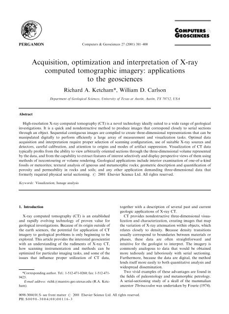
An example <strong>of</strong> the possible utility <strong>of</strong> partial-volume<br />
effects is shown in Fig. 6. A core <strong>of</strong> limestone from the<br />
lower Ismay member <strong>of</strong> the Paradox Formation was<br />
scanned <strong>and</strong> subsequently cut for petrographic analysis.<br />
Individual fractures that appear on the scan were<br />
measured petrographically <strong>and</strong> found to have widths<br />
that were significantly smaller than the pixel dimensions.<br />
The fracture width can be estimated using partialvolume<br />
calculations similar to those used by Johns et al.<br />
(1993), although at least one additional step is required<br />
to take fracture dip into account.<br />
5. Optimization<br />
Medical diagnostic CT has in many instances become<br />
a routine procedure, owing primarily to the fact that<br />
there are a limited number <strong>of</strong> truly different scanning<br />
subjects. Conversely, the wide variety <strong>of</strong> different<br />
materials <strong>and</strong> resolution requirements for geological<br />
investigations generally requires the development <strong>of</strong><br />
specialized scanning procedures, <strong>and</strong> invariably case-bycase<br />
selection <strong>of</strong> scanning parameters. In general, the<br />
objective <strong>of</strong> <strong>optimization</strong> is to maximize the contrast<br />
between features <strong>of</strong> interest while minimizing or<br />
eliminating artifacts that can interfere with analysis.<br />
When planning a scanning project, the first task is to<br />
identify the imaging objectives; these in turn can help<br />
Fig. 6. 100-mm slice through fractured limestone from lower<br />
Ismay member <strong>of</strong> Paradox Formation. Scan field <strong>of</strong> view is<br />
21.5 mm, <strong>and</strong> individual pixels are 42 mm on a side. After<br />
scanning entire volume, sample was cut <strong>and</strong> fractures were<br />
measured in thin section. Fractures are visible despite being<br />
considerably thinner than pixel width, because <strong>of</strong> partial<br />
volume effects. Sample <strong>and</strong> measurements courtesy <strong>of</strong> Dr.<br />
Brenda Kirkl<strong>and</strong>, University <strong>of</strong> Texas at Austin.<br />
define the necessary image resolution, slice thickness,<br />
<strong>and</strong> attenuation discrimination. These requirements<br />
guide decisions about the optimal scanning parameters,<br />
including: source–detector combination; scanning mode;<br />
X-<strong>ray</strong> energy, intensity, <strong>and</strong> spot size; beam filtration, if<br />
any; whether the sample should be scanned in air or<br />
packed; <strong>and</strong> wedge material.<br />
We provide here an example <strong>of</strong> CT as an adaptive<br />
process in which images were progressively improved by<br />
refining scanning technique. The scanned object was a<br />
diamondiferous eclogite from Yakutia, Russia. The<br />
major phases in the rock are clinopyroxene <strong>and</strong> garnet,<br />
with accessory diamond <strong>and</strong> sulfide. Fig. 7a is an initial<br />
50-mm thick scan using the ultra-high-resolution system<br />
with X-<strong>ray</strong>s at 200 keV <strong>and</strong> no other sample preparation<br />
or beam modification; it is essentially a scan with<br />
parameters selected to produce maximum penetration<br />
<strong>and</strong> signal strength, with no regard for mineralogy (i.e.,<br />
with ‘‘everything turned up all the way’’). The scan<br />
suffers from severe beam hardening, <strong>and</strong> although<br />
clinopyroxene, garnet <strong>and</strong> sulfide are fairly easy to<br />
distinguish, one cannot confidently identify diamond,<br />
even though one crystal could be seen on the edge <strong>of</strong><br />
the sample in this plane. By evaluating the relative<br />
attenuation characteristics <strong>of</strong> diamond <strong>and</strong> clinopyroxene,<br />
it was recognized that they have nearly indistinguishable<br />
attenuation coefficients at high X-<strong>ray</strong>
R.A. Ketcham, W.D. Carlson / Computers & Geosciences 27 (2001) 381–400 393<br />
Fig. 7. Three scans <strong>of</strong> diamondiferous eclogite showing effects <strong>of</strong> successive improvements <strong>of</strong> scanning methodology. Phases present,<br />
in order <strong>of</strong> increasing g<strong>ray</strong>scale, are diamond, clinopyroxene, garnet, <strong>and</strong> sulfide. Scanning conditions discussed in text. Sample<br />
approximately 50 mm in long dimension. Sample courtesy <strong>of</strong> Dr. Larry Taylor <strong>and</strong> Dr. R<strong>and</strong>all Keller, University <strong>of</strong> Tennessee at<br />
Knoxville.<br />
energies (>150 keV), but diverge greatly at lower<br />
energies because <strong>of</strong> the low mean atomic number <strong>of</strong><br />
diamond. To help confine the X-<strong>ray</strong> energy spectrum to<br />
a more useful range while attempting to diminish beam<br />
hardening, the scan in Fig. 7b was acquired with X-<strong>ray</strong>s<br />
set at 100 keV <strong>and</strong> pre-filtered with a 1/8 00 brass plate.<br />
The diamond at the edge <strong>of</strong> the sample is now more<br />
easily distinguished, <strong>and</strong> the beam hardening is slightly<br />
diminished by prefiltering. However, these two measures<br />
also produced greater image noise. Optimal imagery was<br />
obtained through use <strong>of</strong> a wedge calibration: the sample<br />
was packed in a cylindrical container with finely ground<br />
garnet powder, the calibration was taken through pure<br />
garnet powder, <strong>and</strong> the brass filter was removed to<br />
increase total X-<strong>ray</strong> intensity (Fig. 7c). The mixture <strong>of</strong><br />
garnet <strong>and</strong> air in the powder make it a close match for<br />
the attenuating characteristics <strong>of</strong> clinopyroxene, <strong>and</strong><br />
thus the calibration was able to eliminate the beamhardening<br />
artifact. Because the wedge material also<br />
allowed the use <strong>of</strong> a higher gain in the video system<br />
during calibration, counting statistics were also improved.<br />
The results are remarkable; not only is the<br />
external diamond even more clearly visible, but previously<br />
invisible diamonds in the center <strong>of</strong> the image have<br />
become apparent, along with a network <strong>of</strong> alteration<br />
zones that may be related to the diamonds’ origin.<br />
6. Visualization <strong>of</strong> CT images<br />
The product <strong>of</strong> a complete X-<strong>ray</strong> CT scan is a threedimensional<br />
matrix <strong>of</strong> relative attenuation values, sometimes<br />
called a ‘‘data brick’’. The most productive way <strong>of</strong><br />
visualizing such data depends on the scientific application.<br />
In many cases, particularly paleontological studies,<br />
two-dimensional slices through the data along three sets<br />
<strong>of</strong> orthogonal planes (coronal, sagittal, horizontal) are<br />
the best way to inspect skeletal structures. The reason<br />
for this is partly traditional, as these planes have long<br />
been used in serial-sectioning studies. In most cases,<br />
however, additional insight can be gained by creating a<br />
three-dimensional representation. We discuss here some<br />
current approaches to 3-D visualization <strong>of</strong> X-<strong>ray</strong> CT<br />
data, <strong>and</strong> in the next section present examples from<br />
various studies.<br />
Two primary techniques exist for creating threedimensional<br />
images from CT data: isocontouring <strong>and</strong><br />
volume rendering. Isocontouring is the more widely<br />
familiar <strong>of</strong> the two; it consists <strong>of</strong> defining one or more<br />
surfaces (isosurfaces) that mark the boundaries <strong>of</strong> the<br />
object(s) <strong>of</strong> interest in the scan. In most cases these<br />
boundaries are assumed to correspond to a certain CT<br />
value, <strong>and</strong> thus the isocontours mark these constant-CTvalue<br />
surfaces in the volume, in much the same manner<br />
as contours on a topographic map mark lines <strong>of</strong><br />
constant elevation. As is analogous to the case <strong>of</strong><br />
constructing topographic maps from gridded data,<br />
isocontour positions are interpolated among the data<br />
points, <strong>and</strong> thus the surfaces are defined on a sub-pixel<br />
scale. In volume rendering each voxel in the volume is<br />
assigned a red–green–blue color (or g<strong>ray</strong>scale) <strong>and</strong> an<br />
opacity, based on its CT number. By setting the opacity<br />
<strong>of</strong> some voxels to zero, they can be made invisible, thus<br />
allowing the observer to ‘‘see through’’ them to<br />
nontransparent materials <strong>of</strong> interest.<br />
These two techniques are useful in different circumstances.<br />
In most cases, we have found volume rendering<br />
to be better suited to CT imagery, because a larger range<br />
<strong>of</strong> information is available to be included in an image. In<br />
particular, if two materials have overlapping CT values<br />
because <strong>of</strong> internal textures, image noise, or artifacts,<br />
defining an isocontour between them becomes arbitrary<br />
<strong>and</strong> hazardous. On the other h<strong>and</strong>, volume rendering<br />
allows the viewer to distinguish the two visually, based<br />
on their changes in average CT value or textural<br />
differences. Also, isocontouring large data sets can be
394<br />
R.A. Ketcham, W.D. Carlson / Computers & Geosciences 27 (2001) 381–400<br />
more dem<strong>and</strong>ing on memory <strong>and</strong> processing power than<br />
volume rendering, <strong>and</strong> in many cases a detailed data set<br />
must be considerably reduced <strong>and</strong> simplified before an<br />
isocontour can be calculated <strong>and</strong> rendered.<br />
Nevertheless, isocontouring is frequently a desirable<br />
goal because <strong>of</strong> the crisper images <strong>and</strong> more detailed<br />
surface information <strong>and</strong> volumetric measurements it can<br />
provide. To be effective for CT data, an additional step<br />
called segmentation is generally necessary to separate<br />
features <strong>of</strong> interest based on more robust criteria than<br />
simply the CT values. Some steps that can aid<br />
segmentation include noise reduction, edge enhancement<br />
<strong>and</strong> tracking, <strong>and</strong> region growing (Glenn et al., 1981).<br />
The field is continuing to develop rapidly, incorporating<br />
a wide variety <strong>of</strong> computational <strong>and</strong> statistical techniques<br />
(Glasbey <strong>and</strong> Horgan, 1995). If quantitative<br />
measurements are desired from X-<strong>ray</strong> CT data, additional<br />
segmentation steps that take partial-volume<br />
effects into account may be required. For example,<br />
Peyton et al. (1992) found that sizes <strong>of</strong> pores in soil as<br />
imaged by medical CT are not directly proportional to<br />
the actual pore sizes, due to blurring <strong>and</strong> partial-volume<br />
effects, which cause different-sized pores to be manifested<br />
with different CT values. Thus, to establish pore<br />
size they use an iterative procedure based on scanned<br />
st<strong>and</strong>ards that link pore sizes to cut<strong>of</strong>f CT values.<br />
Another complication inherent in isosurfacing is that the<br />
CT value marking the boundary <strong>of</strong> an object will change<br />
depending on the character <strong>of</strong> adjacent materials. For<br />
example, if a garnet has a CT value <strong>of</strong> 3000 <strong>and</strong> is<br />
adjacent to both quartz with a CT value <strong>of</strong> 2000 <strong>and</strong> air<br />
with a CT value <strong>of</strong> 0, the appropriate CT value marking<br />
its boundary will be roughly 2500 on the side facing<br />
quartz <strong>and</strong> 1500 on the side facing air. In many cases the<br />
resulting data corruption will be slight, particularly if the<br />
data are high-resolution, but it should be considered<br />
when evaluating data precision.<br />
7. Examples <strong>of</strong> geological applications<br />
In this section we summarize the results from some <strong>of</strong><br />
the geological studies carried out at the UT High-<br />
Resolution X-<strong>ray</strong> CT Facility. Most <strong>of</strong> the figures in this<br />
section have accompanying animations viewable on the<br />
CD that supplements this volume or on the Computers &<br />
Geosciences Internet site.<br />
7.1. Quantitative analysis <strong>of</strong> metamorphic textures<br />
The original stimulus for establishment <strong>of</strong> the UT<br />
high-resolution X-<strong>ray</strong> CT facility was the recognition<br />
that 3-D CT data on the sizes <strong>and</strong> locations <strong>of</strong> crystals<br />
permit quantitative analysis <strong>of</strong> textures. In particular,<br />
such data permit discrimination between competing<br />
hypotheses for the atomic-scale mechanisms controlling<br />
the growth <strong>of</strong> metamorphic porphyroblasts. The result<br />
Fig. 8. Isosurface image <strong>of</strong> garnet (violet) <strong>and</strong> staurolite<br />
(yellow–brown) in schist from Picuris Mountains, New Mexico.<br />
Sizes <strong>and</strong> spatial positions <strong>of</strong> porphyroblasts as revealed by X-<br />
<strong>ray</strong> CT can be used to determine whether there is any ordering<br />
among them, which may indicate that during nucleation <strong>and</strong><br />
growth crystals were surrounded by diffusionally depleted zones<br />
that restricted further nucleation in their vicinity. Image<br />
bounding box is approximately 45 45 42 mm.<br />
<strong>of</strong> initial fo<strong>ray</strong>s into this field demonstrated that reaction<br />
kinetics limited by rates <strong>of</strong> intergranular diffusion were<br />
common for garnet porphyroblasts in regionally metamorphosed<br />
rocks (Carlson <strong>and</strong> Denison, 1992; Denison<br />
<strong>and</strong> Carlson, 1997; Denison et al., 1997). Numerical<br />
simulations <strong>of</strong> such processes (Carlson et al., 1995),<br />
constrained by the X-<strong>ray</strong> CT data, allowed first-ever<br />
estimates <strong>of</strong> critical kinetic parameters such as the<br />
absolute rates <strong>of</strong> garnet nucleation <strong>and</strong> <strong>of</strong> intergranular<br />
diffusion <strong>of</strong> aluminum (the transport <strong>of</strong> which is ratelimiting).<br />
Compositional studies based on serial sectioning<br />
guided by the CT data (Chern<strong>of</strong>f <strong>and</strong> Carlson, 1997, 1999)<br />
demonstrated the importance <strong>of</strong> this reaction control:<br />
sluggish intergranular diffusion appears to account for the<br />
observation that Ca <strong>and</strong> many trace elements in some<br />
metamorphic rocks fail to equilibrate at the thin-section<br />
scale during garnet growth, severely restricting the<br />
confidence that can be placed in thermobarometric<br />
methods that assume otherwise. Further studies are being<br />
conducted in pursuit <strong>of</strong> deeper underst<strong>and</strong>ing <strong>of</strong> reaction<br />
processes at subcrystalline length scales, including analysis<br />
<strong>of</strong> porphyroblasts other than garnet <strong>and</strong> rocks metamorphosed<br />
under a wider range <strong>of</strong> temperature, pressure,<br />
strain rate, <strong>and</strong> fluid-flow conditions.<br />
Fig. 8 shows an isosurface rendering <strong>of</strong> garnet <strong>and</strong><br />
staurolite in a pelitic schist from the Picuris Mountains,
R.A. Ketcham, W.D. Carlson / Computers & Geosciences 27 (2001) 381–400 395<br />
New Mexico. The markedly different attenuation<br />
properties <strong>of</strong> the two phases combined with the<br />
homogeneity <strong>of</strong> the micaceous matrix in this sample<br />
made their separation fairly straightforward, but a<br />
number <strong>of</strong> steps were required to produce the imagery<br />
shown. First, a subset <strong>of</strong> the original data was taken <strong>and</strong><br />
re-scaled from 512 to 128 pixels on a side. This data set<br />
was then used to construct the set <strong>of</strong> isocontours for<br />
garnet, the phase with the highest CT value. Because an<br />
isocontour enclosing the CT value <strong>of</strong> staurolite crystals<br />
would also enclose garnet, in order to render the<br />
staurolite crystals, all <strong>of</strong> the garnets had to be erased<br />
from the original data set. Furthermore, because <strong>of</strong><br />
blurring <strong>and</strong> partial volume effects, the garnet boundaries<br />
have CT values that overlap with the staurolites, so<br />
the material immediately adjacent to the garnets had to<br />
be deleted as well. The staurolite isosurfaces were<br />
calculated from the modified data set, <strong>and</strong> the two sets<br />
<strong>of</strong> isosurfaces were then superposed.<br />
7.2. Metasomatic processes in the Earth’s mantle <strong>and</strong><br />
the origin <strong>of</strong> diamond<br />
The nature <strong>and</strong> extent <strong>of</strong> mantle metasomatism <strong>and</strong><br />
the origin <strong>of</strong> diamond } fundamental enigmas in<br />
mantle petrology } have been addressed by complete<br />
3-D characterization <strong>of</strong> textural relationships in extraordinary<br />
diamondiferous eclogites from Udachnaya,<br />
Siberia (Keller et al., 1999). These rare specimens are<br />
problematic to study by petrographic means, in part<br />
because <strong>of</strong> the extreme difficulty <strong>of</strong> sawing through<br />
diamonds. Furthermore, two-dimensional analysis may<br />
miss crucial textural features relating diamonds to<br />
fractures <strong>and</strong> other phases. CT imagery revealed that<br />
none <strong>of</strong> the diamonds in the specimen are completely<br />
enclosed in either garnet or clinopyroxene, but instead<br />
all are spatially associated with a 3-D network <strong>of</strong><br />
alteration zones that penetrate the specimen (Fig. 9).<br />
The implication is that the diamond postdates formation<br />
<strong>of</strong> the eclogitic assemblage, <strong>and</strong> most likely originated<br />
during a metasomatic event responsible for localized<br />
alteration along subplanar zones; this hypothesis is now<br />
being tested further by means <strong>of</strong> chemical <strong>and</strong> isotopic<br />
analysis <strong>of</strong> the disaggregated specimen.<br />
The images in Fig. 9 were constructed using volume<br />
rendering instead <strong>of</strong> isosurfacing. Each phase is rendered<br />
transparent, partially transparent or opaque to allow<br />
inspection <strong>of</strong> different textural features. Except for<br />
removal <strong>of</strong> the wedge material, this rendering did not<br />
require any processing or enhancement <strong>of</strong> the original<br />
images.<br />
7.3. Melt generation, segregation, <strong>and</strong> migration in a<br />
lodranite<br />
Initial curatorial slabbing <strong>of</strong> the lodranite meteorite<br />
GRA95209 produced the surprising discovery <strong>of</strong> a large<br />
region <strong>of</strong> metal segregated from the mixed silicate–metal<br />
matrix. X-<strong>ray</strong> CT was used to determine, nondestructively,<br />
the geometry <strong>of</strong> this metal segregation (Carlson<br />
Fig. 9. Volume renderings <strong>of</strong> diamondiferous eclogite from Yakutia, Russia, based on scans such as those shown in Fig. 7C. Images<br />
reveal diamond (blue), sulphides (yellow) <strong>and</strong> garnet (red); clinopyroxene that comprises most <strong>of</strong> specimen has been rendered entirely<br />
transparent except for network <strong>of</strong> alteration zones (green) that intersects all <strong>of</strong> the diamonds. A. Portion <strong>of</strong> sample with alteration<br />
zones highlighted. B. Smaller subset with garnet <strong>and</strong> sulphide phases enhanced. Sample courtesy <strong>of</strong> Dr. Larry Taylor <strong>and</strong> Dr. R<strong>and</strong>all<br />
Keller, University <strong>of</strong> Tennessee at Knoxville.
396<br />
R.A. Ketcham, W.D. Carlson / Computers & Geosciences 27 (2001) 381–400<br />
Fig. 10. Volume rendering <strong>of</strong> distribution <strong>of</strong> metal in lodranite<br />
meteorite GRA95209. Long dimension <strong>of</strong> sample is approximately<br />
80 mm. Sample courtesy <strong>of</strong> Dr. Marilyn Lindstrom,<br />
NASA, <strong>and</strong> Dr. Tim McCoy, Smithsonian Institution.<br />
<strong>and</strong> McCoy, 1998; McCoy <strong>and</strong> Carlson, 1998). A large<br />
slab <strong>of</strong> metal was found to be present along the exterior<br />
surface <strong>of</strong> much <strong>of</strong> the specimen (obscured beneath a<br />
fusion crust), extending into its interior, connected to<br />
several veins <strong>and</strong> stringers <strong>of</strong> metal within the matrix;<br />
the matrix was seen to include metal-poor silicaterich<br />
regions (Fig. 10). The coalescing veins <strong>and</strong> dikelets<br />
revealed in 3-D by X-<strong>ray</strong> CT are readily interpreted as<br />
melt-migration pathways, <strong>and</strong> measurements from CT<br />
data <strong>of</strong> the relative volumes <strong>of</strong> metal <strong>and</strong> metal-depleted<br />
matrix permit mass-balance calculations that rule out a<br />
completely local origin for the melt. The images therefore<br />
record a complicated <strong>and</strong> intriguing picture <strong>of</strong> melt<br />
generation <strong>and</strong> migration in the lodranite parent body, a<br />
primitive planetesimal that is one <strong>of</strong> the best existing<br />
models for processes occurring during core–mantle<br />
segregation in the terrestrial planets.<br />
7.4. Characterization <strong>of</strong> porosity in reservoir limestones<br />
X-<strong>ray</strong> CT imagery <strong>of</strong> rock porosity can be used in a<br />
variety <strong>of</strong> applications, from qualitative characterization<br />
<strong>of</strong> porosity types <strong>and</strong> fabrics (Beall et al., 1996; Gournay<br />
<strong>and</strong> Kirkl<strong>and</strong>, 1998) to quantitative description <strong>of</strong> pore<br />
networks for examination <strong>and</strong> modeling <strong>of</strong> fluid-flow<br />
characteristics. Pore geometry, pore dimensions, porethroat<br />
sizes <strong>and</strong> surface roughness all affect flow. Poreto-pore-throat<br />
geometries may affect migration <strong>of</strong><br />
hydrocarbons <strong>and</strong> are certainly significant in production<br />
<strong>of</strong> hydrocarbons from the reservoir. X-<strong>ray</strong> CT data have<br />
been used to examine four types <strong>of</strong> porosity in reservoir<br />
limestones: fracture porosity, which is the most important<br />
for flow <strong>and</strong> interconnection <strong>of</strong> other types <strong>of</strong><br />
porosity; moldic (intra-particle) porosity; inter-particle<br />
porosity; <strong>and</strong> vuggy porosity. The imagery is being used<br />
Fig. 11. Volume rendering image <strong>of</strong> fracture network in<br />
reservoir limestone from lower Ismay formation <strong>of</strong> Paradox<br />
Formation, west Texas. Reconstruction is approximately<br />
15 mm wide. Sample courtesy <strong>of</strong> Dr. Brenda Kirkl<strong>and</strong>,<br />
University <strong>of</strong> Texas at Austin.<br />
to construct three-dimensional permeability-network<br />
maps to help link the geometric characteristics <strong>of</strong> the<br />
pore network to its flow properties. Such maps may help<br />
identify regions <strong>of</strong> suspected laminar versus turbulent<br />
flow that could affect the diagenetic processes governing<br />
the formation <strong>of</strong> porous hydrocarbon reservoirs <strong>and</strong><br />
aquifers.<br />
An example image from a fractured limestone is shown<br />
in Fig. 6, <strong>and</strong> a 3-D volume-rendering reconstruction<br />
<strong>of</strong> the stacked images to reveal the fractures <strong>and</strong> pore<br />
spaces is shown in Fig. 11. An isosurface rendering<br />
<strong>of</strong> the original data to show the fractures would be<br />
problematic, because as discussed above there is no<br />
single appropriate CT value that marks the rock-void<br />
boundary across the size range <strong>of</strong> fractures present.<br />
Volume rendering allowed us to circumvent this difficulty<br />
by showing different-sized fractures in different colors<br />
<strong>and</strong> intensities. In this case, small fractures are shown in<br />
semitransparent red, <strong>and</strong> large, open fractures appear as<br />
bright yellow with red boundaries.<br />
7.5. Structural origin <strong>and</strong> 3-D extent <strong>of</strong> b<strong>and</strong>ing in<br />
scleractinian corals<br />
Annual b<strong>and</strong>ing in scleractinian corals makes them<br />
useful indicators <strong>of</strong> temperature <strong>and</strong> chemical conditions<br />
through time in coral reefs, thus allowing them to
R.A. Ketcham, W.D. Carlson / Computers & Geosciences 27 (2001) 381–400 397<br />
Fig. 12. A. X-<strong>ray</strong> CT image <strong>of</strong> 25-cm cut cube <strong>of</strong> coral Diploria strigosa used to study structural elements responsible for density<br />
b<strong>and</strong>ing. Slice thickness is 100 mm. High-density b<strong>and</strong>s correlate with thickened septa <strong>and</strong> tightly organized <strong>and</strong> thickened columella.<br />
Sample courtesy <strong>of</strong> Dr. Richard Dodge <strong>and</strong> Kevin Helmle <strong>of</strong> Nova Southeastern University. B. Three-dimensional pattern <strong>of</strong> annual<br />
growth b<strong>and</strong>ing in sample <strong>of</strong> coral Montastrea annularis. Sample is approximately 90 mm wide. Sample courtesy <strong>of</strong> Dr. Judy Lang <strong>and</strong><br />
Dr. Lynton L<strong>and</strong>, University <strong>of</strong> Texas at Austin.<br />
serve as precise long-term climate records (Knutson<br />
et al., 1972; Swart et al., 1996). This b<strong>and</strong>ing is commonly<br />
imaged using traditional projection X-radiography,<br />
which can locate the b<strong>and</strong>s in cut slabs <strong>of</strong> coral. X-<strong>ray</strong><br />
CT <strong>of</strong>fers a number <strong>of</strong> opportunities to refine underst<strong>and</strong>ing<br />
<strong>and</strong> implementation <strong>of</strong> these techniques. Ultrahigh-resolution<br />
CT has been used to inspect in detail the<br />
geometrical characteristics <strong>of</strong> the b<strong>and</strong>s in Diploria<br />
strigosa (Fig. 12A) to determine the structural elements<br />
responsible for them (Helmle, 1998). Results indicate that<br />
b<strong>and</strong>ing architecture varies across coral taxa.<br />
High-resolution X-<strong>ray</strong> CT can also be used to ensure<br />
that sampling techniques properly account for the full<br />
three-dimensional variability <strong>of</strong> coral b<strong>and</strong>ing. Sampling<br />
is commonly done using a coring drill positioned normal<br />
to the coral surface. A complete scan <strong>of</strong> coral<br />
Montastrea annularis (Fig. 12B) reveals that these b<strong>and</strong>s<br />
may not remain perpendicular to this surface because <strong>of</strong><br />
variations in the relative growth rates <strong>of</strong> different parts<br />
<strong>of</strong> the coral through time. Visualization <strong>of</strong> these features<br />
can be effectively done through single images <strong>and</strong><br />
animated series <strong>of</strong> digitally resliced data.<br />
7.6. Skull morphology <strong>of</strong>Kryptobataar<br />
The ability <strong>of</strong> high-resolution X-<strong>ray</strong> CT to image<br />
fossil interiors <strong>and</strong> exteriors pr<strong>of</strong>oundly increases the<br />
amount <strong>of</strong> morphological information that can be<br />
nondestructively obtained from them. Furthermore, it<br />
can also fulfill a curatorial function, as once the fossil is<br />
scanned its data can be widely disseminated. For<br />
example, under normal circumstances, to study a certain<br />
species a paleontologist may have to travel to all <strong>of</strong> the<br />
different museums that have relevant specimens. The<br />
potential now exists to catalog museum collections <strong>and</strong><br />
make them available on electronic media or, eventually,<br />
via the Internet.<br />
Although most paleontologists have computers, some<br />
may not have the computational resources <strong>and</strong> expertise<br />
required to efficiently visualize <strong>and</strong> manipulate the tens<br />
to hundreds <strong>of</strong> megabytes <strong>of</strong> data that CT can produce<br />
for a single specimen. Thus, distribution <strong>of</strong> CT data<br />
generally includes preparation <strong>of</strong> images <strong>and</strong> animations<br />
for trouble-free viewing <strong>and</strong> analysis. Examples <strong>of</strong> such<br />
data processing include reslicing the data along the<br />
traditional orthogonal planes used for morphological<br />
analysis <strong>and</strong> constructing three-dimensional renderings.<br />
Fig. 13 shows an example <strong>of</strong> some rendering <strong>and</strong> datadistribution<br />
techniques for a high-resolution scan <strong>of</strong> a<br />
specimen <strong>of</strong> Kryptobataar dashzevegi, from the late<br />
Cretaceous Djadokhta Formation, Ukhaa Tolgod,<br />
Mongolia. Fig. 13A shows a volume rendering <strong>of</strong> the<br />
Kryptobataar skull with part <strong>of</strong> it digitally removed to<br />
reveal the internal features at the slice plane. The ability<br />
to simultaneously display the internal structure <strong>and</strong><br />
external form <strong>of</strong> the skull helps in the identification <strong>of</strong><br />
individual skeletal elements <strong>and</strong> their relation to each<br />
other. Figs. 13B <strong>and</strong> C are three-dimensional images <strong>of</strong><br />
the entire skull created using volume rendering <strong>and</strong><br />
isosurfacing, respectively. The isosurface shows the<br />
details <strong>of</strong> the skull surface more clearly, but the volume<br />
rendering allows for better differentiation between skull<br />
<strong>and</strong> matrix, <strong>and</strong> reproduces more faithfully the state <strong>of</strong><br />
the original specimen.
398<br />
R.A. Ketcham, W.D. Carlson / Computers & Geosciences 27 (2001) 381–400<br />
The University <strong>of</strong> Texas at Austin High-Resolution<br />
X-<strong>ray</strong> CT Facility was created with support from the<br />
W.M. Keck Foundation, the National Science Foundation<br />
(NSF EAR-9406258; NSF EAR-9816020), <strong>and</strong> the<br />
Geology Foundation <strong>of</strong> the University <strong>of</strong> Texas. Many<br />
<strong>of</strong> the images <strong>and</strong> animations presented here were<br />
created by Dr. Cambria Denison, who also participated<br />
in the development <strong>of</strong> many <strong>of</strong> the scanning techniques<br />
described <strong>and</strong> employed. We thank two anonymous<br />
referees for constructive comments, as well as editors<br />
R. Marschallinger <strong>and</strong> S. Johnson.<br />
References<br />
Fig. 13. Three-dimensional reconstructions based on X-<strong>ray</strong> CT<br />
data <strong>of</strong> skull <strong>of</strong> Kryptobataar. Skull is 29 mm long. (A) volume<br />
rendering <strong>of</strong> skull from above, with cutaway showing internal<br />
structure; (B) volume rendering <strong>of</strong> skull; (C) isosurface<br />
rendering <strong>of</strong> skull. Sample courtesy <strong>of</strong> Dr. Guillermo W.<br />
Rougier, University <strong>of</strong> Louisville.<br />
8. Summary<br />
High-resolution X-<strong>ray</strong> CT is ideally suited to many<br />
geological applications, <strong>and</strong> its potential is only beginning<br />
to be explored. We hope that the overview <strong>and</strong><br />
examples given here can provide inspiration for new<br />
investigations that will benefit from further utilization <strong>of</strong><br />
this breakthrough technology.<br />
Acknowledgements<br />
Amos, C.L., Sutherl<strong>and</strong>, T.F., Radzijewski, B., Doucette, M.,<br />
1996. A rapid technique to determine bulk density <strong>of</strong> finegrained<br />
sediments by X-<strong>ray</strong> <strong>computed</strong> tomography. Journal<br />
<strong>of</strong> Sedimentary Research 66 (5), 1023–1024.<br />
Anderson, S.H., Peyton, R.L., Wigger, J.W., Gantzer, C.J., 1992.<br />
Influence <strong>of</strong> aggregate size on solute transport as measured<br />
using <strong>computed</strong> tomography. Geoderma 53, 387–398.<br />
Arnold, J.R., Testa, J.P.J., Friedman, P.J., Kambic, G.X., 1982.<br />
Computed tomographic analysis <strong>of</strong> meteorite inclusions.<br />
Science 219, 383–384.<br />
ASTM, 1992. St<strong>and</strong>ard Guide for Computed Tomography<br />
(CT) Imaging, ASTM Designation E 1441-92a. In: 1992<br />
Annual Book <strong>of</strong> ASTM St<strong>and</strong>ards, Section 3 Metals Test<br />
Methods <strong>and</strong> Analytical Procedures. ASTM, Philadelphia,<br />
pp. 690–713.<br />
Bauer, R.L., Ketcham, R.A., Denison, C., Carlson, W.D.,<br />
1998. X-<strong>ray</strong> <strong>computed</strong> tomography (CT) imaging <strong>of</strong> spiral<br />
inclusion trails <strong>and</strong> the external morphology <strong>of</strong> garnet<br />
porphyroblasts. Eos 79 (17), 357.<br />
Beall, J.L., Gordon, I.T., Gournay, J.P., Kirkl<strong>and</strong>, B.L., White,<br />
J.V., 1996. Analysis <strong>of</strong> porosity in lower Ismay phylloid algal<br />
packstone using high-resolution <strong>computed</strong> X-<strong>ray</strong> tomography.<br />
American Association <strong>of</strong> Petroleum Geologists, Annual<br />
Meeting Abstracts 5, 13.<br />
Brown, M.A., Brown, M., Carlson, W.D., Denison, C., 1999.<br />
Topology <strong>of</strong> syntectonic melt flow networks in the deep crust:<br />
inferences from three-dimensional images <strong>of</strong> leucosome geometry<br />
in migmatites. American Mineralogist 84 1793–1818.<br />
Carlson, W.D., Denison, C., 1992. Mechanisms <strong>of</strong> porphyroblast<br />
crystallization: results from high-resolution <strong>computed</strong><br />
X-<strong>ray</strong> tomography. Science 257, 1236–1239.<br />
Carlson, W.D., Denison, C., Ketcham, R.A., 1995. Controls on<br />
the nucleation <strong>and</strong> growth <strong>of</strong> porphyroblasts: kinetics from<br />
natural textures <strong>and</strong> numerical models. Geological Journal<br />
30, 207–225.<br />
Carlson, W.D., McCoy, T.J., 1998. High-resolution X-<strong>ray</strong><br />
<strong>computed</strong> tomography <strong>of</strong> lodranite GRA 95209. In: Lunar<br />
<strong>and</strong> Planetary Science, Vol. XXIX, CD-ROM. Lunar <strong>and</strong><br />
Planetary Science Institute, Houston.<br />
Chen, M.-R., Hinkley, R.E., Killough, J.E., 1996. Computed<br />
tomography imaging <strong>of</strong> air sparging in porous media. Water<br />
Resources Research 32 (10), 3013–3024.<br />
Chern<strong>of</strong>f, C.B., Carlson, W.D., 1997. Disequilibrium for Ca<br />
during growth <strong>of</strong> pelitic garnet. Journal <strong>of</strong> Metamorphic<br />
Geology 15 (4), 421–438.<br />
Chern<strong>of</strong>f, C.B., Carlson, W.D., 1999. Trace element zoning as a<br />
record <strong>of</strong> chemical disequilibrium during garnet growth.<br />
Geology 27 (6), 555–558.
R.A. Ketcham, W.D. Carlson / Computers & Geosciences 27 (2001) 381–400 399<br />
Cifelli, R.L., Muizon, C.D., 1998a. Marsupial mammal from<br />
the Upper Cretaceous North Horn Formation, central Utah.<br />
Journal <strong>of</strong> Paleontology 72, 532–537.<br />
Cifelli, R.L., Muizon, C.D., 1998b. Tooth eruption <strong>and</strong><br />
replacement pattern in early marsupials. Comptes Rendus,<br />
Académie des Sciences 326, 215–220.<br />
Cifelli, R.L., Rowe, T.B., Luckett, W.P., Banta, J., Reyes, R.,<br />
Howes, R.I., 1996. Fossil evidence for the origin <strong>of</strong> the<br />
marsupial pattern <strong>of</strong> tooth replacement. Nature 379, 715–718.<br />
Coker, D.A., Torquato, S., Dunsmuir, J.H., 1996. Morphology<br />
<strong>and</strong> physical properties <strong>of</strong> Fontainebleau s<strong>and</strong>stone via a<br />
tomographic analysis. Journal <strong>of</strong> Geophysical Research 101<br />
(B8), 17,497–17,506.<br />
Conroy, G.C., Vannier, M.W., 1984. Noninvasive three-dimensional<br />
computer imaging <strong>of</strong> matrix-filled fossil skulls by highresolution<br />
<strong>computed</strong> tomography. Science 226, 456–458.<br />
Denison, C., Carlson, W.D., 1997. Three-dimensional quantitative<br />
textural analysis <strong>of</strong> metamorphic rocks using high-resolution<br />
<strong>computed</strong> X-<strong>ray</strong> tomography: Part II. Application to natural<br />
samples. Journal <strong>of</strong> Metamorphic Geology 15 (1), 45–57.<br />
Denison, C., Carlson, W.D., Ketcham, R.A., 1997. Threedimensional<br />
quantitative textural analysis <strong>of</strong> metamorphic<br />
rocks using high-resolution <strong>computed</strong> X-<strong>ray</strong> tomography:<br />
Part I. Methods <strong>and</strong> techniques. Journal <strong>of</strong> Metamorphic<br />
Geology 15 (1), 29–44.<br />
Flannery, B.P., Deckman, H.W., Roberge, W.G., D’Amico,<br />
K.L., 1987. Three-dimensional X-<strong>ray</strong> microtomography.<br />
Science 237, 1439–1444.<br />
Fourie, S., 1974. The cranial morphology <strong>of</strong> Thrinaxodon<br />
liorhinus Seeley. Annals <strong>of</strong> the South African Museum 65,<br />
337–400.<br />
Glasbey, C.A., Horgan, G.W., 1995. Image Analysis for the<br />
Biological Sciences. Wiley, New York, 218 pp.<br />
Glenn, W.V., Harlow, C.A., Dwyer, S.J.I., Rhodes, M.L.,<br />
Parker, D.L., 1981. Image manipulation <strong>and</strong> pattern<br />
recognition. In: Newton, T.H., Potts, D.G. (Eds.), Technical<br />
Aspects <strong>of</strong> Computed Tomography. Mosby, St. Louis, pp.<br />
4326–4354.<br />
Gournay, J.P., Kirkl<strong>and</strong>, B.L., 1998. Fabric development in<br />
phylloid algal bioherms. American Association <strong>of</strong> Petroleum<br />
Geologists, Annual Meeting Abstracts 7.<br />
Haubitz, B., Prokop, M., Dohring, W., Ostrom, J.H., Wellnh<strong>of</strong>er,<br />
P., 1988. Computed tomography <strong>of</strong> Archaeopteryx.<br />
Paleobiology 14 (2), 206–213.<br />
Heijs, A.W.J., De Lange, J., Schoute, J.F.T., Bouma, J., 1995.<br />
Computed tomography as a tool for non-destructive analysis<br />
<strong>of</strong> flow patterns in macroporous clay soils. Geoderma 64,<br />
183–196.<br />
Helmle, K., 1998. Relationships between skeletal architecture<br />
<strong>and</strong> density b<strong>and</strong>ing in the reef coral Diploria strigosa from<br />
X-radiography, X-<strong>ray</strong> <strong>computed</strong> tomography, <strong>and</strong> image<br />
analysis techniques. Oceanographic Center, M.S. Nova<br />
Southeastern University, Dania, FL, 86 pp.<br />
Hendee, W.R., 1979. Medical Radiation Physics. 2nd edn. Year<br />
Book Medical Publishers, Chicago, 517 pp.<br />
Johns, R.A., Steude, J.S., Castanier, L.M., Roberts, P.V., 1993.<br />
Nondestructive measurements <strong>of</strong> fracture aperture in crystalline<br />
rock cores using X-<strong>ray</strong> <strong>computed</strong> tomography. Journal<br />
<strong>of</strong> Geophysical Research 98 (B2), 1889–1900.<br />
Keller, R.A., Taylor, L.A., Snyder, G.A., Sobolev, V.N.,<br />
Carlson, W.D., Bezborodov, S.M., Sobolev, N.V., 1999.<br />
Detailed pull-apart <strong>of</strong> a diamondiferous eclogite xenolith:<br />
Implications for mantle processes during diamond genesis.<br />
In: Gurney, J.J., Gurney, J.L., Pascoe, M.D., Richardson,<br />
S.H. (Eds.), Proc. 7th International Kimberlite Conf., Vol. 1,<br />
National Printers, So. Afr., pp. 397–412.<br />
Kinney, J.H., Breuning, T.M., Starr, T.L., Haupt, D., Nichols,<br />
M.C., Stock, S.R., Butts, M.D., Saroyan, R.A., 1993. X-<strong>ray</strong><br />
tomographic study <strong>of</strong> chemical vapor infiltration processing<br />
<strong>of</strong> ceramic deposits. Science 260, 782–789.<br />
Knutson, D.W., Buddemeier, R.W., Smith, S.V., 1972. Coral<br />
chronometers: seasonal growth b<strong>and</strong>s in reef corals. Science<br />
177, 270–272.<br />
Kretz, R., 1966. Grain size distribution for certain metamorphic<br />
minerals in relation to nucleation <strong>and</strong> growth. Journal <strong>of</strong><br />
Geology 74, 147–173.<br />
Kretz, R., 1969. On the spatial distribution <strong>of</strong> crystals in rocks.<br />
Lithos 2, 39–66.<br />
Kretz, R., 1993. A garnet population in Yellowknife schist,<br />
Canada. Journal <strong>of</strong> Metamorphic Geology 11, 101–120.<br />
Kuebler, K.E., McSween, H.Y., Carlson, W.D., Hirsch, D.,<br />
1999. Sizes <strong>and</strong> masses <strong>of</strong> chondrules <strong>and</strong> metal-troilite grains<br />
in ordinary chondrites: possible implications for nebular<br />
sorting. Icarus 141, 96–106.<br />
Le Calvez, J., Vendeville, B.C., 1999. X-<strong>ray</strong> tomographic<br />
analysis <strong>of</strong> a physical experiment <strong>of</strong> salt tongue remobilization<br />
during sedimentation. AAPG International Conference<br />
<strong>and</strong> Exhibition Program, 31.<br />
Markowicz, A.A., 1993. X-<strong>ray</strong> physics. In: Van Grieken, R.E.,<br />
Markowicz, A.A. (Eds.), H<strong>and</strong>book <strong>of</strong> X-<strong>ray</strong> Spectrometry.<br />
Marcel Dekker, New York, pp. 1–28.<br />
McCoy, T.J., Carlson, W.D., 1998. Opaque minerals in GRA<br />
95209 lodranite: a snapshot <strong>of</strong> metal segregation. In: Lunar<br />
<strong>and</strong> Planetary Science XXIX, CD-ROM. Lunar <strong>and</strong> Planetary<br />
Science Institute, Houston.<br />
Moline, G.R., Knight, C.R., Ketcham, R.A., 1997. Laboratory<br />
measurement <strong>of</strong> transport processes in a fractured limestone/<br />
shale saprolite using solute <strong>and</strong> colloid tracers. Geological<br />
Society <strong>of</strong> America, Abstracts with Programs 29, 370.<br />
Newton, T.H., Potts, D.G., 1981. Technical Aspects <strong>of</strong><br />
Computed Tomography, Vol. 5. Mosby, St. Louis, 615 pp.<br />
Ogasawara, Y., Ando, T., Carlson, W.D., Ketcham, R.A.,<br />
1998. Serial-grinding <strong>and</strong> X-<strong>ray</strong> CT techniques for 3D<br />
observation <strong>of</strong> rock textures } an example <strong>of</strong> magmamingled<br />
rock from Colorado. Eos 79 (17), 356.<br />
Orsi, T.H., Anderson, A.L., 1995. X-<strong>ray</strong> <strong>computed</strong> tomography<br />
<strong>of</strong> macroscale variability in sediment physical properties,<br />
<strong>of</strong>fshore Louisiana. In: John, C.J., Byrnes, M.R. (Eds.),<br />
Transactions <strong>of</strong> the Forty-Fifth Annual Convention <strong>of</strong> the<br />
Gulf Coast Association <strong>of</strong> Geological Societies. Gulf Coast<br />
Association <strong>of</strong> Geological Societies, New Orleans, pp. 475–480.<br />
Orsi, T.H., Edwards, C.M., Anderson, A.L., 1994. X-<strong>ray</strong><br />
<strong>computed</strong> tomography: a nondestructive method for quantitative<br />
analysis <strong>of</strong> sediment cores. Journal <strong>of</strong> Sedimentary<br />
Research, Section A: Sedimentary Petrology <strong>and</strong> Processes<br />
64 (3), 690–693.<br />
Peyton, R.L., Haeffner, B.A., Anderson, S.H., Gantzer, C.J.,<br />
1992. Applying X-<strong>ray</strong> CT to measure macropore diameters in<br />
undisturbed soil cores. Geoderma 53, 329–340.<br />
Proussevitch, A., Ketcham, R.A., Carlson, W.D., Sahagian, D.,<br />
1998. Preliminary results <strong>of</strong> X-<strong>ray</strong> CT analysis <strong>of</strong> Hawaiian<br />
vesicular basalts. Eos 79 (17), 360.
400<br />
R.A. Ketcham, W.D. Carlson / Computers & Geosciences 27 (2001) 381–400<br />
Pullan, B.R., Ritchings, R.T., Isherwood, I., 1981. Accuracy<br />
<strong>and</strong> meaning <strong>of</strong> <strong>computed</strong> tomography attenuation<br />
values. In: Newton, T.H., Potts, D.G. (Eds.), Technical<br />
Aspects <strong>of</strong> Computed Tomography. Mosby, St. Louis, pp.<br />
3904–3917.<br />
Ramach<strong>and</strong>ran, G.N., Lakshmina<strong>ray</strong>anan, A.V., 1970. Threedimensional<br />
reconstruction from radiographs <strong>and</strong> electron<br />
micrographs: application <strong>of</strong> convolutions instead <strong>of</strong> Fourier<br />
transforms. Proceedings <strong>of</strong> the National Academy <strong>of</strong> Science<br />
68, 2236–2240.<br />
Rowe, T., 1996. Coevolution <strong>of</strong> the mammalian middle ear <strong>and</strong><br />
neocortex. Science 273, 651–654.<br />
Rowe, T.B., Carlson, W.D., Bottdorf, W.W., 1993. Thrinaxodon<br />
Digital Atlas <strong>of</strong> the Skull, CD-ROM, 623 Mb.<br />
University <strong>of</strong> Texas Press, Austin.<br />
Schreurs, G., Hänni, R., 1998. 4-D analysis <strong>of</strong> analogue model<br />
experiments. American Association <strong>of</strong> Petroleum Geologists<br />
Bulletin 82 (10), 1965.<br />
Shepp, L.A., Logan, B.F., 1974. Reconstructing interior head<br />
tissue from X-<strong>ray</strong> transmissions. IEEE Transactions on<br />
Nuclear Science NS-21, 228–236.<br />
Silver, M.D., 1994. Target self-attenuation extension to the<br />
Desorby <strong>and</strong> Boyer thick-target bremsstrahlung spectrum.<br />
Medical Physics 21 (4), 577–579.<br />
Swart, P.K., Healy, G.F., Dodge, R.E., Kramer, P., Hudson,<br />
J.H., Halley, R.B., Robblee, M.B., 1996. The stable oxygen<br />
<strong>and</strong> carbon isotopic record from a coral growing in Florida<br />
Bay: a 160 year record <strong>of</strong> climatic <strong>and</strong> anthropogenic change.<br />
Palaeogeography, Palaeoclimatology, Palaeoecology 123,<br />
219–237.<br />
Wellington, S.L., Vinegar, H.J., 1987. X-<strong>ray</strong> computerized<br />
tomography. Journal <strong>of</strong> Petroleum Technology 39 (8),<br />
885–898.<br />
Withjack, E.M., 1988. Computed tomography for rockproperty<br />
determination <strong>and</strong> fluid-flow visualization. SPE<br />
Formation Evaluation 3 (4), 696–704.<br />
Zatz, L.M., 1981. Basic principles <strong>of</strong> <strong>computed</strong> tomography<br />
scanning. In: Newton, T.H., Potts, D.G. (Eds.), Technical<br />
Aspects <strong>of</strong> Computed Tomography. Mosby, St. Louis, pp.<br />
3853–3876.<br />
Zeng, Y., Gantzer, C.J., Peyton, R.L., Anderson, S.H., 1996.<br />
Fractal dimension <strong>and</strong> lacunarity <strong>of</strong> bulk density determined<br />
with X-<strong>ray</strong> <strong>computed</strong> tomography. Soil Science Society <strong>of</strong><br />
America Journal 60, 1718–1724.<br />
Zinsmeister, W.J., De Nooyer, C., 1996. Computed tomography;<br />
non-destructive techniques for visualizing internal<br />
morphology <strong>of</strong> invertebrates. Geological Society <strong>of</strong> America,<br />
Abstracts with Programs 28 (7), 294.


