Lab7-cardiovascular system.pdf
Lab7-cardiovascular system.pdf
Lab7-cardiovascular system.pdf

Create successful ePaper yourself
Turn your PDF publications into a flip-book with our unique Google optimized e-Paper software.
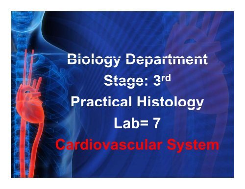
Biology Department<br />
Stage: 3<br />
rd<br />
Practical Histology<br />
Lab= 7<br />
Cardiovascular System
Students should be able to:<br />
1) Describe the histological structure of the heart<br />
wall.<br />
2) Classify arteries and veins, identify and<br />
describe their histological structure.<br />
3) Describe and identify different types of<br />
capillaries.
Cardiovascular <strong>system</strong><br />
The circulatory <strong>system</strong> consists of blood and lymphatic vascular<br />
<strong>system</strong>s.<br />
The blood vascular <strong>system</strong> composed of:<br />
■ Heart: an organ whose function is to pump the blood.<br />
■ Arteries: carry the blood, with its nutrients<br />
and oxygen, to the tissues.<br />
■ Veins: they convey the blood to heart to be pumped again.<br />
■ Capillaries: through whose walls the interchange between blood and<br />
tissues takes place.<br />
Cardiovascular <strong>system</strong> act to distribute hormones and<br />
nutrients to cells and tissue and transport waste material to<br />
excretory organs.
The Heart Chambers:<br />
1-The right and left<br />
ventricles pump<br />
blood to the lungs and<br />
the rest of the body<br />
respectively<br />
2- Right and left atria<br />
receive blood from the<br />
body and the<br />
pulmonary veins<br />
respectively.
Heart Wall<br />
The heart is a muscular organ, its<br />
wall consists of 3 layers, the<br />
internal (endocardium), the<br />
middle (myocardium) and the<br />
external (epicardium).<br />
1 Endocardium<br />
consists of a lining endothelium<br />
( single layer of squamous cells )<br />
& subendothelial layer of C.T<br />
containing (Purkinje) fibers in<br />
the ventricles the conducting
Heart Wall Cont.<br />
2 Myocardium:<br />
Is the thickest layer,<br />
consists of cardiac<br />
muscle cells.<br />
The myocardium in<br />
ventricles is much<br />
thickerthaninatrium?<br />
than in atrium ?<br />
M
Heart Wall Cont.<br />
3 Epicardium<br />
simple squamous epithelium<br />
(mesothelium) supported by a<br />
thin layer of connective tissue<br />
contains considerable adipose<br />
tissue.<br />
A- Fibrous layer<br />
B- Parietal layer<br />
C- Visceral layer (Pericardium),<br />
the serous membrane in which<br />
the heart lies.<br />
In the space between the pericardium's<br />
visceral layer (epicardium) and its<br />
parietal layer is a small amount of<br />
lubricant fluid that facilitates the<br />
heart's movements.<br />
Epicardium or visceral pericardium.
Epicardium
Epicardium or visceral pericardium.
Purkinje fibers<br />
Are modified cardiac<br />
muscle fibers located<br />
under the endocardium,<br />
-have larger diameter than<br />
cardiac muscle fibers & the<br />
sarcoplasm has a large<br />
amount of glycogen so<br />
they are less intensely<br />
stained.<br />
Purkinje fibers have fewer<br />
myofibrils than cardiac<br />
muscle fibers & distributed<br />
peripherally.<br />
Function: transmit<br />
impulses to the cardiac<br />
muscle cells located at the<br />
apex of the heart.
Arteries<br />
artery has a relatively thick wall & a<br />
small lumen. The wall of artery<br />
consists of the following layers.<br />
1- Tunica intima :<br />
• an inner layer of endothelium, a<br />
subendothelial layer of delicate<br />
fibro-elastic connective tissue<br />
• internal elastic lamina of<br />
membrane.<br />
2- Tunica media : consists chiefly<br />
of smooth muscle fibers, circularly<br />
arranged. A loose network of fine<br />
elastic fibers is interspersed<br />
among the smooth muscle cells,<br />
the amount of elastic fiber increase<br />
as the diameter of the artery<br />
increases.<br />
3- Tunica adventitia : composed<br />
3 Tunica adventitia : composed<br />
of C.T which contains nerves &<br />
blood vessels .
1 Arterioles They are less than 0.5 mm in diameter,<br />
their wall consist of:<br />
Tunica intima: Consist of endothelium ,subendothelial layer<br />
is very thin and internal elastic lamina is lacking except<br />
in largest arterioles.<br />
Tunica media composed of 1-5 layers of smooth. m. cells.<br />
T. adventitia is thinner than T. media
2 Muscular arteries :<br />
Include small & medium-sized arteries,<br />
The wall of the muscular arteries are relatively thick, having large<br />
amount of muscle in T. media.<br />
A-Small artery:
2-Muscular artery cont.<br />
B- Medium sized artery:<br />
•Tunica media is composed of smooth muscle.<br />
• Internal elastic lamina:Single, fenestrated, elastic sheet; lies internal to the<br />
smooth muscle of the tunica media.<br />
• External elastic lamina: Multiple elastic sheets; lie external to the smooth<br />
muscle of the tunica media.<br />
•Vasa vasorum are confined to the adventitial layer.
3 Large elastic arteries<br />
They are also called conducting arteries,<br />
which include aorta and its large branches.<br />
T. intima is thicker than the muscular art. The endothelial cells are polygonal<br />
in shape, a small bundle of smooth muscle cells are present in deeper portion<br />
of the intima. A distinct internal elastic lamina is difficult to seen.<br />
Tunica media is characterized by numerous distinct elastic lamina 40-60 in<br />
number, arranged concentrically, between these lamina are smooth muscle,<br />
fibroblasts, and affine elastic network.<br />
IEL<br />
T.adventitia is a thin coat & cannot be<br />
sharply distinguished from surrounding<br />
connective tissue.<br />
Vasa vasorum : are blood vessels of<br />
TM<br />
blood vessels that present in adventitia<br />
of large vessels.<br />
Vasa vasorum
Veins<br />
Have relatively thin walls and large lumens. The wall of vein consists<br />
of the following layers:-<br />
Tunica intima : comosed of endothelium & thin layer of fine<br />
collagenous & elastic fibers.<br />
Tunica media : consists of<br />
a thin layer of circularly arranged<br />
smooth muscle & is much<br />
Thinner than in arteries.<br />
Tunica adventitia : consists of<br />
a wide layer of connective tissue<br />
& is much thicker than<br />
the tunica media.
1 Venules<br />
T. intima composed of endothelium only.<br />
T. media: may contain only a few layers<br />
of smooth m. cells.<br />
T. adventitia is the thickest layer and composed of C.T.
2 Small and medium sized veins<br />
With the exception of the main trunks, most veins are small or medium<br />
sized.<br />
• T. intima consists of endothelium , the sub-endothelial tissue is very thin,<br />
which may be some times absent.<br />
• T. media consists of small bundles of smooth m. cells.<br />
• T. adventitia is a well developed C.T layer.<br />
Small vein
3 Large veins<br />
T. intimai is little l thicker than medium-sized i d veins.<br />
T. media is poorly developed, smooth muscle fibers are much reduced or<br />
absent.<br />
the adventitia is the thickest layer & consists of collagen & elastic fibers &<br />
many longitudinal muscle fibers.
Capillaries:<br />
Small blood vessels with<br />
average diameter of 7-9<br />
micrometer<br />
subdivided into three<br />
types<br />
Continuous capillaries<br />
most common type, found<br />
in connective tissue, muscle<br />
tissue & nervous tissue<br />
Fenestrated capillaries<br />
:found in endocrine glands,<br />
small intestine &<br />
glomerulus's l of kidney.<br />
Sinusoids are large<br />
capillaries (30-40 microns)<br />
found in liver, spleen and<br />
bone marrow
References<br />
‣ Junqueira, L.C and Carneiro, J. (2010) Basic<br />
Histology, text and atlas, 12 th Edition.<br />
McGraw-Hill, Inc.<br />
‣ Gartner and Hiatt: Color Textbook of<br />
Histology, 3rd ed. Philadelphia, W.B.<br />
Saunders, 2007.<br />
‣ Victor p. Eroschenko. (1993). diFiore’s Atlas<br />
of Histology with functionalcorrelations,7th<br />
ed. (Egyptian Edition);Elias Modern press.<br />
‣ http://biology.about.com/library/organs/heart/<br />
p gy y g<br />
blpurkinje.htm