Rapid evolutionary divergence of Photosystem I core subunits PsaA ...
Rapid evolutionary divergence of Photosystem I core subunits PsaA ...
Rapid evolutionary divergence of Photosystem I core subunits PsaA ...

You also want an ePaper? Increase the reach of your titles
YUMPU automatically turns print PDFs into web optimized ePapers that Google loves.
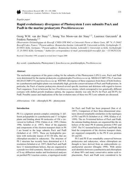
Photosynthesis Research 65: 131–139, 2000.<br />
© 2000 Kluwer Academic Publishers. Printed in the Netherlands.<br />
131<br />
Regular paper<br />
<strong>Rapid</strong> <strong>evolutionary</strong> <strong>divergence</strong> <strong>of</strong> <strong>Photosystem</strong> I <strong>core</strong> <strong>subunits</strong> <strong>PsaA</strong> and<br />
PsaB in the marine prokaryote Prochlorococcus<br />
Georg W.M. van der Staay 1,2 , Seung Yeo Moon-van der Staay 1,3 , Laurence Garczarek 1 &<br />
Frédéric Partensky 1,∗<br />
Observatoire Océanologique de Rosc<strong>of</strong>f, CNRS-UPR 9042 et Université Pierre et Marie Curie, BP 74, F-29682<br />
Rosc<strong>of</strong>f Cedex, France; 2 Present address: Botanisches Institut, Lehrstuhl III, Universität zu Köln, Gyrh<strong>of</strong>straße 15,<br />
D-50931 Köln, Germany; 3 Present address: Botanisches Institut, Lehrstuhl I, Universität zu Köln, Gyrh<strong>of</strong>straße<br />
15, D-50931 Köln, Germany; ∗ Author for correspondence (e-mail: partensky@sb-rosc<strong>of</strong>f.fr; fax: +33-98292324)<br />
Received 2 December 1999; accepted in revised form 9 August 2000<br />
Key words: cyanobacteria, <strong>Photosystem</strong> I, Synechococcus, prochlorophyte, Prochlorococcus<br />
Abstract<br />
The nucleotide sequences <strong>of</strong> the genes coding for the <strong>subunits</strong> <strong>of</strong> the <strong>Photosystem</strong> I (PS I) <strong>core</strong>, <strong>PsaA</strong> and PsaB<br />
were determined for the marine prokaryotic oxyphototrophs Prochlorococcus sp. MED4 (CCMP1378), P. marinus<br />
SS120 (CCMP1375) and Synechococcus sp. WH7803. Divergence <strong>of</strong> these sequences from those <strong>of</strong> both freshwater<br />
cyanobacteria and higher plants was remarkably high, given the conserved nature <strong>of</strong> <strong>PsaA</strong> and PsaB proteins. In<br />
particular, the <strong>PsaA</strong> <strong>of</strong> marine prokaryotes showed several specific insertions and deletions with regard to known<br />
<strong>PsaA</strong> sequences. Even in between the two Prochlorococcus strains, which correspond to two genetically different<br />
ecotypes with shifted growth irradiance optima, the sequence identity was only 80.2% for <strong>PsaA</strong> and 88.9% for<br />
PsaB. Possible causes and implications <strong>of</strong> the fast evolution rates <strong>of</strong> these two PS I <strong>core</strong> <strong>subunits</strong> are discussed.<br />
Abbreviations: Chl – chlorophyll; PS – photosystem<br />
Introduction<br />
PS I is a pigment–protein complex containing 11 different<br />
polypeptides in cyanobacteria and 17 in higher<br />
plants and binding about 90 molecules <strong>of</strong> Chl a (reviewed<br />
in Golbeck 1994; Chitnis et al. 1995; Chitnis<br />
1996; Scheller et al. 1997). Most <strong>of</strong> the pigments<br />
and components <strong>of</strong> the electron-transport chain in PS<br />
I are bound to the large <strong>subunits</strong> <strong>PsaA</strong> and PsaB<br />
(Schubert et al. 1997). These are hydrophobic proteins<br />
with molecular masses <strong>of</strong> 83–84 kDa, that are<br />
chloroplast-encoded in eukaryotes. The structure <strong>of</strong><br />
PS I structure has recently been determined at 4 Å<br />
resolution (Schubert et al. 1997). Both <strong>PsaA</strong> and PsaB<br />
have 11 transmembrane helices, that are organized in<br />
a pseudo two-fold symmetry. Based on a combination<br />
<strong>of</strong> X-ray diffraction studies and biochemical analyses<br />
on purified PS I reaction centers, a topological model<br />
for <strong>PsaA</strong> and PsaB has been proposed (Sun et al.<br />
1997). Comparison <strong>of</strong> their three-dimensional structures<br />
showed remarkable similarities in the topologies<br />
<strong>of</strong> PS I and PS II (Schubert et al. 1998; Klukas et al.<br />
1999). The six N-terminal helices <strong>of</strong> <strong>PsaA</strong> and PsaB,<br />
the antenna binding domain, are organized like the six<br />
helices <strong>of</strong> the inner PS II antenna proteins CP43 and<br />
CP47. The 5 C-terminal helices <strong>of</strong> <strong>PsaA</strong> and PsaB, that<br />
bind the components <strong>of</strong> the electron transport chain,<br />
are organized comparably to the PS II <strong>core</strong> proteins<br />
D1 and D2.<br />
PS I and PS II are remarkably conserved between<br />
prokaryotes and eukaryotes, whose chloroplasts are<br />
thought to have derived from an endosymbiotic cyanobacterial<br />
ancestor (Douglas 1998). With amino<br />
acid identities between cyanobacteria and chloroplasts<br />
<strong>of</strong> about 80% and many conservative amino acid replacements,<br />
<strong>PsaA</strong> and PsaB were considered to be
132<br />
very conserved proteins (Cantrell and Bryant 1987).<br />
This point <strong>of</strong> view has recently been challenged by the<br />
determination <strong>of</strong> the psaA and psaB sequences from<br />
the din<strong>of</strong>lagellate Heterocapsa triquetra (Zhang et al.<br />
1999), that showed a much lower degree <strong>of</strong> sequence<br />
conservation. This surprising result suggests that the<br />
available sequences are not sufficiently representative<br />
<strong>of</strong> the diversity <strong>of</strong> <strong>PsaA</strong> and PsaB proteins.<br />
Prochlorococcus (Chisholm et al. 1992) is a genus<br />
<strong>of</strong> planktonic photosynthetic prokaryotes very abundant<br />
in the ocean [see Partensky et al. (1999) for a<br />
review]. In contrast to other cyanobacteria, that use<br />
phycobiliproteins to collect light, the major light harvesting<br />
complexes in Prochlorococcus are membrane<br />
intrinsic proteins that bind mainly divinyl chlorophylls<br />
a and b. This genus is closely related to the marine<br />
AandBSynechococcus species, as shown by analysis<br />
<strong>of</strong> 16S rRNA, rpoC1, psbA, psbB, petB and petD<br />
gene sequences (Hess et al. 1995; Palenik and Swift<br />
1996; Urbach et al. 1998). PS I in Prochlorococcus<br />
shows several peculiar features. Recently, we have<br />
shown that the PS I <strong>subunits</strong> PsaF and PsaL in Prochlorococcus<br />
are longer and less conserved than in<br />
other organisms (van der Staay et al. 1998; van der<br />
Staay and Partensky 1999). Additionally, PS I in Prochlorococcus<br />
contains divinyl Chl b, which seems to<br />
be linked to the PS I <strong>core</strong> itself (Garczarek et al. 1998).<br />
To investigate the sequence conservation <strong>of</strong> the<br />
two large PS I <strong>subunits</strong> <strong>PsaA</strong> and PsaB, we have<br />
isolated and cloned these genes from two strains <strong>of</strong><br />
Prochlorococcus. P. marinus SS120 (CCMP1375) is<br />
a low light adapted strain which has a divinyl chlorophyll<br />
a to b ratio lower than 1 (Moore et al. 1995).<br />
This strain also contains some ‘normal’ (monovinyl)<br />
chlorophyll b (Partensky et al. 1993) and low amounts<br />
<strong>of</strong> phycoerythrin (Hess et al. 1996). Prochlorococcus<br />
sp. MED4 (CCMP1378) is adapted to grow at higher<br />
photon fluxes and has a divinyl chlorophyll a to b ratio<br />
<strong>of</strong> about 10 (Moore et al. 1995). Phylogenetic analyses<br />
using 16S rRNA consistently show MED4 belongs<br />
to the most evolved Prochlorococcus clade which includes<br />
all high light-adapted strains, whereas SS120<br />
and other low-light adapted strains belong to separate<br />
clusters genetically closer to the marine Synechococcus<br />
(Moore et al. 1998; Urbach et al. 1998; Moore and<br />
Chisholm 1999). As, prior to our study, there were<br />
no psaA and psaB gene sequences known from typical<br />
marine cyanobacteria, we have also determined their<br />
sequences from Synechococcus WH 7803. This strain<br />
is closely related to the other marine cyanobacteria<br />
and Prochlorococcus, as determined e.g. by analysis<br />
<strong>of</strong> DNA dependent RNA polymerase gene sequences<br />
(Palenik and Swift 1996). Our data confirm the close<br />
relatedness between Prochlorococcus and the marine<br />
Synechococcus. We show that in these three marine<br />
oxyphototrophs, the psaA and psaB genes evolved<br />
very rapidly.<br />
Materials and methods<br />
Isolation <strong>of</strong> clones<br />
The isolation <strong>of</strong> Lambda vectors containing the psaIpsaL<br />
gene clusters from Prochlorococcus sp. MED4<br />
and P. marinus SS120 has been previously described<br />
(van der Staay et al. 1998). The same vectors also<br />
contained the psaA-psaB genes (see ‘Results’).<br />
A genomic library <strong>of</strong> Synechococcus WH7803<br />
in Charon 35 was kindly provided by Dr D.<br />
Scanlan. A 499 bp psaB gene fragment from P.<br />
marinus SS120 was amplified by PCR with the<br />
primers CCGCTACATATTCGCCCAA and AGTGC-<br />
CGAAAACAGCATC. An initial denaturation at 94<br />
◦ C for 4 min was followed by 35 cycles <strong>of</strong> 94 ◦ C, 50<br />
◦ C and 72 ◦ C for 1 min each, with a final elongation at<br />
72 ◦ C for 7 min, in the presence <strong>of</strong> 1.5 mM Mg 2+ .The<br />
PCR product was cloned into the pCR 2.1 vector (In-<br />
Vitrogen). For probe labeling, the insert was excised<br />
with Eco RI. About 25 pg total DNA, corresponding to<br />
about 3 pg insert, was amplified by PCR in a volume<br />
<strong>of</strong> 50 µl as described above, except that the nucleotide<br />
mix was replaced by 5 µl <strong>of</strong> a fluorescein nucleotide<br />
mix (DuPont NEN). The labeled PCR product was<br />
used as a probe. Plaque hybridization proceeded as<br />
described in van der Staay et al. (1998). Hybridization<br />
occurred at 45 ◦ C, blots were washed with 5 ×<br />
SSC/0.1%SDS and 1 × SSC/0.1% SDS at 50 ◦ C. All<br />
other methods are described in van der Staay et al.<br />
(1998).<br />
Sequences used in this study<br />
The sequences determined in this study were deposited<br />
in the EMBL databank under the accession numbers<br />
AJ133190 for Synechococcus WH7803 psaA/B,<br />
AJ133191 for Prochlorococcus sp. MED4 psaA/B,<br />
AJ133192 for P. marinus SS120 psaA/B.<br />
The other sequences are available at GenBank under<br />
the following accession numbers: Anabaena variabilis:<br />
L26326, Mastigocladus laminosus (Fischerella<br />
PCC7605): AF038558, Synechococcus elongatus:
133<br />
X63768, Synechococcus PCC7002: M18165; Synechocystis<br />
PCC6803: D90906; Cyanophora paradoxa:<br />
U30821, Porphyra purpurea: U38804, Guillardia<br />
theta AF041468, Odontella sinensis Z67753, Heterocapsa<br />
triquetra: psaA AF130031, psaB AF130032;<br />
Euglena gracilis: X70810; Nephroselmis olivacea:AF<br />
137379; Chlamydomonas reinhardtii: psaA X05845,<br />
psaB X05848; Chlorella vulgaris: AB001684;<br />
Marchantia polymorpha: X04465; Pinus thunbergii<br />
(pine): D17510; Zea mays (maize): M11203; Oryza<br />
sativa (rice): X15901, Spinacia oleracea (spinach):<br />
X04131; Nicotiana tabacum (tobacco): Z00044.<br />
Protein alignments<br />
<strong>PsaA</strong> and PsaB sequences were aligned with the program<br />
ClustalX 1.64 (Thompson et al. 1994) using<br />
the PAM 250 matrix (Dayh<strong>of</strong>f et al. 1978). Alignments<br />
made with this matrix were very similar to the<br />
ones obtained using other matrices. Alignments were<br />
manually refined, keeping the number <strong>of</strong> gaps caused<br />
by insertions and deletions to a minimum. This program<br />
was also used to calculate the percentages <strong>of</strong><br />
sequence identity.<br />
Phylogenetic analysis<br />
Phylogenetic analysis used the parsimony and the<br />
neighbor-joining distance algorithms. PHYLIP Version<br />
3.5c (Felsenstein 1992) was used for all analyses.<br />
Distance matrices were constructed with the program<br />
PROTDIST using the PAM 250 matrix (Dayh<strong>of</strong>f et al.<br />
1978). Neighbor-joining distance trees were determined<br />
with the neighbor-joining (NEIGHBOR) method<br />
(Saitou and Nei 1987). Bootstrap analyses (SEQ-<br />
BOOT) with 100 replicates were applied to test the<br />
stability <strong>of</strong> the tree topology.<br />
Results<br />
Further upstream sequencing <strong>of</strong> a genomic clone containing<br />
the previously described genes coding for the<br />
PS I <strong>subunits</strong> PsaL and PsaI <strong>of</strong> Prochlorococcus sp.<br />
MED4 (van der Staay et al. 1998) revealed that this<br />
genomic fragment also contained the genes for the<br />
PS I <strong>core</strong> <strong>subunits</strong> <strong>PsaA</strong> and PsaB. The latter were<br />
separated from psaL and psaI by a gene encoding a<br />
protein that showed the closest similarity to a dolicholphosphate<br />
mannosyltransferase from Aquifex aeolicus<br />
(GenBank accession number AE000762), as determined<br />
by partial sequencing. Analogously, psaA and<br />
psaB from P. marinus SS120 were located on a genomic<br />
clone containing psaL and psaI. The psaA and<br />
psaB genes <strong>of</strong> the marine Synechococcus sp. WH<br />
7803 were isolated from a genomic library in lambda<br />
Charon 35, using a psaB probe from P. marinus<br />
SS120. As found in a study on the psbB and petB/D<br />
genes <strong>of</strong> several strains <strong>of</strong> Prochlorococcus and marine<br />
Synechococcus (Urbach et al. 1998), Prochlorococcus<br />
shows a third codon bias towards A and T<br />
(not shown), with a more pronounced bias in Prochlorococcus<br />
sp. MED4 than SS120. In contrast, Synechococcus<br />
WH7803 has a bias towards G and C at<br />
the third codon position, like reported for several other<br />
marine Synechococcus strains (Urbach et al. 1998).<br />
An alignment <strong>of</strong> the deduced amino acid sequences<br />
is shown in Figure 1. The corresponding proteins<br />
<strong>of</strong> the cyanobacterium Synechocystis PCC 6803, for<br />
which a topological model <strong>of</strong> the PS I <strong>core</strong> complex<br />
has been presented (Sun et al. 1997), are included in<br />
the alignment. The numbering <strong>of</strong> the putative membrane<br />
spanning helices and the extramembrane loops<br />
is done according to the model proposed for Synechocystis.<br />
The deduced <strong>PsaA</strong> proteins from P. marinus<br />
SS120, Prochlorococcus sp. MED4 and Synechococcus<br />
WH7803 are, respectively, 773, 767 and 767<br />
amino acids long. This is longer than <strong>PsaA</strong> sequences<br />
reported for other organisms, which vary between 732<br />
and 761 amino acids, with most species having a <strong>PsaA</strong><br />
750–755 amino acids long. None <strong>of</strong> the insertions or<br />
deletions are situated in the 11 potential membrane<br />
spanning regions, nor in regions known to be involved<br />
in the binding <strong>of</strong> components <strong>of</strong> the electron transport<br />
chain (Chitnis 1996). The <strong>PsaA</strong> proteins <strong>of</strong> all three<br />
marine prokaryotes contain a 13 amino acid insertion<br />
between the transmembrane helices III and IV (extramembrane<br />
loop D), the least conserved area <strong>of</strong> this<br />
protein (Figure 1A). A conserved insertion <strong>of</strong> three<br />
amino acids (F-P-A) is located between the helices IV<br />
and V (loop E). This loop also shows a low degree <strong>of</strong><br />
sequence identity. <strong>PsaA</strong> proteins from both Prochlorococcus<br />
strains have an additional insertion between<br />
helices VII and VIII and a deletion <strong>of</strong> 10 amino acids<br />
between the helices XI and X (loop J) not found in any<br />
other species.<br />
PsaB from P. marinus SS120 is with 747 amino<br />
acids the longest known PsaB sequence with the exception<br />
<strong>of</strong> the one <strong>of</strong> Heterocapsa triquetra, which<br />
contains 776 amino acids (Zhang et al. 1999). The<br />
PsaB proteins from Prochlorococcus sp. MED4 and<br />
Synechococcus sp. WH7803 fall, with 742 and 738<br />
amino acids, respectively, within the usual range <strong>of</strong>
134<br />
Figure 1. Alignment <strong>of</strong> the <strong>PsaA</strong> (A) and PsaB (B) from the marine prokaryotes Prochlorococcus sp. MED4, Prochlorococcus marinus SS120<br />
and Synechococcus WH7803. For comparison, the corresponding sequences <strong>of</strong> the freshwater cyanobacterium Synechocystis PCC6803 is<br />
included. The putative membrane spanning helices I–XI and the extramembrane loops A–L, determined and numbered based on the analogy<br />
to the model presented by Sun et al. (1997) are indicated. The cysteines that ligate the F x electron acceptor are indicated with a block. For<br />
positions where amino acids were identical, only the amino acid for Prochlorococcus sp. MED4 is given. Gaps are indicated by -.<br />
731–743 amino acids reported for most other organisms.<br />
Most diverse is loop E between the helices IV<br />
and V and loop H between helices VII and VIII, the<br />
areas that are the least conserved in all PsaB pro-
135<br />
Figure 1. Continued.<br />
teins. The C-terminal domains <strong>of</strong> <strong>PsaA</strong> and PsaB, from<br />
helices VIII to XI, that are thought to bind the components<br />
<strong>of</strong> the electron transport chain including the<br />
F x -binding domain, are the most conserved regions.<br />
In all phylogenetic analyses using the concatenated<br />
<strong>PsaA</strong> and PsaB sequences, the Prochlorococcus<br />
strains grouped together with the marine Synechococcus<br />
strain (Figure 2), separately from the other cyanobacteria.<br />
Analyses <strong>of</strong> either <strong>PsaA</strong> or PsaB lead<br />
to comparable results. Inclusion <strong>of</strong> the sequences<br />
from the din<strong>of</strong>lagellate Heterocapsa triquetra resulted<br />
in a destabilization <strong>of</strong> the trees, due to the high<br />
<strong>divergence</strong> <strong>of</strong> these sequences. Depending on small<br />
differences in the alignments and the inclusion or<br />
omission <strong>of</strong> gaps, Heterocapsa grouped together with<br />
Guillardia and Odontella or with the Prochlorococcus/Synechococcus<br />
cluster (not shown).
136<br />
Figure 2. Phylogenetic analysis <strong>of</strong> concatenated <strong>PsaA</strong> and PsaB proteins using the neighbor-joining distance method. For this analysis, all<br />
gaps created by the alignment were excluded. The first number at nodes represent the bootstrap percentage from 100 replicates for the<br />
neighbor-joining distance method, the second number indicates the bootstrap support for this node found by the maximum parsimony method.<br />
Values below 50% are indicated by – or not shown. Synechocystis PCC6803 was arbitrarily chosen as an outgroup. The scale bar indicates<br />
substitutions per amino acid position.<br />
The neighbor-joining <strong>PsaA</strong>/B tree exhibits unexpectedly<br />
long branches within the Synechocococcus/Prochlorococcus<br />
cluster (Figure 2), despite the<br />
fact that these species are phylogenetically close. This<br />
suggests considerable differences in the <strong>evolutionary</strong><br />
rates <strong>of</strong> the psaA/B operon both in between marine<br />
cyanobacteria and between these and the other phototrophs.<br />
The high <strong>divergence</strong> in PS I protein sequences<br />
between these three strains is confirmed by the comparison<br />
<strong>of</strong> sequence identities (Table 1). The <strong>PsaA</strong> sequences<br />
are more divergent than the PsaB sequences.<br />
Prochlorococcus sp. SS120 shows the highest deviation<br />
<strong>of</strong> both the <strong>PsaA</strong> and PsaB sequences. For<br />
both <strong>PsaA</strong> and PsaB, Heterocapsa showed by far the<br />
highest deviation (not shown). Since the larger number<br />
<strong>of</strong> insertions and deletions in these proteins from Heterocapsa<br />
complicated the alignment, leading to more<br />
gaps, they were excluded from the Table. Compared<br />
to the species indicated in Table 1, <strong>PsaA</strong> from H. triquetra<br />
showed sequence identities between 51.4% to<br />
P. marinus SS120 and 56.6% to Cyanophora paradoxa.<br />
For Heterocapsa PsaB, the sequence identity<br />
ranged between 51.3% to Prochlorococcus sp. MED4<br />
and 55.9% to Cyanophora paradoxa.<br />
Discussion<br />
The amino acid sequence analysis <strong>of</strong> the PS I <strong>subunits</strong><br />
<strong>PsaA</strong> and PsaB from one Synechococcus strain<br />
(WH 7803), representative <strong>of</strong> the Marine-Cluster A<br />
(Waterbury and Rippka 1989), and two Prochlorococcus<br />
strains, representative <strong>of</strong> the low-light (SS120)<br />
and high-light adapted (MED4) ecotypes (Moore et al.<br />
1995, 1998; Urbach et al. 1998), are in agreement with<br />
previous molecular studies showing that these marine<br />
oxyphototrophs are closely related and probably<br />
evolved from a common ancestor (Hess et al. 1995;<br />
Urbach et al. 1998; Honda et al. 1999; Turner et al.<br />
1999).<br />
The high <strong>evolutionary</strong> rate <strong>of</strong> the <strong>PsaA</strong> and PsaB<br />
sequences from all the three marine oxyphototrophs
137<br />
Table 1. Sequence identity <strong>of</strong> <strong>PsaA</strong> and PsaB <strong>of</strong> cyanobacteria and chloroplasts. Sequences were aligned with the program<br />
ClustalX and manually refined. The percentage sequence identity was calculated from a distance matrix constructed with the<br />
program ClustalX. Positions with gaps were excluded from the alignment. Upper triangle: <strong>PsaA</strong>, lower triangle: PsaB<br />
<strong>PsaA</strong><br />
1 2 3 4 5 6 7 8 9 10 11 12 13 14<br />
1 Prochlorococcus sp. MED4 – 80.2 75.7 69.9 70.3 70.9 69.1 69.7 70.9 69.4 67.2 70.2 69.9 68.8<br />
2 Prochlorococcus marinus SS120 88.9 – 70.8 68.2 67.1 68.9 65.8 66.4 67.9 66.8 65.4 67.5 67.4 65.8<br />
3 Synechococcus WH7803 81.5 80.6 – 77.6 77.7 78.4 75.8 77.7 77.3 75.1 71.3 76.1 77.7 74.7<br />
4 Anabaena variabilis 76.1 74.5 81.9 – 94.4 87.6 82.9 85.0 82.6 80.9 77.2 82.6 83.2 80.6<br />
5 Mastigocladus 74.4 74.5 80.3 90.6 – 87.4 81.4 84.4 82.6 80.7 76.7 81.9 82.6 80.9<br />
6 Synechococcus elongatus 76.8 76.0 81.6 89.6 87.7 – 84.3 87.9 84.3 85.0 79.3 85.1 86.1 85.6<br />
7 Synechococcus PCC7002 79.0 78.5 87.6 87.0 85.5 87.1 – 88.4 81.5 82.1 75.9 81.8 81.5 79.0<br />
8 Synechocystis PCC6803 76.8 76.8 84.1 86.4 85.6 87.1 92.6 – 83.1 84.2 75.7 83.7 83.6 80.9<br />
9 Cyanophora paradoxa 74.6 74.8 82.0 82.3 82.4 84.8 86.4 84.8 – 85.6 79.9 85.4 85.0 82.4<br />
10 Porphyra purpurea 74.4 74.1 80.4 80.5 81.1 81.7 84.4 83.5 83.5 – 78.9 86.2 85.1 82.3<br />
11 Euglena gracilis 70.9 71.3 76.4 77.4 76.7 79.2 80.5 78.3 80.4 79.0 – 82.5 82.2 79.3<br />
12 Chlamydomonas reinhardtii 73.0 73.0 79.7 79.0 79.0 81.6 82.7 80.9 82.2 81.5 80.4 – 89.8 86.8<br />
13 Marchantia polymorpha 73.8 73.6 81.2 79.3 79.3 81.8 84.1 82.3 83.8 84.5 83.2 85.0 – 93.2<br />
14 Spinacia oleracea 72.7 72.2 79.8 77.9 77.5 80.5 81.8 79.8 81.7 83.2 82.3 83.8 92.4 –<br />
PsaB<br />
is unexpected. Most striking is the low percent identity<br />
(80.2%) between the <strong>PsaA</strong> proteins <strong>of</strong> the two<br />
Prochlorococcus strains. This percent identity is as<br />
low as the identity between freshwater cyanobacteria<br />
and chloroplasts, that are thought to have separated<br />
about one billion years ago. By comparison, the respective<br />
16S rRNA sequences <strong>of</strong> these ecotypes are<br />
98.4% identical (Urbach et al. 1998). One may wonder<br />
which <strong>evolutionary</strong> constraint led to this rapid <strong>divergence</strong><br />
<strong>of</strong> <strong>PsaA</strong> (and to a lesser extent PsaB). It has<br />
been observed for other proteins that their <strong>evolutionary</strong><br />
rate differs among taxa. (Lockhart et al. 1996;<br />
Lopez et al. 1999). The underlying mechanism is not<br />
well understood yet. For Prochlorococcus, themost<br />
dramatic difference (with regard to photosynthesis)<br />
between the respective niches <strong>of</strong> these ecotypes in the<br />
field is certainly the amount <strong>of</strong> available light (Campbell<br />
and Vaulot 1993; Moore et al. 1998; Moore and<br />
Chisholm 1999). Besides having shifted growth irradiance<br />
optima and divinyl Chl a to divinyl Chl b ratios<br />
(Partensky et al. 1993, Moore et al. 1995), the Prochlorococcus<br />
ecotypes MED4 and SS120 also have<br />
dissimilar thylakoid protein pr<strong>of</strong>iles (Partensky et al.<br />
1997; Garczarek et al. 1998). The antenna system<br />
is probably the component <strong>of</strong> the photosynthetic apparatus<br />
the most differentiated between these strains<br />
(LaRoche et al. 1996). We recently discovered that<br />
SS120 possesses multiple pcb genes encoding seven<br />
different antenna proteins whereas MED4 possesses<br />
a single pcb gene (Garczarek et al. 2000). Another<br />
major difference between SS120 and MED4 is the<br />
presence <strong>of</strong> a large gene cluster implicated in the<br />
biosynthesis <strong>of</strong> phycoerythrin and its associated phycobilins<br />
in the latter but not the former strain (Hess et<br />
al. 1999). Thus, it seems that light has been a major<br />
driving force in the evolution <strong>of</strong> several key photosynthetic<br />
proteins in Prochlorococcus and this might be<br />
the case for the PS I <strong>core</strong> as well. In the open ocean,<br />
the wavelengths <strong>of</strong> photons found at depths below<br />
100 m (i.e. in the environment where the low light adapted<br />
Prochlorococcus ecotype thrives), are narrowly<br />
centered around 470 nm (Morel 1978). These photons<br />
are most efficiently captured by the divinyl Chl b and<br />
much less by divinyl Chl a. So to cope with these low<br />
blue light levels, the low-light adapted ecotypes must<br />
have evolved to optimize the capture <strong>of</strong> photons by PS<br />
I, e.g. either by binding Chl b molecules (Garczarek et<br />
al. 1998) or by recruiting as an antenna one or several<br />
<strong>of</strong> the multiple divinyl Chl a/b-binding Pcb proteins<br />
present in the cell. Comparative analysis <strong>of</strong> <strong>PsaA</strong>/B<br />
and other proteins associated with the photosystems<br />
from other representatives <strong>of</strong> the low-light and high<br />
light ecotypes should help us to get more insight in the<br />
underlying adaptation processes.<br />
The analysis <strong>of</strong> PS I-enriched fractions from<br />
MED4 and SS120 strains previously showed that they
138<br />
have similar PS I protein pr<strong>of</strong>iles, but both possess<br />
two proteins with apparent molecular mass <strong>of</strong> 21 and<br />
25 kDa, which have no equivalent in cyanobacteria,<br />
including the marine Synechococcus WH 8103 (Garczarek<br />
et al. 1998). These proteins were identified as<br />
the PS I <strong>subunits</strong> PsaF and PsaL, respectively, and<br />
their anomalous length was found to be due to specific<br />
gene insertions (van der Staay et al. 1998; van<br />
der Staay and Partensky 1999). Here we demonstrate<br />
that the two large <strong>core</strong> proteins <strong>PsaA</strong> and PsaB in<br />
Prochlorococcus show some specific insertions, too,<br />
but also deletions. Not surprisingly, the C-terminal<br />
part <strong>of</strong> both these proteins is the most conserved. This<br />
part is thought to bind the components <strong>of</strong> the electron<br />
transport chain. Therefore, a differentiation <strong>of</strong> this region<br />
is severely restricted. In the remaining part <strong>of</strong> the<br />
regions, variations are more tolerated, allowing insertions<br />
and deletions. The main insertions in <strong>PsaA</strong> <strong>of</strong> the<br />
three marine prokaryotes occur in loop D, located in<br />
the lumen [compared to topographic model <strong>of</strong> the PS<br />
I <strong>core</strong> proteins proposed by Sun et al. (1997)] and the<br />
cytoplasmic loop E. Both these loops contain less conserved<br />
regions in all species. Whereas no function has<br />
been assigned to loop D, loop E might interact with the<br />
subunit PsaE. An insertion in <strong>PsaA</strong> <strong>of</strong> Prochlorococcus,<br />
but not <strong>of</strong> other species, is located in the luminal<br />
loop H. This loop is thought to interact with PsaF. It is<br />
tempting to speculate that both this insertion in <strong>PsaA</strong><br />
and the ones in PsaF from Prochlorococcus might be<br />
involved in the interaction between these proteins. A<br />
significant deletion <strong>of</strong> 10 amino acids is present in<br />
the loop J <strong>of</strong> <strong>PsaA</strong> from Prochlorococcus. Thecorresponding<br />
loop in PsaB was shown to be involved in<br />
the interaction with soluble electron transporters (Sun<br />
et al. 1999). Assuming a pseudo tw<strong>of</strong>old symmetry <strong>of</strong><br />
<strong>PsaA</strong> and PsaB in the PS I complex, loop J might have<br />
a similar function in <strong>PsaA</strong>. In PsaB, major insertions<br />
occur in the cytoplasmic loop E and the luminal loop<br />
H. Insertions in both <strong>of</strong> these loops, that are the least<br />
conserved in all species, are found in PsaB from other<br />
species, too.<br />
Our results on marine cyanobacteria, in addition to<br />
the ones obtained with the din<strong>of</strong>lagellate Heterocapsa<br />
triquetra (Zhang et al. 1999), show that PS I <strong>core</strong> proteins<br />
can be more variable than previously assumed.<br />
We demonstrated that <strong>PsaA</strong> <strong>of</strong> marine cyanobacteria<br />
has characteristic features that distinguish them from<br />
the corresponding proteins <strong>of</strong> all other groups. In addition,<br />
based on the characteristic insertion and deletion<br />
in the <strong>PsaA</strong> sequence, and the much lower GC content<br />
in the psaA and psaB genes, representatives <strong>of</strong> the<br />
genus Prochlorococcus can probably be distinguished<br />
from marine Synechococcus. Therefore, these genes<br />
might constitute useful genetic markers for studies on<br />
the biodiversity <strong>of</strong> natural picoplanktonic communities.<br />
To get a more complete picture, sequences from<br />
other organisms should be obtained. Of special interest<br />
would be the cyanobacterium Gloeobacter violaceus.<br />
It is considered as a representative <strong>of</strong> the most primitive<br />
group <strong>of</strong> cyanobacteria (Honda et al. 1999; Turner<br />
et al. 1999) and, like Prochlorococcus (Garczarek et<br />
al. 1998), its PS I lacks the characteristic fluorescence<br />
at 77 K (Koenig and Schmidt 1995). PS I from Prochlorococcus<br />
appears to be unique, because it binds a<br />
divinyl form <strong>of</strong> Chl a, and probably divinyl Chl b as<br />
well (Garczarek et al. 1998). Since Chl b has also been<br />
reported in the PS I <strong>of</strong> Prochloron and Prochlorothrix<br />
(Hiller and Larkum 1985; van der Staay et al. 1992),<br />
obtaining sequences from the <strong>PsaA</strong> and PsaB <strong>of</strong> these<br />
two organisms might cast some light on the potential<br />
effect at the protein sequence level <strong>of</strong> the kind <strong>of</strong><br />
bound pigment. Also <strong>of</strong> particular interest in this context<br />
is Acaryochloris marina, an oxygenic prokaryote,<br />
the PS I <strong>of</strong> which contains almost exclusively Chl d<br />
(Hu et al. 1998).<br />
References<br />
Campbell L and Vaulot D (1993) Photosynthetic picoplankton community<br />
structure in the subtropical North Pacific Ocean near<br />
Hawaii (station ALOHA) Deep-Sea Res 40: 2043–2060<br />
Cantrell A and Bryant DA (1987) Molecular cloning and nucleotide<br />
sequence <strong>of</strong> the psaA and psaB genes <strong>of</strong> the cyanobacterium<br />
Synechococcus sp PCC 7002. Plant Mol Biol 9: 453–468<br />
Chisholm SW, Frankel SL, Goericke R, Olson RJ, Palenik B,<br />
Waterbury JB, West-Johnsrud L and Zettler ER (1992) Prochlorococcus<br />
marinus nov. gen. nov. sp.: An oxyphototrophic<br />
marine prokaryote containing divinyl chlorophyll a and b. Arch<br />
Microbiol 157: 297–300<br />
Chitnis PR (1996) <strong>Photosystem</strong> I – update on photosynthetic electron<br />
transport. Plant Physiol 111:<br />
Chitnis PR, Xu Q, Chitnis VP and Nechushtai R (1995) Function<br />
and organization <strong>of</strong> <strong>Photosystem</strong> I polypeptides. Photosynth Res<br />
44: 23–40 661–669<br />
Dayh<strong>of</strong>f MO, Schwartz RM and Orcutt BC (1978) A model <strong>of</strong> <strong>evolutionary</strong><br />
change in proteins. In Dayh<strong>of</strong>f MO (ed) Atlas <strong>of</strong> Protein<br />
Sequence and Structure, pp 345–352. Natural Biomedical<br />
Research Foundation, Washington, DC<br />
Douglas SE (1998) Plastid evolution: Origins, diversity, trends. Curr<br />
Opin Genet Develop 8: 655–661<br />
Felsenstein J (1992) PHYLIP (phylogeny interference package).<br />
University <strong>of</strong> Washington, Seattle<br />
Garczarek L, van der Staay GWM, Thomas JC and Partensky F<br />
(1998) Isolation and characterization <strong>of</strong> <strong>Photosystem</strong> I from<br />
two strains <strong>of</strong> the marine oxychlorobacterium Prochlorococcus.<br />
Photosynth Res 56: 131–141
139<br />
Garczarek L, Hess W, Holtzendorff J, van der Staay, GWM and<br />
Partensky, F (2000) Multiplication <strong>of</strong> antenna genes as a major<br />
adaptation process to growth at low light in a marine prokaryote.<br />
Proc Natl Acad Sci USA 97: 4098–4101<br />
Hess WR, Weihe A, Loiseaux-de Goer S, Partensky F and Vaulot D<br />
(1995) Characterization <strong>of</strong> the single psbA gene <strong>of</strong> Prochlorococcus<br />
marinus CCMP1375 (Prochlorophyta). Plant Mol Biol 27:<br />
1189–1196<br />
Hess WR, Partensky F, van der Staay GWM, Garcia-Fernandez JM,<br />
Börner T and Vaulot D (1996) Coexistence <strong>of</strong> phycoerythrin and<br />
a chlorophyll a/b antenna in a marine prokaryote. Proc Natl Acad<br />
Sci USA 93: 11126–11130<br />
Hess WR, Steglich C, Lichtlé C and Partensky F (1999) Phycoerythrins<br />
<strong>of</strong> the oxyphotobacterium Prochlorococcus marinus<br />
are associated to the thylakoid membrane and are encoded by<br />
a single large gene cluster. Plant Mol Biol 40: 507–521<br />
Hiller RD and Larkum AWD (1985) The chlorophyll–protein complexes<br />
<strong>of</strong> Prochloron sp. (Prochlorophyta). Biochim Biophys<br />
Acta 806: 107–115<br />
Honda D, Yokota A and Sugiyama J (1999) Detection <strong>of</strong> seven<br />
major <strong>evolutionary</strong> lineages in cyanobacteria based on the 16S<br />
rRNA gene sequence analysis with new sequences <strong>of</strong> five marine<br />
Synechococcus strain. J Mol Evol 48: 723–739<br />
Hu Q, Miyashita H, Iwasaki I, Kurano N, Miyachi S, Iwaki M and<br />
Itoh S (1998) A <strong>Photosystem</strong> I reaction center driven by chlorophyll<br />
d in oxygenic photosynthesis. Proc Natl Acad Sci USA 95:<br />
13319–13323<br />
Klukas O, Schubert W-D, Jordan P, Krauß N, Fromme P, Witt HAT<br />
and Saenger W (1999) <strong>Photosystem</strong> I, an improved model <strong>of</strong> the<br />
stromal <strong>subunits</strong> PsaC, PsaD and PsaE. J Biol Chem 274: 7351–<br />
7360<br />
Koenig F and Schmidt M (1995) Gloeobacter violaceus –investigation<br />
<strong>of</strong> an unusual photosynthetic apparatus – absence <strong>of</strong> the<br />
long wavelength emission <strong>of</strong> <strong>Photosystem</strong> I in 77 K fluorescence<br />
spectra. Physiol Plant 94: 621–628<br />
Lockhart PJ, Larkum AWD, Steel MA, Waddell PJ and Penny D<br />
(1996) Evolution <strong>of</strong> chlorophyll and bacteriochlorophyll: The<br />
problem <strong>of</strong> invariant sites in sequence analysis. Proc Natl Acad<br />
Sci USA 93: 1930–1934<br />
Lopez P, Forterre P and Philippe H (1999) The root <strong>of</strong> the tree <strong>of</strong><br />
life in the light <strong>of</strong> the covarion model. J Mol Evol 49: 496–508<br />
Moore LR and Chisholm SW (1999) Photophysiology <strong>of</strong> the marine<br />
cyanobacterium Prochlorococcus: Ecotypic differences among<br />
cultured isolates. Limnol Ocean 44: 628–638<br />
Moore LR, Goericke R and Chisholm SW (1995) Comparative<br />
physiology <strong>of</strong> Synechococcus and Prochlorococcus: Influence<br />
<strong>of</strong> light and temperature on growth, pigments, fluorescence and<br />
absorptive properties. Mar Ecol Prog Ser 116: 259–275<br />
Moore LR, Rocap G and Chisholm SW (1998) Physiology and<br />
molecular phylogeny <strong>of</strong> coexisting Prochlorococcus ecotypes.<br />
Nature 393: 464–467<br />
Morel A (1978) Available, usable and stored radiant energy in<br />
relation to marine photosynthesis. Deep-Sea Res 25: 673–688<br />
Palenik B and Swift H (1996) Cyanobacterial evolution and prochlorophyte<br />
diversity as seen in DNA-dependent RNA polymerase<br />
gene sequences. J Phycol 32: 638–646<br />
Partensky F, Hoepffner N, Li WKW, Ulloa O and Vaulot D<br />
(1993) Photoacclimation <strong>of</strong> Prochlorococcus sp. (Prochlorophyta)<br />
strains isolated from the North Atlantic and the Mediterranean<br />
Sea. Plant Physiol 101: 285–296<br />
Partensky F, LaRoche J, Wyman K and Falkowski P (1997) The<br />
divinyl chlorophyll a/b-protein complexes <strong>of</strong> two strains <strong>of</strong> the<br />
oxyphototrophic marine prokaryote Prochlorococcus – characterization<br />
and response to changes in growth irradiance. Photosynth<br />
Res 51: 209–222<br />
Partensky F, Hess WR, and Vaulot D (1999) Prochlorococcus, a<br />
marine photosynthetic prokaryote <strong>of</strong> global significance. Microbiol<br />
Mol Biol Rev 63: 106–127<br />
Saitou N and Nei M (1987) The neighbor-joining method: A new<br />
method for reconstructing phylogenetic trees. Mol Biol Evol 4:<br />
406–425<br />
Scheller HV, Naver H and Møller BL (1997) Molecular aspects <strong>of</strong><br />
<strong>Photosystem</strong> I. Physiol Plant 100: 842–851<br />
Schubert W-D, Klukas O, Krauß N, Saenger W, Fromme P and Witt<br />
HT (1997) <strong>Photosystem</strong> I <strong>of</strong> Synechococcus elongatus at 4 Å<br />
resolution: Comprehensive structure analysis. J Mol Biol 272:<br />
741–769<br />
Schubert W-D, Klukas O, Saenger W, Witt HAT, Fromme P and<br />
Krauss N (1998) A common ancestor for oxygenic and anoxygenic<br />
photosynthetic systems – a comparison based on the<br />
structural model <strong>of</strong> <strong>Photosystem</strong> I. J Mol Biol 280: 297–314<br />
Sun J, Xu Q, Chitnis VP, Jin P and Chitnis PR (1997) Topography<br />
<strong>of</strong> the <strong>Photosystem</strong> I <strong>core</strong> proteins <strong>of</strong> the cyanobacterium<br />
Synechocystis sp. PCC 6803. J Biol Chem 272: 21793–21802<br />
SunJ,XuW,HervásM,NavarroJA,DeLaRosamandChitnis<br />
PR (1999) Oxidizing side <strong>of</strong> the cyanobacterial <strong>Photosystem</strong> –<br />
Evidence for interaction between the electron donor proteins and<br />
a luminal surface helix <strong>of</strong> the PsaB subunit. J Biol Chem 274:<br />
19048–19054<br />
Thompson JD, Higgins DG and Gibson DJ (1994) Clustal W: Improving<br />
the sensitivity <strong>of</strong> multiple sequence alignment through<br />
sequence weighting, position specific gap penalties and matrix<br />
choice. Nucleic Acids Res 22: 4673–4680<br />
Turner S, Pryer KM, Miao VPW and Palmer JD (1999) Investigating<br />
deep phylogenetic relationships among cyanobacteria and<br />
plastids by small subunit rRNA sequence analysis. J Eukaryot<br />
Microbiol 46: 327–338<br />
Urbach E, Scanlan DJ, Distel DL, Waterbury JB and Chisholm SW<br />
(1998) <strong>Rapid</strong> diversification <strong>of</strong> marine picophytoplankton with<br />
dissimilar light harvesting structures inferred from sequences <strong>of</strong><br />
Prochlorococcus and Synechococcus (cyanobacteria). J Mol Evol<br />
46: 188–201<br />
van der Staay GWM, Brouwer A, Baard RL, van Mourik F and<br />
Matthijs HCP (1992) Separation <strong>of</strong> <strong>Photosystem</strong>s I and II from<br />
the oxychlorobacterium (prochlorophyte) Prochlorothrix hollandica<br />
and association <strong>of</strong> chlorophyll b binding antennae with<br />
<strong>Photosystem</strong> II. Biochim Biophys Acta 1102: 220–228<br />
van der Staay GWM, Moon-van der Staay SY, Garczarek L and<br />
Partensky F (1998) Characterization <strong>of</strong> the <strong>Photosystem</strong> I <strong>subunits</strong><br />
PsaI and PsaL from two strains <strong>of</strong> the marine oxyphototrophic<br />
prokaryote Prochlorococcus. Photosynth Res 57: 183–<br />
192<br />
van der Staay GWM and Partensky F (1999) The 21 kDa protein<br />
associated with <strong>Photosystem</strong> I in Prochlorococcus marinus is<br />
the PsaF protein (Accession No. AJ131438). (PGR99-067). Plant<br />
Physiol 120: 339<br />
Waterbury JB and Rippka R (1989) Order Chroococcales Wettstein<br />
1924, Emend. Rippka et al. 1979. In: Kreig NR and Holt JB (eds)<br />
Bergey’s Manual <strong>of</strong> Systematic Bacteriology, pp 1728-01746.<br />
Williams and Wilkins, Baltimore<br />
Zhang Z, Green BR and Cavalier-Smith T (1999) Single gene circles<br />
in din<strong>of</strong>lagellate chloroplast genomes. Nature 400: 155–159