Unn Ljøstad and Åse Mygland Jone Furlund Owe and Nils ... - ACNR
Unn Ljøstad and Åse Mygland Jone Furlund Owe and Nils ... - ACNR
Unn Ljøstad and Åse Mygland Jone Furlund Owe and Nils ... - ACNR

You also want an ePaper? Increase the reach of your titles
YUMPU automatically turns print PDFs into web optimized ePapers that Google loves.
ISSN 1473-9348 Volume 8 Issue 5 November/December 2008<br />
<strong>ACNR</strong>www.acnr.co.uk<br />
Advances in Clinical Neuroscience & Rehabilitation<br />
<strong>Unn</strong> Ljøstad <strong>and</strong> Åse Mygl<strong>and</strong><br />
Lyme Neuroborreliosis in Adults: Clinical Aspects<br />
<strong>Jone</strong> <strong>Furlund</strong> <strong>Owe</strong> <strong>and</strong> <strong>Nils</strong> Erik Gilhus<br />
Myasthenia Gravis <strong>and</strong> the Heart<br />
Justin Cross<br />
NEW NEURORADIOLOGY SECTION<br />
Computed Tomography in Neurology<br />
Win a copy of<br />
Greenfield’s<br />
Neuropathology<br />
worth £475<br />
Turn to page 29<br />
for details<br />
News Review • Conference News • Journal Reviews • Book Reviews • Diary of Events
Same... yet different<br />
Up to 16 weeks between injections <strong>and</strong> ready-to-use 1<br />
Adds efficiency to efficacy. That’s smart.<br />
ABBREVIATED PRESCRIBING INFORMATION<br />
NeuroBloc ® (Botulinum toxin Type B)<br />
Please refer to the SPC before prescribing.<br />
Presentation: 0.5ml, 1ml <strong>and</strong> 2ml vials containing 2500U, 5000U <strong>and</strong> 10000U of Botulinum Toxin<br />
Type B solution for injection.<br />
Indication: Treatment of cervical dystonia (torticollis).<br />
Dose <strong>and</strong> administration: For intramuscular (IM) administration only. Must only be administered by<br />
experienced physicians. When low doses are required, it must be diluted before use with preservativefree<br />
0.9% sodium chloride solution for injection. Dosage units are specific to botulinum Toxin Type<br />
B only <strong>and</strong> are not relevant to preparations of Botulinum Toxin Type A. See SPC for instructions for<br />
use <strong>and</strong> h<strong>and</strong>ling.<br />
Adults <strong>and</strong> elderly: 5000U or 10000U divided between two to four affected muscles. 10000U may<br />
increase the clinical benefit. The dose <strong>and</strong> frequency of administration should be adjusted for each<br />
patient depending on the clinical response.<br />
Patients with renal or hepatic impairment: No dose adjustment required. (see SPC)<br />
Children <strong>and</strong> adolescents under 18 years: Not recommended<br />
Contra-Indications: Hypersensitivity to Botulinum Toxin Type B or any excipient. Individuals with<br />
other neuromuscular diseases or neuromuscular junctional disorders.<br />
Pregnancy: Do not use during pregnancy unless clearly necessary. Studies in animals are insufficient<br />
<strong>and</strong> potential risk in humans is unknown.<br />
Lactation: Do not use during lactation unless clearly necessary as it is unknown whether Botulinum<br />
Toxin Type B is excreted in breast milk.<br />
Warnings <strong>and</strong> Precautions: Caution should be exercised to prevent administration into a blood<br />
vessel. Caution should be used in patients with bleeding disorders or receiving anticoagulant therapy.<br />
Neuromuscular side effects due to toxin spread have been reported. Development of an immune response <strong>and</strong><br />
subsequent tolerance can occur after repeated administration. Spontaneous reports of dysphagia, aspiration<br />
pneumonia <strong>and</strong>/or potentially fatal respiratory disease, after treatment with Botulinum Toxin Type A/B have<br />
been reported. There is an increased risk of side effects in patients with underlying neuromuscular disease <strong>and</strong><br />
swallowing disorders. Close medical supervision is advised in patients with neuromuscular disorders or history of<br />
dysphagia <strong>and</strong> aspiration. Seeking medical attention for respiratory difficulties, choking or any new or worsening<br />
dysphagia is advised. Dysphagia has been reported following injection to sites other than the cervical musculature.<br />
Botulinum Toxin Type B contains human albumin <strong>and</strong> therefore the possibility of transmitting infectious agents<br />
cannot be totally excluded. Dosage units are specific to Botulinum Toxin Type B only <strong>and</strong> are not relevant to<br />
preparations of Botulinum Toxin Type A.<br />
Drug Interactions: No specific interaction studies. Effect of co-administration with other botulinum toxin<br />
types is unknown. Co-administration of Botulinum Toxin Type B <strong>and</strong> aminoglycosides or agents interfering with<br />
neuromuscular transmission (e.g. curare-like compounds) should be considered with caution.<br />
Side effects: Adverse reactions reported with Botulinum Toxin Type B (toxin-naïve <strong>and</strong> toxin-responsive) are:<br />
Very common (m1/10): dry mouth, dysphagia, headache <strong>and</strong> injection site pain. Common (m1/100 to
NEW NEURORADIOLOGY SECTION<br />
Editorial<br />
Lyme disease of the central nervous system is relatively<br />
rare, although not uncommonly considered in the differential<br />
diagnosis of patients with an array of neurological<br />
problems including facial palsies <strong>and</strong> a radiculitis. In this clear<br />
article by <strong>Unn</strong> Ljøstad <strong>and</strong> Åse Mygl<strong>and</strong>, we are taken through<br />
the common clinical features of this condition <strong>and</strong> how it can<br />
be diagnosed. This article contains a couple of really useful<br />
tables summarising the main points, which overall makes for<br />
an extremely succinct, up to date account, of this condition.<br />
<strong>Jone</strong> Furl<strong>and</strong> <strong>Owe</strong> <strong>and</strong> <strong>Nils</strong> Erik Gilhus discuss an unusual<br />
aspect of myasthenia gravis – namely how it affects the heart.<br />
They conclude that “There is good evidence for myocardial pathology in<br />
MG patients, including a diversity of heart-reactive autoantibodies <strong>and</strong><br />
focal inflammation with cellular infiltrates <strong>and</strong> myocardial necrosis. On<br />
the other h<strong>and</strong>, any firm clinical correlates to these findings have yet to be<br />
established”. Interesting.<br />
On the theme of epilepsy, Paul Johns <strong>and</strong> Maria Thom discuss in our<br />
neuropathology article the relationship between hippocampal sclerosis<br />
(HS) <strong>and</strong> epilepsy. It is well known that the two are associated but it is not<br />
clear whether one causes the other. In this article, the competing theories<br />
are discussed in particular whether seizures, especially early in life, cause<br />
HS or whether HS is consequent to a developmental problem which then<br />
causes seizures.<br />
Plaha <strong>and</strong> colleagues in their article for the Neurosurgery series consider<br />
chronic subdural haematoma. They begin by discussing how such<br />
lesions come about <strong>and</strong> how the patient may present, before considering<br />
how to optimally treat it. An issue that is far from clearcut! This is an easy<br />
to read account which represents a measured <strong>and</strong> pragmatic approach to<br />
this relatively common neurological problem.<br />
Michael Turner reports on the guidelines <strong>and</strong> stipulations for jockeys<br />
engaged in different types of horse racing <strong>and</strong> the consequences to them of<br />
a fall <strong>and</strong> a head injury. This is not an uncommon event as there are approximately<br />
110,00 rides a year <strong>and</strong> “Amateur jockeys fall every 7.9 rides, Jump<br />
jockeys fall every 15.8 rides <strong>and</strong> Flat jockeys fall every 200-250 rides. It is<br />
therefore not surprising that horse racing has some of the highest<br />
rates of injury seen in any sport, including the highest rates of<br />
concussion <strong>and</strong> fatal accidents in the published literature.<br />
Concussion rates are in fact six times greater than in Australian<br />
Rules Football – the sport with the closest incidence of concussion.”<br />
I think I will continue to walk <strong>and</strong> ride my bike.<br />
In the first of our new series on Neuroradiology edited by<br />
Justin Cross we are treated to a sumptuously illustrated<br />
account on CT. In this first article by Justin we are introduced<br />
to the Compston effect <strong>and</strong> Hounsfield Units as well as having<br />
partial volume effects explained. This is a wonderful article<br />
that gives just the right amount of background physics <strong>and</strong> technical<br />
detail whilst also illustrating the role <strong>and</strong> value of this imaging modality<br />
to neurological practice in 2008.<br />
In our Neurology <strong>and</strong> Literature series, Dr Pearce takes us through the<br />
Charles Bonnet syndrome <strong>and</strong> how it came to be called this <strong>and</strong> what exactly<br />
is meant by it. This article with a liberal sprinkling of original quotations<br />
from Dr Bonnet follows on from the earlier article we featured by Dominic<br />
Ffytche in <strong>ACNR</strong> 4.2 on visual hallucinations <strong>and</strong> optical illusions.<br />
Heather Angus-Leppan gives us a picture of where the ABN is going (as<br />
opposed to where it has come from (see <strong>ACNR</strong> 7.5).We are also pleased to<br />
announce that Heather will be joining the editorial team of <strong>ACNR</strong>, helping<br />
to keep us abreast of developments within the ABN. Finally we have<br />
all our usual journal, conference <strong>and</strong> book reviews. Do enjoy <strong>and</strong> if you<br />
would like to become a part of the reviewing team just let us know by<br />
email to Rachael@acnr.co.uk.<br />
Roger Barker, Co-Editor, Email: roger@acnr.co.uk<br />
Sadly we report that Dr Raymond D Adams passed away on the 18th October,<br />
aged 97. He made a colossal contribution to neurology. We were fortunate<br />
enough to be the recipient of some of that knowledge in the article he wrote for<br />
<strong>ACNR</strong> in issue 6:5 (Nov/Dec 2006).<br />
<strong>ACNR</strong> is published by Whitehouse Publishing, 1 The Lynch, Mere, Wiltshire, BA12 6DQ.<br />
Publisher: Rachael Hansford • E. rachael@acnr.co.uk<br />
Advertising: Rachael Hansford, T. 01747 860168, M. 07989 470278, E. rachael@acnr.co.uk<br />
Course Advertising: Nathalie Fricker, E. events@acnr.co.uk<br />
Editorial: Anna Phelps, E. editorial@acnr.co.uk<br />
Design & Production E. design.dept@sky.com<br />
Printed by: Warners Midl<strong>and</strong>s PLC, T. 01778 391000<br />
<strong>ACNR</strong><br />
ISSN 1473-9348 Volume 8 Issue 5 November/December 2008<br />
www.acnr.co.uk<br />
Advances in Clinical Neuroscience & Rehabilitation<br />
Copyright: All rights reserved; no part of this publication may be reproduced, stored in a retrieval system or transmitted in any form or<br />
by any means, electronic, mechanical, photocopying, recording or otherwise without either the prior written permission of the publisher<br />
or a license permitting restricted photocopying issued in the UK by the Copyright Licensing Authority.<br />
Disclaimer: The publisher, the authors <strong>and</strong> editors accept no responsibility for loss incurred by any person acting or refraining from<br />
action as a result of material in or omitted from this magazine. Any new methods <strong>and</strong> techniques described involving drug usage<br />
should be followed only in conjunction with drug manufacturers' own published literature.<br />
This is an independent publication - none of those contributing are in any way supported or remunerated by any of the companies<br />
advertising in it, unless otherwise clearly stated. Comments expressed in editorial are those of the author(s) <strong>and</strong> are not necessarily<br />
endorsed by the editor, editorial board or publisher. The editor's decision is final <strong>and</strong> no correspondence will be entered into.<br />
<strong>Unn</strong> Ljøstad <strong>and</strong> Åse Mygl<strong>and</strong><br />
Lyme Neuroborreliosis in Adults: Clinical Aspects<br />
<strong>Jone</strong> <strong>Furlund</strong> <strong>Owe</strong> <strong>and</strong> <strong>Nils</strong> Erik Gilhus<br />
Myasthenia Gravis <strong>and</strong> the Heart<br />
Justin Cross<br />
Computed Tomography in Neurology<br />
Win a copy of<br />
Greenfield’s<br />
Neuropathology<br />
worth £475<br />
Turn to page 29<br />
for details<br />
News Review • Conference News • Journal Reviews • Book Reviews • Diary of Events<br />
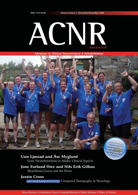
Cover picture shows participants<br />
in the first ever ABN Triathlon,<br />
held at the Autumn meeting of<br />
the ABN in Aviemore recently.<br />
For a full report see page 35.<br />
Avoid Misdiagnosis<br />
Make chromosomal testing<br />
routine for your refractory<br />
epilepsy patients<br />
r(20) epilepsy syndrome<br />
can only be identified<br />
through chromosome analysis<br />
For more information about r(20)<br />
syndrome <strong>and</strong> to download research<br />
forms please visit our website<br />
www.ring20.org/research<br />
Foundation Contact Information<br />
(888) 313 2025<br />
(TollFree – US, Canada <strong>and</strong> Mexico Only)<br />
(212) 860 2668<br />
(US <strong>and</strong> International)<br />
+44 (0)20 7710 6920<br />
(UK <strong>and</strong> International)<br />
Email: info@ring20.org<br />
Call for Patients – Please ask your r(20) patients to participate in our ongoing<br />
genetic <strong>and</strong> clinical research studies.<br />
<strong>ACNR</strong> • VOLUME 8 NUMBER 5 • NOVEMBER/DECEMBER 2008 I 3
Contents<br />
Contents<br />
November/December 2008<br />
3 Editorial<br />
7 Review Article<br />
Lyme Neuroborreliosis in Adults: Clinical Aspects<br />
Dr <strong>Unn</strong> Ljøstad <strong>and</strong> Dr Åse Mygl<strong>and</strong><br />
9 Review Article<br />
Myasthenia Gravis <strong>and</strong> the Heart<br />
Dr <strong>Jone</strong> <strong>Furlund</strong> <strong>Owe</strong> <strong>and</strong> Dr <strong>Nils</strong> Erik Gilhus<br />
12 Neurosurgery Article<br />
Management of Chronic Subdural Haematoma<br />
Dr Puneet Plaha, Dr Malhotra, Dr Heuer, Dr Peter Whitfield<br />
16 Neuropathology Article<br />
Epilepsy <strong>and</strong> Hippocampal Sclerosis: Cause or Effect?<br />
Dr Paul Johns <strong>and</strong> Dr Maria Thom<br />
19 Neurology <strong>and</strong> Literature<br />
Charles Bonnet’s Syndrome<br />
Dr JMS Pearce<br />
20 Rehabilitation Article<br />
Fitness to Ride – Horse Racing in Great Britain<br />
Dr Michael Turner<br />
22 Neuroradiology Article<br />
Computed Tomography in Neurology<br />
Dr Justin Cross<br />
28 Book Reviews<br />
30 ABN<br />
Future directions of the Association of British Neurologists<br />
Dr Heather Angus-Leppan<br />
32 Events Diary<br />
33 Courses <strong>and</strong> Conferences<br />
34 Conference News<br />
39 Sponsored Feature<br />
Recent Advances in the Treatment of Focal Dystonia<br />
42 Journal Reviews<br />
44 Awards <strong>and</strong> Appointments<br />
45 News Review<br />
BrainVision UK Ltd was established<br />
in 2004. We are a distribution<br />
company dedicated to the provision<br />
of technical <strong>and</strong> software solutions to<br />
universities <strong>and</strong> hospitals in the UK<br />
<strong>and</strong> Irel<strong>and</strong>. We represent specialist<br />
manufacturers from around the<br />
world to offer novel <strong>and</strong> unique<br />
product lines for Neurophysiological<br />
<strong>and</strong> psychophysiological research<br />
<strong>and</strong> clinical applications.<br />
The home in the UK<br />
<strong>and</strong> Irel<strong>and</strong> for<br />
Medoc Thermal Stimulators<br />
MagVenture Magnetic Stimulators<br />
Supplies <strong>and</strong> consumables<br />
020 8543 0022<br />
sales@brainvision.co.uk<br />
www.brainvision.co.uk<br />
For more information please<br />
contact: Brain Vision (UK) Ltd<br />
Suite 4, Zeal House<br />
8 Deer Park Rd.<br />
London SW19 3GY<br />
MagVenture<br />
MagPro Series of<br />
magnetic<br />
stimulators<br />
For use in Neurophysiology, Neurology,<br />
rehabilitation, psychiatry & research<br />
Applications:<br />
• Motor Evoked Potentials with built in<br />
MEP monitor on selected models<br />
• Transcranial magnetic stimulation (TMS)<br />
• Repetitive Transcranial Magnetic<br />
Stimulation (rTMS)<br />
• Functional Magnetic Stimulation (FMS)<br />
• Dynamic Coils, Static coils (fluid filled) &<br />
fMRI Compatible coils<br />
TSA II Neuro<br />
Sensory Analyzer<br />
The Essential Tool in Sensory Nerve<br />
Evaluation<br />
“<br />
QST of the thermal<br />
modalities is the only<br />
clinical test for<br />
quantitative assessment<br />
of small caliber sensory<br />
nerve fiber function, the<br />
primary transmitters of<br />
pain sensation<br />
”<br />
4 I <strong>ACNR</strong> • VOLUME 8 NUMBER 5 • NOVEMBER/DECEMBER 2008
As soon as first line oral<br />
dopamine agonists start to fail<br />
Go directly to APO-go<br />
Trying subsequent oral medication once your PD patient’s<br />
first line dopamine agonist begins to fail, resulting in increasing<br />
motor complications, can be a time of frustration <strong>and</strong><br />
disappointment for you <strong>and</strong> your patient - compromising<br />
their optimum quality of life.<br />
For responsive patients with Early Complex PD, APO-go CDS<br />
is a highly effective, 1,2 rapid-acting 3 drug that, combined with<br />
Britannia’s extensive Package of Care, can maintain your<br />
PD patients’ independence. 4<br />
Prescribing information can be found overleaf<br />
Version Number: APG.API.V7<br />
Britannia<br />
Pharmaceuticals<br />
Britannia Pharmaceuticals is a trading name of Genus Pharmaceuticals Ltd
ABBREVIATED PRESCRIBING INFORMATION<br />
Consult Summary of Product Characteristics before prescribing. Uses: The treatment of disabling<br />
motor fluctuations (“on-off” phenomena) in patients with Parkinson’s disease which persist<br />
despite individually titrated treatment with levodopa (with a peripheral decarboxylaze inhibitor)<br />
<strong>and</strong>/or other dopamine agonists. Dosage <strong>and</strong> Administration: Apomorphine hydrochloride<br />
is administered subcutaneously either as an intermittent bolus injection or by continuous<br />
subcutaneous infusion. Its rapid onset (5-10 mins) <strong>and</strong> duration of action (about 1 hour) may<br />
prevent an “off” episode which is refractory to other treatments. Hospital admission under<br />
appropriate specialist supervision is necessary during patient selection <strong>and</strong> when establishing<br />
a patient’s therapeutic regime. Please refer to the Summary of Product Characteristics for full<br />
details before initiating therapy. Treatment with domperidone (typical dosage 20mg three times a day)<br />
before <strong>and</strong> during apomorphine HCl therapy is essential. The optimal dosage of apomorphine<br />
HCl has to be determined on an individual patient bases; individual bolus injections should not<br />
exceed 10mg <strong>and</strong> the total daily dose should not exceed 100mg. Contraindications: Children<br />
<strong>and</strong> adolescents (up to 18 years of age). Known sensitivity to apomorphine or any other ingredients<br />
of the product. Respiratory depression, dementia, psychotic disease or hepatic insufficiency.<br />
Intermittent apomorphine HCl treatment is not suitable for patients who have an “on” response<br />
to levodopa which is marred by severe dyskinesia or dystonia. Pregnancy <strong>and</strong> lactation:<br />
Caution should be exercised if prescribing apomorphine to pregnant women <strong>and</strong> women<br />
of childbearing age. Breast-feeding should be avoided during apomorphine HCl therapy.<br />
Interactions: Patients should be monitored for potential interactions during initial<br />
stages of apomorphine therapy. Particular caution should be given when apomorphine<br />
is used with other medications that have a narrow therapeutic window. It should be<br />
noted that there is potential for interaction with neuroleptic <strong>and</strong> antihypertensive agents.<br />
Precautions: Use with caution in patients with renal, pulmonary or cardiovascular disease, or who<br />
are prone to nausea or vomiting. Extra caution is recommended during initiation of therapy in elderly<br />
<strong>and</strong>/or debilitated patients. Since apomorphine may produce hypotension, care should be<br />
exercised in patients with cardiac disease or who are taking vasoactive drugs,<br />
particularly when pre-existing postural hypotension is present. Neuropsychiatric disturbances<br />
are common in Parkinsonian patients. APO-go should be used with special caution in these<br />
patients. Apomorphine has been associated with somnolence <strong>and</strong> other dopamine agonists<br />
can be associated with sudden sleep onset episodes, particularly in patients with Parkinson’s<br />
disease. Patients must be informed of this <strong>and</strong> advised to exercise caution whilst driving or<br />
operating machines during treatment with apomorphine. Haematology tests should be undertaken<br />
at regular intervals as with levodopa with given concomitantly with apomorphine. Pathological<br />
gambling, increased libido <strong>and</strong> hypersexuality have been reported in patients treated with dopamine<br />
agonists, including apomorphine. Side Effects: Local induration <strong>and</strong> nodules (usually<br />
asymptomatic) often develop at subcutaneous site of injection leading to areas of erythema,<br />
tenderness, induration <strong>and</strong> (rarely) ulceration. Pruritus may occur at the site of injection.<br />
Drug-induced dyskinesias during “on” periods can be severe, <strong>and</strong> in a few patients may<br />
result in cessation of therapy. Postural hypotension is seen infrequently <strong>and</strong> is usually<br />
intransient. Transient sedation following each dose of apomorphine may occur at the start of<br />
therapy, but this usually resolves after a few weeks of treatment. Nausea <strong>and</strong> vomiting may occur,<br />
particularly when APO-go treatment is initiated, usually as a result of the omission of domperidone.<br />
Neuropyschiatric disturbances (including transient mild confusion <strong>and</strong> visual hallucinations)<br />
have occurred during apomorphine therapy <strong>and</strong> neuropsychiatric disturbances may be<br />
exacerbated by apomorphine. Positive Coombs’ tests <strong>and</strong> haemolytic anaemia have been<br />
reported in patients receiving apomorphine <strong>and</strong> levodopa. Local <strong>and</strong> generalised rashes<br />
have been reported. Eosinophilia has occurred in only a few patients during treatment with<br />
apomorphine HCl. Patients treated with dopamine agonists, including apomorphine, have been<br />
reported as exhibiting signs of pathological gambling, increased libido <strong>and</strong> hypersexuality<br />
(especially at high doses). Apomorphine is associated with somnolence. Breathing difficulties have<br />
been reported. Prescribers should consult the Summary of Product Characteristics in relation to<br />
other side effects. Presentation <strong>and</strong> Basic NHS Cost: Apo-go ampoules contain apomorphine<br />
hydrochloride 10mg/ml, as follows: 20mg in 2ml – basic NHS cost £37.96 per carton of 5 ampoules.<br />
50mg in 5ml – basic NHS cost £73.11 per carton of 5 ampoules. APO-go opens (disposable<br />
multiple dosage injector system) contain apomorphine hydrochloride 10mg/ml, as follows:<br />
30mg in 3ml – basic NHS cost £123.91 per carton of 5 pens. APO-go Pre-filled syringes<br />
contain apomorphine hydrochloride 5mg/ml, as follows: 50mg in 10ml – basic NHS cost<br />
£73.11 per carton of 5 syringes.<br />
References: 1. Pietz K, Hagell P, Odin P, 1998. Subcutaneous apomorphine in late stage<br />
Parkinson’s disease: a long term follow up. J Neurol Neurosurg Psychiatry. 65:709–716.<br />
2. Lees A, Turner K, 2002. Apomorphine for Parkinson’s Disease. Practical Neurology,<br />
2:280-287. 3. Deleu D, Hanssens Y, Northway M G, 2004. Subcutaneous Apomorphine:<br />
An Evidence-Based Review of its Use in Parkinson’s Disease. Drugs Aging, 21(11), 687-709.<br />
4. Ellis C, Lemmens Get al 1997. Use of Apomorphine in Parkinsonian Patients with Neurosychiatric<br />
Complications to Oral Tretment. Parkinsonism & Related Disorders, 3 (2), 103-107.<br />
Marketing Authorisation Numbers:<br />
APO-go Ampoules: PL04483/0064<br />
APO-go Pens: PL04483/0065<br />
APO-go Pre filled syringes: PL05928/0025<br />
Legal Category: POM<br />
Date of last revision: July 2008<br />
For further information please contact: Britannia Pharmaceuticals Limited, 41-51 Brighton Road, Redhill,<br />
Surrey RH1 6YS.<br />
Adverse events should be reported.<br />
Reporting forms <strong>and</strong> information can be found at www.yellowcard.gov.uk.<br />
Adverse events should also be reported to Medical Information at Britannia at the above<br />
address or telephone 01737 773741 or e-mail: drugsafety@britannia-pharm.com<br />
Version Number: APG.API.V7<br />
Editorial board <strong>and</strong> contributors<br />
Roger Barker is co-editor of <strong>ACNR</strong>, <strong>and</strong> is Honorary Consultant in<br />
Neurology at The Cambridge Centre for Brain Repair. His main area of<br />
research is into neurodegenerative <strong>and</strong> movement disorders, in particular<br />
parkinson's <strong>and</strong> Huntington's disease. He is also the university lecturer<br />
in Neurology at Cambridge where he continues to develop his<br />
clinical research into these diseases along with his basic research into<br />
brain repair using neural transplants.<br />
Alasdair Coles is co-editor of <strong>ACNR</strong>. He is a University Lecturer in<br />
Neuroimmuniology at Cambridge University. He works on experimental<br />
immunological therapies in multiple sclerosis.<br />
Stephen Kirker is the editor of the Rehabilitation Section of <strong>ACNR</strong><br />
<strong>and</strong> Consultant in Rehabilitation Medicine in Addenbrooke's NHS<br />
Trust, Cambridge. He trained in neurology in Dublin, London <strong>and</strong><br />
Edinburgh before moving to rehabilitation in Cambridge <strong>and</strong><br />
Norwich. His main research has been into postural responses after<br />
stroke. His particular interests are in prosthetics, orthotics, gait training<br />
<strong>and</strong> neurorehabilitation.<br />
David J Burn is the editor of our Conference News Section <strong>and</strong> is<br />
Professor in Movement Disorder Neurology & Honorary Consultant,<br />
Newcastle General Hospital. He runs Movement Disorders clinics in<br />
Newcastle-upon-Tyne. Research interests include progressive supranuclear<br />
palsy <strong>and</strong> dementia with Lewy bodies. He is also involved in several<br />
drugs studies for Parkinson's Disease.<br />
Andrew Larner is the editor of our Book Review Section. He is a<br />
Consultant Neurologist at the Walton Centre for Neurology <strong>and</strong><br />
Neurosurgery in Liverpool, with a particular interest in dementia <strong>and</strong><br />
cognitive disorders. He is also an Honorary Apothecaries' Lecturer in<br />
the History of Medicine at the University of Liverpool.<br />
Alastair Wilkins is our Case Report Co-ordinator. He is Senior<br />
Lecturer in Neurology <strong>and</strong> Consultant Neurologist, University of<br />
Bristol. He trained in Neurology in Cambridge, Norwich <strong>and</strong> London.<br />
His research interests are the basic science of axon degeneration <strong>and</strong><br />
developing treatments for progressive multiple sclerosis.<br />
Nicki Cohen is <strong>ACNR</strong>’s Neuropathology Editor. She is a Specialist<br />
Registrar in Neuropathology at Southampton <strong>and</strong> has a DPhil in<br />
Neuroscience. Her research interests lie in CNS stem cell biology, <strong>and</strong><br />
the brain’s response to injury.<br />
Peter Whitfield is <strong>ACNR</strong>’s Neurosurgery Editor. He is a Consultant<br />
Neurosurgeon at the South West Neurosurgery Centre, Plymouth. His<br />
clinical interests are wide including neurovascular conditions, head<br />
injury, stereotactic radiosurgery, image guided tumour surgery <strong>and</strong><br />
lumbar microdiscectomy. He is an examiner for the MRCS <strong>and</strong> is a<br />
member of the SAC in neurosurgery.<br />
Heather Angus-Leppan is <strong>ACNR</strong>'s ABN representative on the Editorial<br />
Board. She is Head of the Neurology Department at Barnet Hospital <strong>and</strong><br />
Consultant Neurologist, Honorary Senior Lecturer <strong>and</strong> Epilepsy Lead at<br />
the Royal Free Hospital, London, UK. She is the Honorary Assistant<br />
Secretary of the Association of British Neurologists, Honorary Secretary<br />
of the Neurosciences Section of the Royal Society of Medicine <strong>and</strong> current<br />
Chair of the Map of Medicine Epilepsy Group, UK.<br />
International editorial liaison committee<br />
Professor Riccardo Soffietti, Italy: Chairman of the Neuro-Oncology Service, Dept of<br />
Neuroscience <strong>and</strong> Oncology, University <strong>and</strong> S. Giovanni Battista Hospital.<br />
Professor Klaus Berek, Austria: Head of the Neurological Department of the KH Kufstein.<br />
Professor Hermann Stefan, Germany: Professor of Neurology /Epileptology in the<br />
Department of Neurology, University Erlangen-Nürnberg.<br />
Professor <strong>Nils</strong> Erik Gilhus, Norway: Professor of Neurology at the University of Bergen<br />
<strong>and</strong> Haukel<strong>and</strong> University Hospital.<br />
6 I <strong>ACNR</strong> • VOLUME 8 NUMBER 5 • NOVEMBER/DECEMBER 2008
Review Article<br />
Lyme Neuroborreliosis in Adults: Clinical Aspects<br />
Lyme neuroborreliosis is caused by the tick-borne<br />
spirochete Borrelia burgdorferi (Bb) sensu lato. The<br />
Bb sensu lato complex consists of at least three<br />
human pathogenic species; Bb sensu stricto, B. garinii <strong>and</strong><br />
B. afzelii. All are endemic in Europe, whereas Bb sensu<br />
stricto is the only species detected in North America. This<br />
may account for clinical differences in American <strong>and</strong><br />
European neuroborreliosis. In this short review we convey<br />
basic facts, our own experience <strong>and</strong> some new knowledge<br />
of clinical manifestations, diagnostics, treatment <strong>and</strong><br />
prognosis of Lyme neuroborreliosis in European adult<br />
patients.<br />
Clinical manifestations<br />
At least 80% of European patients with neuroborreliosis<br />
present with facial weakness due to facial neuritis, or<br />
lancinating pain, numbness <strong>and</strong> sometimes weakness in<br />
an extremity, the chest or abdomen due to spinal radiculitis,<br />
or a combination of these (Bannwarths syndrome).<br />
1,2 More unusual symptoms are diplopia due to<br />
oculomotor, trochlear- or abducens neuritis, 3 visual loss<br />
due to retrobulbar optic neuritis, 4 hearing loss due to<br />
acoustic neuritis, 5 dizziness due to vestibular neuritis, 6<br />
shortness of breath due to diaphragmatic paralysis, 7 <strong>and</strong><br />
isolated muscle weakness <strong>and</strong> fasciculations due to selective<br />
involvement of motor neurons <strong>and</strong> motor roots. 8 A<br />
recent report describes a patient with an autoimmunemediated<br />
motor neuropathy with conduction blocks <strong>and</strong><br />
GM1 antibodies concomitant to, <strong>and</strong> probably triggered<br />
by infection with Bb. 9 Symptoms from the central nervous<br />
system are rare, but some patients may present with<br />
weakness <strong>and</strong> clumsiness of both legs due to myelitis, 10<br />
or memory loss, confusion, unsteadiness, parkinsonism,<br />
11 or opsoclonus myoclonus due to focal encephalitis.<br />
12 Vasculitis may account for acute stroke like or<br />
relapsing symptoms with hemiparesis <strong>and</strong> aphasia. 13,14<br />
Most patients with Lyme neuroborreliosis suffer from<br />
Table 1: Suggested case definitions<br />
Definite neuroborreliosis<br />
All three criteria fulfilled<br />
constitutional symptoms in addition to their neurological<br />
complaints, but it should be emphasized that<br />
headache, fatigue, paresthesias or malaise alone are not<br />
typical symptoms of Lyme neuroborrelisis.<br />
The onset of Lyme neuroborreliosis is usually subacute<br />
with progression over weeks, so-called early or<br />
stage II Lyme neuroborreliosis. Some cases are self-limiting,<br />
even without treatment. Less than 10% of<br />
untreated cases progress slowly over months <strong>and</strong> years,<br />
so-called late, or stage III, Lyme neuroborreliosis. 1<br />
Laboratory confirmation<br />
Due to the low yield from culture <strong>and</strong> polymerase chain<br />
reaction (PCR) in Lyme neuroborreliosis, we are left<br />
with indirect laboratory diagnostic methods. Both an<br />
elevated cell count in the cerebrospinal fluid (CSF) <strong>and</strong><br />
elevated CSF-to-serum Bb antibody index, indicating<br />
intrathecal Bb antibody production, have to be present<br />
to confirm definite Lyme neuroborreliosis.<br />
In day to day practice, however, diagnostics may be a<br />
challenge because the CSF Bb antibody index has a low<br />
sensitivity (about 75%) in patients with a symptom<br />
duration of less than six weeks, 15 <strong>and</strong> the CSF cell count<br />
may be normal in rare patients infected with certain<br />
genotypes of Bb. 16 It therefore seems reasonable to<br />
introduce the term possible Lyme neuroborreliosis.<br />
Suggested case definitions with two levels of diagnostic<br />
accuracy are presented in Table 1.<br />
Different other laboratory tests such as CSF<br />
CXCL 13,17,18 LTT MELISA, 19 <strong>and</strong> Bb specific immune<br />
complexes, 20 may in the future be helpful diagnostic<br />
markers, but they need further validation.<br />
Antibiotic treatment<br />
All patients with Lyme neuroborreliosis should be treated<br />
with antibiotics to achieve rapid resolution of symptoms,<br />
<strong>and</strong>, theoretically, to prevent further dissemination<br />
Possible neuroborreliosis<br />
Criterion 1 <strong>and</strong> one of a-d<br />
1. Neurological symptoms suggestive of Lyme neuroborreliosis without 1. Neurological symptoms suggestive of Lyme neuroborreliosis<br />
other obvious reasons<br />
without other obvious reasons<br />
2. CSF pleocytosis (>5 leucocytes/mm 3 ) a. CSF pleocytosis (> 5 leucocytes/mm 3 )<br />
3. Intrathecal Bb antibody production b. Intrathecal Bb antibody production<br />
c. Bb antibodies in serum<br />
d. Erythema migrans during the last four months<br />
Dr <strong>Unn</strong> Ljøstad is a consultant<br />
clinical neurologist based at<br />
Sørl<strong>and</strong>et Hospital, Kristians<strong>and</strong>,<br />
Norway. She will present her doctoral<br />
thesis on ‘Lyme neuroborreliosis,<br />
diagnostics <strong>and</strong> treatment in<br />
European adult patients’ later this<br />
year.<br />
Dr Åse Mygl<strong>and</strong> is Consultant<br />
Neurologist at Sørl<strong>and</strong>et Hospital<br />
Kristians<strong>and</strong> <strong>and</strong> Assistant<br />
Professor at the Institute of<br />
Clinical Medicin, University of<br />
Bergen, Norway. She has a special<br />
interest in managing patients with<br />
neuromuscular <strong>and</strong> inflammatory<br />
neurological disorders.<br />
Correspondence to:<br />
<strong>Unn</strong> Ljøstad,<br />
Department of Neurology,<br />
Sørl<strong>and</strong>et Hospital,<br />
Servicebox 416,<br />
4604 Kristians<strong>and</strong>, Norway.<br />
Email. unn.ljostad@sshf.no<br />
Table 2: Suggested treatment guidelines for European adult patients with Lyme neuroborreliosis<br />
Drug • First choice:<br />
o Oral doxycycline 200 mg daily<br />
• Alternative choices:<br />
o IV ceftriaxone 2 g daily<br />
o IV penicillin G 3 g X 3-4<br />
o IV cefotaxime 2 g X 3<br />
Treatment duration<br />
Repeated course with a different drug<br />
Post Lyme disease syndrome<br />
2-4 weeks<br />
Should be restricted to patients with definite Lyme neuroborreliosis<br />
who do not improve after first course (exceptionally rare)<br />
Symptomatic treatment<br />
<strong>ACNR</strong> • VOLUME 8 NUMBER 5 • NOVEMBER/DECEMBER 2008 I 7
Review Article<br />
<strong>and</strong> persistence of the infection. European Concerted Action on Lyme<br />
Borreliosis (EUCALB) suggests 14-30 days courses of IV ceftriaxone,<br />
IV penicillin, oral amoxicillin or oral doxycyline for early subacute<br />
Lyme neuroborreliosis, <strong>and</strong> 30 days courses of IV ceftriaxone or IV<br />
penicillin for late Lyme neuroborreliosis. 21 IV regimens with penicillin<br />
<strong>and</strong> ceftriaxone have similar efficacy, <strong>and</strong> it has recently been shown<br />
that oral doxycycline for 14 days is as efficient as IV ceftriaxone for 14<br />
days in adult European patients. 2 We therefore suggest oral doxycyline<br />
as a first treatment choice for Lyme neuroborreliosis in adults, due to<br />
lower cost <strong>and</strong> easier administration. Suggested treatment guidelines<br />
are presented in Table 2. Enhanced therapeutic efficacy of extended<br />
treatment beyond two to three weeks is not well documented. The issue<br />
of longer treatment duration in all, or subgroups of, patients with<br />
Lyme neuroborreliosis, thus remains to be answered.<br />
How to h<strong>and</strong>le ‘possible’ Lyme neuroborreliosis<br />
In our opinion patients who fulfill the criteria for possible Lyme neuroborreliosis<br />
should be offered one, but not repeated courses of antibiotic<br />
treatment. If one course does not lead to lasting improvement, a<br />
search for other causes of the symptoms should be sought.<br />
Outcome after treatment<br />
Outcome after treatment for Lyme neuroborreliosis is debated. A<br />
Swedish questionnaire follow-up study found persistent complaints 2.5<br />
years post-treatment in 50% of patients who had experienced neuroborreliosis,<br />
as compared to 16% in control patients who had experienced<br />
erythema migrans. 22 Most of the complaints were subjective such<br />
as headache, attention problems, memory difficulties, depression <strong>and</strong><br />
paresthesia. The real burden of remaining symptoms <strong>and</strong> neurological<br />
abnormalities after treatment for Lyme neuroborreliosis, as well as the<br />
pathophysiological mechanisms of post-Lyme disease syndrome,<br />
should be better charted in well designed studies. It seems clear that<br />
additional prolonged antibiotic treatment does not give any benefit in<br />
post-Lyme disease syndrome, <strong>and</strong> it should be remembered that treatments<br />
continued for months carry substantial risk for the patients. 23<br />
Anecdotal reports of better response to long-term or repeated antibiotic<br />
treatment exist, but this could be due to placebo effect, which is<br />
known to occur in at least 30% of cases. A positive Bb antibody index<br />
may last for years <strong>and</strong> is not a suitable marker for treatment response.<br />
Conclusion<br />
We suggest two levels of diagnostic accuracy for clinical practice; definite<br />
<strong>and</strong> possible Lyme neuroborreliosis. Treatment with 2-4 weeks<br />
courses of IV ceftriaxone, penicilline or oral doxycycline are effective.<br />
We recommend oral doxycycline for two weeks as the first treatment<br />
choice. Patients with possible Lyme neuroborreliosis should be offered<br />
one, but not repeated courses of antibiotic treatment. Extensive antibiotic<br />
treatment is not recommended under any circumstances. The<br />
prognosis of neuroborreliosis <strong>and</strong> pathophysiology of post-treatment<br />
conditions need further studies.<br />
We recommend oral doxycycline for two weeks as the first treatment choice.<br />
Enhanced therapeutic efficacy of extended treatment beyond two to three weeks is<br />
not well documented<br />
References<br />
1. Oschmann P, Dorndorf W, Hornig C, Schafer C,<br />
Wellensiek HJ, Pflughaupt KW. Stages <strong>and</strong> syndromes of<br />
neuroborreliosis. J.Neurol. 1998;245:262-72.<br />
2. Ljostad U, Skogvoll E, Eikel<strong>and</strong> R, Midgard R, Skarpaas<br />
T, Berg A, Mygl<strong>and</strong> A. Oral doxycycline versus intravenous<br />
ceftriaxone for European Lyme neuroborreliosis: a<br />
multicentre, non-inferiority, double-blind, r<strong>and</strong>omised<br />
trial. Lancet Neurol. 2008;7(8):690-5.<br />
3. Lell M, Schmid A, Stemper B, Maihofner C, Heckmann<br />
JG, Tom<strong>and</strong>l BF. Simultaneous involvement of third <strong>and</strong><br />
sixth cranial nerve in a patient with Lyme disease.<br />
Neuroradiology. 2003;45:85-7.<br />
4. Krim E, Guehl D, Burbaud P, Lagueny A. Retrobulbar<br />
optic neuritis: a complication of Lyme disease?<br />
J.Neurol.Neurosurg.Psychiatry. 2007;78:1409-10.<br />
5. Walther LE, Hentschel H, Oehme A, Gudziol H, Beleites<br />
E. Lyme disease--a reason for sudden sensorineural hearing<br />
loss <strong>and</strong> vestibular neuronitis?. Laryngorhinootologie.<br />
2003;82:249-57.<br />
6. Hyden D, Roberg M, Odkvist L. Borreliosis as a cause of<br />
sudden deafness <strong>and</strong> vestibular neuritis in Sweden. Acta<br />
Otolaryngol.Suppl. 1995;520 Pt 2:320-2.:320-2.<br />
7. Winterholler M, Erbguth FJ. Tick bite induced respiratory<br />
failure. Diaphragm palsy in Lyme disease. Intensive Care<br />
Med. 2001;27:1095.<br />
8. Charles V, Duprez TP, Kabamba B, Ivanoiu A, Sindic CJ.<br />
Poliomyelitis-like syndrome with matching magnetic resonance<br />
features in a case of Lyme neuroborreliosis.<br />
J.Neurol.Neurosurg.Psychiatry. 2007;78:1160-1.<br />
9. Rupprecht TA, Elstner M, Weil S, Pfister HW.<br />
Autoimmune-mediated polyneuropathy triggered by borrelial<br />
infection? Muscle Nerve. 2008;37:781-5.<br />
10. Blanc F, Froelich S, Vuillemet F, Carre S, Baldauf E, de<br />
MS, Jaulhac B, Maitrot D, Tranchant C, de SJ. Acute<br />
myelitis <strong>and</strong> Lyme disease. Rev.Neurol.(Paris).<br />
2007;163:1039-47.<br />
11. Kohlhepp W, Kuhn W, Kruger H. Extrapyramidal features<br />
in central Lyme borreliosis. Eur.Neurol.<br />
1989;29:150-5.<br />
12. Skeie GO, Eldoen G, Skeie BS, Midgard R, Kristoffersen<br />
EK, Bindoff LA. Opsoclonus myoclonus syndrome in two<br />
cases with neuroborreliosis. Eur.J.Neurol. 2007;14:e1-e2.<br />
13. Romi F, Krakenes J, Aarli JA, Tysnes OB. Neuroborreliosis<br />
with vasculitis causing stroke-like manifestations.<br />
Eur.Neurol. 2004;51:49-50.<br />
14. May EF, Jabbari B. Stroke in neuroborreliosis. Stroke.<br />
1990;21:1232-5.<br />
15. Ljostad U, Skarpaas T, Mygl<strong>and</strong> A. Clinical usefulness of<br />
intrathecal antibody testing in acute Lyme neuroborreliosis.<br />
Eur.J.Neurol. 2007;14:873-6.<br />
16. Strle F, Ruzic-Sabljic E, Cimperman J, Lotric-Furlan S,<br />
Maraspin V. Comparison of findings for patients with<br />
Borrelia garinii <strong>and</strong> Borrelia afzelii isolated from cerebrospinal<br />
fluid. Clin.Infect.Dis. 2006;43:704-10.<br />
17. Rupprecht TA, Pfister HW, Angele B, Kastenbauer S,<br />
Wilske B, Koedel U. The chemokine CXCL13 (BLC): a<br />
putative diagnostic marker for neuroborreliosis. Neurology<br />
2005;65:448-50.<br />
18. Ljostad U, Mygl<strong>and</strong> A. CSF B - Lymphocyte<br />
Chemoattractant (CXCL13) in the early diagnosis of acute<br />
Lyme Neuroborreliosis. J.Neurol. 2007.<br />
19. Valentine-Thon E, Ilsemann K, S<strong>and</strong>kamp M. A novel<br />
lymphocyte transformation test (LTT-MELISA) for Lyme<br />
borreliosis. Diagn.Microbiol.Infect.Dis. 2007;57:27-34.<br />
20. Brunner M, Sigal LH. Use of serum immune complexes in<br />
a new test that accurately confirms early Lyme disease <strong>and</strong><br />
active infection with Borrelia burgdorferi.<br />
J.Clin.Microbiol. 2001;39:3213-21.<br />
21. Smith M, Gray J, Granstrom, M., Crawford R, <strong>and</strong><br />
Gettinby G. European Concerted Action on Lyme<br />
Borreliosis (EUCALB). Stanek, G. <strong>and</strong> Gray, J. ©<br />
EUCALB 1997-2007. European Union Concerted Action<br />
on Lyme Borreliosis . 2008. Ref Type: Electronic<br />
Citation.<br />
22. Vrethem M, Hellblom L, Widlund M, Ahl M, Danielsson<br />
O, Ernerudh J, Forsberg P. Chronic symptoms are common<br />
in patients with neuroborreliosis -- a questionnaire<br />
follow-up study. Acta Neurol.Sc<strong>and</strong>. 2002;106:205-8.<br />
23. Krupp LB, Hyman LG, Grimson R, Coyle PK, Melville P,<br />
Ahnn S, Dattwyler R, Ch<strong>and</strong>ler B. Study <strong>and</strong> treatment of<br />
post Lyme disease (STOP-LD): a r<strong>and</strong>omized double<br />
masked clinical trial. Neurology 2003;60:1923-30.<br />
8 I <strong>ACNR</strong> • VOLUME 8 NUMBER 5 • NOVEMBER/DECEMBER 2008
Review Article<br />
Myasthenia Gravis <strong>and</strong> the Heart<br />
Myasthenia gravis (MG) is well established as an<br />
autoimmune disorder with specific autoantibodies<br />
directed against the nicotinic acetylcholine receptor<br />
(AChR) in the neuromuscular junction. Involvement of<br />
the heart has been claimed <strong>and</strong> reported, but a causal connection<br />
between MG <strong>and</strong> altered cardiac function has not<br />
been found. This review summarises what is known about<br />
the possibility of heart involvement in MG.<br />
Immunology<br />
Whereas the AChR-antibody is the mainstay of MG, several<br />
other antibodies can be found. Some of these antibodies<br />
have been shown to react with cardiac muscle, thus being<br />
termed skeletal <strong>and</strong> heart reactive autoantibodies, 1 or striational<br />
antibodies. A proportion of this striational reactivity<br />
is due to antibodies against titin <strong>and</strong> the ryanodine receptor<br />
(RyR) found in the sarcoplasmic reticulum, 2 whereas<br />
other antibodies are claimed to react with myosin, 3 α-actin<br />
<strong>and</strong> actinin. 4 Non-striational antibodies have also been<br />
demonstrated in MG, directed against beta-adrenergic<br />
receptors 5 <strong>and</strong> against AChR of the muscarinic type. 6<br />
Mygl<strong>and</strong> demonstrated that the existence of heart muscle<br />
antibodies was related to type of MG, in finding such antibodies<br />
in MG-patients with thymoma <strong>and</strong> late-onset MG,<br />
but not in early onset MG. These heart muscle antibodies<br />
were shown to be specific for MG. 7 Lately, the discovery of<br />
antibodies to the muscle specific tyrosine kinase (MuSK) 8<br />
has identified a subgroup of MG patients with rather distinct<br />
clinical characteristics. Antibodies to MuSK inhibit<br />
agrin-induced AChR-clustering, indicating a potential<br />
effect on agrin-dependent maintenance of AChRs at the<br />
neuromuscular junction. Cardiac function has not been<br />
examined specifically in MuSK-positive MG-patients.<br />
The existence of all these antibodies – <strong>and</strong> antibodies<br />
not yet characterised – offers theoretical mechanisms for<br />
cardiac involvement in MG, by interfering with contractility,<br />
cardiac conduction, autonomous regulation or by<br />
an immunological attack on the myocardium, involving<br />
complement activation, infiltrates of inflammatory cells<br />
<strong>and</strong> with consequent necrosis.<br />
Giant-cell myocarditis (GCM) is a condition showing a<br />
clear association to thymoma <strong>and</strong> in some cases to MG.<br />
GCM is characterised by degeneration <strong>and</strong> necrosis of<br />
heart muscle, <strong>and</strong> has been linked to IgG anti-cardiomyocyte<br />
antibodies. 9 GCM is however extremely rare, with<br />
only a few reported cases.<br />
However, looking beyond the rare giant-cell myocarditis,<br />
the potential immunlogical mechanisms are at the<br />
present insufficiently correlated to clinical cardiological<br />
findings in MG patients.<br />
Clinical findings<br />
ECG<br />
Changes in cardiac function as registered by changes in<br />
the ECG have been taken as a marker for MG-related cardiac<br />
disease. Hofstad found ECG abnormalities in 14 of<br />
87 patients (16%), regarding the findings in twelve of<br />
these patients to be directly MG-related. 10 A larger study<br />
by Luomanmäki reported 97 MG-patients where 11% had<br />
abnormalities from concomitant heart disease, 9% had<br />
minor alterations (same rate as normal population) <strong>and</strong><br />
15% showed terminal notching of the QRS complex. 11<br />
Later, Büyükötürk reported a higher frequency of QTprolongation,<br />
right bundle branch block, sinus tachycardia<br />
<strong>and</strong> arrythmias than found in the normal population.<br />
Ashok found in 4 of 10 MG-patients flat to inverted T-<br />
waves, ST-depression <strong>and</strong> poor progression of R-waves in<br />
three precordial leads, all abnormalities reverting to normal<br />
following oral neostigmine. 12 On the other h<strong>and</strong>, a<br />
clinical study by Kornfeld 13 could not demonstrate any<br />
convincing ECG–changes in MG-patients. In conclusion,<br />
no distinct features of the ECG can be assigned to MG<br />
alone, <strong>and</strong> the significance of ECG in evaluation of MGrelated<br />
cardiac disease is uncertain.<br />
Clinical cardiac function<br />
Few studies have clinically investigated cardiac function<br />
other than ECG in MG-patients. Johannessen found a<br />
reduced peak diastolic filling rate in 25 MG-patients without<br />
any known cardiovascular disease, hypertension, diabetes<br />
mellitus or pulmonary disease. 14 Ejection fraction was<br />
similar in patients <strong>and</strong> controls. Another study demonstrated<br />
a reduced global heart ejection fraction in 40% of MG<br />
patients without known cardiac disease after exercise, interpreted<br />
as a true association between MG <strong>and</strong> heart disease.<br />
15 We have recently studied cardiac function in MGpatients<br />
without known cardiovascular disease, <strong>and</strong> found<br />
a reduced cardiac tissue velocity <strong>and</strong> strain (<strong>Owe</strong> et al, in<br />
press). These findings indicate subclinical alterations in<br />
cardiac function of some MG-patients. No modality of<br />
examination has been proven to be of value in routine<br />
examination of MG patients without symptoms of heart<br />
disease, <strong>and</strong> MG-patients do not have an increased risk of<br />
heart-related deaths. 16 There is a need for larger-scale clinical<br />
studies examining cardiac function in MG patients.<br />
Post-mortem examinations <strong>and</strong> thymoma MG<br />
The most convincing evidence for cardiac involvement in<br />
MG comes from autopsies. As early as 1901, Weigert<br />
demonstrated myocardial abnormalities in MG-patients.<br />
Later studies have confirmed the pathological findings in<br />
the hearts of MG-patients, focal inflammation <strong>and</strong> necrosis,<br />
<strong>and</strong> in the absence of atherosclerotic disease. The focal<br />
(spotty) appearance of the inflammation <strong>and</strong> necrosis bears<br />
resemblance to findings in skeletal muscles of MG-patients,<br />
<strong>and</strong> is believed to be due to lymphocytic infiltration of the<br />
myocardium. In a review from 1975, Gibson 17 found<br />
myocarditic changes in 28 of 75 patients with MG, the<br />
majority being associated with thymomas. Hofstad. 10<br />
reported focal myocarditis in all three examined thymoma-<br />
MG patients. Among non-thymoma MG-patients, focal<br />
myocarditis or non-specific myocardial changes were found<br />
in three of five autopsies. Thus, thymomas are usually associated<br />
with myocardial involvement, but this can also occur<br />
in non-thymoma MG-patients.<br />
Thymoma MG-patients are in addition prone to local<br />
thymoma- infiltration <strong>and</strong> invasion of the pericardium,<br />
myocardium, large vessels <strong>and</strong> other neighbouring structures,<br />
with the possible result of altered cardiac function.<br />
The observed high frequency of myocardial pathology in<br />
thymoma MG-patients could mean that an immunological<br />
attack with resulting inflammation <strong>and</strong> myocardial necrosis<br />
represents a paraneoplastic phenomenon related to the thymoma.<br />
Furthermore, thymoma patients have a broad range<br />
of autoantibodies, providing a theoretical mechanism for<br />
altered cardiac function, whether it is due to altered contractile<br />
force or altered conduction.<br />
Effect of acetylcholine esterase inhibitors<br />
The effect of acetylcholine esterase inhibitors (ACh-I) on<br />
cardiac function has been highlighted in several studies.<br />
Ashok found that non-specific ECG abnormalities in MGpatients<br />
reverted towards normal following neostigmine. 12<br />
Johannesen found an increase in the peak diastolic filling<br />
rate in MG-patients following pyridostigmine. 14 We have<br />
demonstrated a normalizing of cardiac tissue velocity <strong>and</strong><br />
<strong>Jone</strong> <strong>Furlund</strong> <strong>Owe</strong>, MD, works in<br />
the Department of Clinical<br />
Medicine, Section for Neurology,<br />
University of Bergen, Norway.<br />
<strong>Nils</strong> Erik Gilhus, MD, works in<br />
the Department of Neurology,<br />
Haukel<strong>and</strong> University Hospital,<br />
Bergen, Norway.<br />
Correspondence to:<br />
Department of Clinical Medicine,<br />
University of Bergen; <strong>and</strong><br />
Department of Neurology,<br />
Haukel<strong>and</strong> University Hospital,<br />
Bergen, Norway.<br />
<strong>ACNR</strong> • VOLUME 8 NUMBER 5 • NOVEMBER/DECEMBER 2008 I 9
Review Article<br />
strain following oral administration of pyridostigmine. It is unclear<br />
whether the effect of ACh-Is is a direct pharmacological effect on the<br />
myocardium, if it is an indirect effect altering the autonomous control of<br />
cardiac function or if it is due to down-modulation of receptors in MGpatients<br />
continuously treated with ACh-I, when the medication is withdrawn<br />
prior to the study. Anyhow, the effect of ACh-I on cardiac function<br />
in MG-patients, but not in controls, indicates that there is an ACh-I<br />
responsive alteration of cardiac function in some MG-patients.<br />
Conclusions<br />
When evaluating cardiac involvement in MG, a distinction has to be made<br />
between laboratory findings <strong>and</strong> clinical cardiac function. There is good<br />
evidence for myocardial pathology in MG patients, including a diversity of<br />
heart-reactive autoantibodies <strong>and</strong> focal inflammation with cellular infiltrates<br />
<strong>and</strong> myocardial necrosis. On the other h<strong>and</strong>, any firm clinical correlates<br />
to these findings have yet to be established. Clinical studies indicating<br />
a functional alteration in cardiac function are either too small, have conflicting<br />
results or cannot reliably distinguish any altered cardiac function<br />
caused by MG from altered function of other causes such as atherosclerotic<br />
disease. The indirect effect of MG, due to respiratory impairment <strong>and</strong><br />
low muscle tone, can reduce venous return, altering cardiac haemodynamics.<br />
Hypoxia, hypercapnia, acidosis <strong>and</strong> associated respiratory infections in<br />
a myasthenic crisis could give rise to persisting myocardial changes.<br />
Suspected MG-related cardiac disease often occurs late in life, at a time<br />
when there is an increased risk of atherosclerotic disease. This creates a<br />
dilemma: excluding from the studies MG-patients with the slightest symptoms<br />
of cardiac disease may produce false negative findings, whereas inclusion<br />
of all MG-patients may obscure results due to presence of coexisting<br />
cardiovascular disease.<br />
From the present evidence, one should regard thymoma MG-patients as<br />
a group at special risk for myocardial pathology. Heart symptoms in thymoma<br />
MG-patients should always be suspected as being caused by<br />
myocarditis (or pericarditis) <strong>and</strong> related to MG <strong>and</strong> autoimmunity.<br />
However, with available modalities of heart examination, there is, in our<br />
opinion, no need for MG-patients, regardless of any thymic pathology, to<br />
be examined routinely for heart disease unless cardiac symptoms arise.<br />
References<br />
1. Connor RI, Lefvert AK, Benes SC, Lang RW. Incidence <strong>and</strong> reactivity patterns of skeletal<br />
<strong>and</strong> heart (SH) reactive autoantibodies in the sera of patients with myasthenia gravis. J<br />
Neuroimmunol 1990;26(2):147-57.<br />
2. Romi F, Skeie GO, Gilhus NE, Aarli JA. Striational antibodies in myasthenia gravis: reactivity<br />
<strong>and</strong> possible clinical significance. Arch Neurol 2005;62(3):442-6.<br />
3. Penn AS, Schotl<strong>and</strong> DL, Rowl<strong>and</strong> LP. Antibody to human myosin in man. Trans Am<br />
Neurol Assoc 1969;94:48-53.<br />
4. Ohta M, Ohta K, Itoh N, Kurobe M, Hayashi K, Nishitani H. Anti-skeletal muscle antibodies<br />
in the sera from myasthenic patients with thymoma: identification of anti-myosin,<br />
actomyosin, actin, <strong>and</strong> alpha-actinin antibodies by a solid-phase radioimmunoassay <strong>and</strong> a<br />
western blotting analysis. Clin Chim Acta 1990;187(3):255-64.<br />
5. Xu BY, Pirskanen R, Lefvert AK. Antibodies against beta1 <strong>and</strong> beta2 adrenergic receptors<br />
in myasthenia gravis. J Neuroimmunol 1998;91(1-2):82-8.<br />
6. Murie-Fern<strong>and</strong>ez M, Gurpide A, de la CS, de CP. Total remission of thymus carcinoma<br />
after treatment with intravenous immunoglobulin. Clin Transl Oncol 2006;8(9):697-9.<br />
7. Mygl<strong>and</strong> A, Aarli JA, Hofstad H, Gilhus NE. Heart muscle antibodies in myasthenia<br />
gravis. Autoimmunity 1991;10(4):263-7.<br />
8. Hoch W, McConville J, Helms S, Newsom-Davis J, Melms A, Vincent A. Auto-antibodies<br />
to the receptor tyrosine kinase MuSK in patients with myasthenia gravis without acetylcholine<br />
receptor antibodies. Nat Med 2001;7(3):365-8.<br />
9. HooKim K, deRoux S, Igbokwe A, Stanek A, Koo J, Hsu J, et al. IgG anti-cardiomyocyte<br />
antibodies in giant cell myocarditis. Ann Clin Lab Sci 2008;38(1):83-7.<br />
10. Hofstad H, Ohm OJ, Mork SJ, Aarli JA. Heart disease in myasthenia gravis. Acta<br />
Neurol Sc<strong>and</strong> 1984;70(3):176-84.<br />
11. Luomanmaki K, Hokkanen E, Heikkila J.<br />
Electrocardiogram in myasthenia gravis. Analysis of a series of 97 patients. Ann Clin Res<br />
1969;1(4):236-45.<br />
12. Ashok PP, Ahuja GK, Manch<strong>and</strong>a SC, Jalal S. Cardiac involvement in myasthenia gravis.<br />
Acta Neurol Sc<strong>and</strong> 1983;68(2):113-20.<br />
13. Kornfeld P, Osserman KE. Studies in myasthenia gravis. Evaluation of cardiac conduction.<br />
J Mt Sinai Hosp N Y 1962;29:330-3.<br />
14. Johannessen KA, Mygl<strong>and</strong> A, Gilhus NE, Aarli J, Vik-Mo H. Left ventricular function in<br />
myasthenia gravis. Am J Cardiol 1992;69(1):129-32.<br />
15. Rakocevic-Stojanovic V, Pavlovic S, Apostolski S, Lavrnic D, Vidakovic A, Kozarevic N.<br />
Importance of radionuclide exercise ventriculography in patients with myasthena gravis.<br />
1996. Medicinska Istrazivanja 1996;30:17-19.<br />
16. <strong>Owe</strong> JF, Daltveit AK, Gilhus NE. Causes of death among patients with myasthenia gravis<br />
in Norway between 1951 <strong>and</strong> 2001. J Neurol Neurosurg Psychiatry 2006;77(2):203-7.<br />
17. Gibson TC. The heart in myasthenia gravis. Am Heart J 1975;90(3):389-96.<br />
PRESCRIBING INFORMATION – UK AND ROI<br />
REBIF® 8.8 MICROGRAMS AND 22 MICROGRAMS SOLUTION FOR INJECTION<br />
REBIF® 22 MICROGRAMS SOLUTION FOR INJECTION<br />
REBIF® 44 MICROGRAMS SOLUTION FOR INJECTION<br />
Interferon beta-1a<br />
Presentation Rebif 8.8 <strong>and</strong> 22: Pre-filled glass syringe containing 8.8 μg or 22 μg of Interferon<br />
beta-1a in respectively 0.2 or 0.5 ml. Rebif 22 or 44: Pre-filled glass syringe containing 22 μg or<br />
44 μg Interferon beta-1a in 0.5ml. Indication Treatment of relapsing multiple sclerosis. Efficacy<br />
has not been demonstrated in patients with secondary progressive multiple sclerosis without<br />
ongoing relapse activity. Dosage <strong>and</strong> administration Initiate under supervision of a physician<br />
experienced in the treatment of multiple sclerosis. Administer by subcutaneous injection<br />
Recommended dose: Weeks 1 <strong>and</strong> 2: 8.8 μg three times per week (TIW); weeks 3 <strong>and</strong> 4: 22 μg<br />
TIW; week 5 onwards: 44 μg TIW (22 μg TIW if patients cannot tolerate higher dose). Limited<br />
published data suggest that the safety profile in adolescents aged 12-16 years receiving Rebif 22<br />
TIW is similar to that in adults. Do not use in patients under 12 years of age. Prior to injection <strong>and</strong><br />
for 24 h afterwards, an antipyretic analgesic is advised to decrease flu-like symptoms. Evaluate<br />
patients at least every second year of the treatment period. Contraindications History of<br />
hypersensitivity to natural or recombinant interferon beta, or to any of the excipients; treatment<br />
initiation in pregnancy; current severe depression <strong>and</strong>/or suicidal ideation. Precautions Inform<br />
patients of most common adverse reactions. Use with caution in patients with previous or current<br />
depressive disorders <strong>and</strong> those with antecedents of suicidal ideation. Advise patients to report<br />
immediately any symptoms of depression <strong>and</strong>/or suicidal ideation. Closely monitor patients<br />
exhibiting depression <strong>and</strong> treat appropriately. Consider cessation of therapy. Administer with<br />
caution in patients with a history of seizures <strong>and</strong> those receiving anti-epileptics, particularly if<br />
epilepsy is not adequately controlled. Closely monitor patients with cardiac disease for worsening<br />
of their condition during initiation of therapy. Patients should use an aseptic injection technique<br />
<strong>and</strong> rotate injection sites to minimise risk of injection site necrosis. If breaks in skin occur, patients<br />
should consult their doctor before continuing injections. If multiple lesions occur, discontinue<br />
Rebif until healed. Use with caution in patients with history of significant liver disease, active liver<br />
disease, alcohol abuse or increased serum ALT. Monitor serum ALT prior to the start of therapy<br />
at months 1, 3 <strong>and</strong> 6 <strong>and</strong> periodically thereafter. Stop treatment if icterus or symptoms of liver<br />
dysfunction appear. Treatment has potential to cause severe liver injury including acute hepatic<br />
failure. Full haematological monitoring is recommended at months 1, 3 <strong>and</strong> 6 <strong>and</strong> periodically<br />
thereafter. All monitoring should be more frequent when initiating Rebif 44. New or worsening<br />
thyroid abnormalities may occur. Thyroid function testing is recommended at baseline <strong>and</strong> if<br />
abnormal every 6 – 12 months. Use with caution in, <strong>and</strong> closely monitor patients with, severe<br />
renal <strong>and</strong> hepatic failure or severe myelosuppression. Serum neutralising antibodies may develop<br />
<strong>and</strong> are associated with reduced efficacy. If a patient responds poorly <strong>and</strong> has neutralising<br />
antibodies, reassess treatment. Use with caution in patients receiving medicines with a narrow<br />
therapeutic index cleared by cytochrome P450. Women of childbearing potential should use effective<br />
contraception. Limited data suggest a possible increased risk of spontaneous abortion. During<br />
lactation, either discontinue Rebif or nursing. If overdose occurs, hospitalise patient <strong>and</strong> give<br />
supportive treatment. Side effects In the case of severe or persistent undesirable effects, consider<br />
temporarily lowering or interrupting dose. Very common: flu-like symptoms, injection site<br />
inflammation/reaction, headache, asymptomatic transaminase increase, neutropenia, lymphopenia<br />
leucopenia, thrombocytopenia, anaemia. Common: injection site pain, myalgia, arthralgia, fatigue<br />
rigors, fever, pruritus, rash, erythematous/maculo-papular rash, diarrhoea, vomiting, nausea<br />
depression, insomnia. Serious side effects include: injection site necrosis, hepatitis with or without<br />
icterus, severe liver injury, anaphylactic reactions, angioedema, erythema multiforme, erythema<br />
multiforme-like skin reactions, seizures, thromboembolic events, suicide attempt. Consult the<br />
Summary of Product Characteristics for more information relating to side effects.<br />
Legal category POM<br />
Price<br />
Rebif 8.8 <strong>and</strong> 22: 6 (0.2 ml) + 6 (0.5 ml) syringes - £586.19<br />
Rebif 22: 12 syringes (0.5 ml) - £650.13<br />
Rebif 44: 12 syringes (0.5 ml) - £863.28<br />
For prices in Irel<strong>and</strong>, consult distributors Allphar Services Ltd.<br />
Marketing Authorisation Holder <strong>and</strong> Numbers:<br />
Serono Europe Ltd, 56 Marsh Wall, London, E14 9TP; EU/1/98/063/007; 003 & 006<br />
For further information contact:<br />
UK: Merck Serono, Bedfont Cross, Stanwell Road, Feltham, Middlesex, TW14 8NX<br />
Tel: 020 8818 7373.<br />
Republic of Irel<strong>and</strong>: Merck Serono, 3013 Lake Drive, Citywest Business Campus, Dublin 24<br />
Tel: 01 4661910<br />
Date of Preparation: September 2007<br />
Adverse events should be reported to Merck Serono - see contact<br />
details within PI. Information about adverse event reporting in the UK<br />
can be found at www.yellowcard.gov.uk.<br />
Date of preparation: October 2007<br />
REB07-0146<br />
10 I <strong>ACNR</strong> • VOLUME 8 NUMBER 5 • NOVEMBER/DECEMBER 2008
Rebif ® New Formulation<br />
Balancing Performance & Safety<br />
Prescribing information is available on the adjacent page<br />
Date of preparation: October 2007<br />
REB07-0146<br />
A balanced choice in MS
Neurosurgery Article<br />
Management of Chronic Subdural Haematoma<br />
Chronic subdural haematoma (CSDH) is one of<br />
the most common clinical entities encountered<br />
in daily neurosurgical practice. It generally<br />
occurs in the elderly population in whom age related<br />
reductions in brain volume with a corresponding<br />
increase in the size of the subdural space increase the<br />
vulnerability to this disease. Cerebral atrophy is also<br />
important in increasing the risk of CSDH in patients<br />
with epilepsy, alcoholism, Huntington’s disease <strong>and</strong><br />
those with overdrainage from a ventriculo-peritoneal<br />
shunt. Patients with a coagulopathy, including<br />
antiplatelet <strong>and</strong> antithrombotic therapy (e.g. aspirin,<br />
dypyridamole, warfarin <strong>and</strong> heparin) are also at an<br />
increased risk of CSDH.<br />
Pathogenesis (Figure 1)<br />
Two mechanisms, either alone or in combination,<br />
appear to play an aetiological role in the development<br />
of a CSDH.<br />
1. An acute subdural haematoma that has not been<br />
evacuated may evolve into a CSDH. As the acute<br />
haematoma matures an inflammatory membrane<br />
forms <strong>and</strong> envelopes the clot. Repeated minor haemorrhages<br />
from neovascular structures in the membrane<br />
may contribute to haematoma expansion. 1 In<br />
addition the acute haematoma liquefies within days<br />
of the initial bleed. Fluid ingress, driven by an<br />
osmotic gradient generated by fibrinolytic products<br />
within the haematoma has been postulated to cause<br />
expansion during the conversion of an acute to a<br />
chronic subdural haematoma. 2,3<br />
2. Subdural hygroma formation secondary to a traumatic<br />
tear of the cortical arachnoid membrane<br />
allowing egress of CSF into the subdural space.<br />
The further expansion of the hygroma with conversion<br />
to a CSDH is attributed to repeated minor<br />
haemorrhages into the subdural collection from<br />
the membrane that surrounds the collection. 4 The<br />
demonstration of Beta–trace protein, a highly specific<br />
CSF marker, in the subdural fluid of the vast<br />
majority of patients with CSDH <strong>and</strong> all of the<br />
patients with a subdural hygroma offers support<br />
for this hypothesis. 5<br />
Presentation<br />
Patients can present with one or more of the following<br />
clinical scenarios:<br />
1. Symptoms <strong>and</strong> signs of raised intracranial pressure:<br />
headache, nausea, vomiting, impaired level of consciousness,<br />
papilloedema.<br />
2. Focal neurological deficit secondary to compression<br />
of neuronal pathways: This depends on the location<br />
of the subdural haematoma (e.g hemiplegia with a<br />
posterior frontal haematoma, dysphasia with a dominant<br />
temporal haematoma, <strong>and</strong> sensory inattention<br />
with a parietal haematoma). In clinical practice<br />
deficits, including altered level of consciousness can<br />
fluctuate in severity leading to a delay in diagnosis.<br />
3. Seizures: Focal or generalised.<br />
Investigations<br />
Computed tomography remains the preferred imaging<br />
modality <strong>and</strong> CSDH is classically described as a hypodense<br />
sickle-shaped extra axial fluid collection with evidence<br />
of surrounding mass effect. Where the<br />
haematoma evolves as a result of an acute bleed its density<br />
<strong>and</strong> appearance change with time in relation to the<br />
surrounding cortical surface. Three phases are<br />
described:<br />
1. Hyperdense (0-7 days)<br />
2. Isodense (1-3 weeks)<br />
3. Hypodense (>3 weeks)<br />
Puneet Plaha is a<br />
Specialist Registrar in<br />
Neurosurgery. He has<br />
completed three years<br />
of his South West<br />
Neurosurgical rotation<br />
at Derriford<br />
Hospital, Plymouth<br />
<strong>and</strong> is currently at Frenchay Hospital,<br />
Bristol. He graduated from JIPMER,<br />
University Hospital, Pondicherry in<br />
India. His main research interests are in<br />
surgery for movement disorders.<br />
Dr Malhotra received<br />
his medical degree<br />
from the University<br />
of Virginia, where he<br />
was the J. Collins<br />
Scholar. He is currently<br />
serving as a resident<br />
in the<br />
Department of Neurosurgery at the<br />
Hospital of the University of<br />
Pennsylvania. Dr. Malhotra's primary<br />
research interests focus on restorative<br />
approaches to treat degenerative disc<br />
disease. More specifically, his interests<br />
focus on a tissue engineering approach<br />
to the development of treatments to<br />
restore native tissue mechanics of the<br />
spine while delivering therapeutic<br />
agents <strong>and</strong> supporting tissue regeneration.<br />
Dr Heuer received his<br />
medical degree from<br />
the University of<br />
Pennsylvania in 2003.<br />
He has a Ph.D. in Cell<br />
<strong>and</strong> Molecular<br />
Biology. He is currently<br />
serving as a<br />
senior trainee in the Department of<br />
Neurosurgery at the Hospital of the<br />
University of Pennsylvania. Dr Heuer’s<br />
research interests include the molecular<br />
mechanisms underlying genetic<br />
forms of epilepsy, surgery for movement<br />
disorders <strong>and</strong> epilepsy, foetal<br />
neurosurgery, <strong>and</strong> paediatric neurooncology.<br />
Peter Whitfield is a<br />
Consultant Neurosurgeon<br />
at the South<br />
West Neurosurgery<br />
Centre, Plymouth.<br />
His clinical interests<br />
are wide including<br />
neurovascular conditions,<br />
head injury, stereotactic radiosurgery,<br />
image guided tumour surgery<br />
<strong>and</strong> lumbar microdiscectomy. He is an<br />
examiner for the MRCS <strong>and</strong> is a member<br />
of the SAC in neurosurgery.<br />
Correspondence to:<br />
Peter Whitfield,<br />
South West Neurosurgery Centre,<br />
Derriford Hospital,<br />
Plymouth PL6 8DH.<br />
Email. Peter.whitfield@<br />
phnt.swest.nhs.uk<br />
Figure 1: Diagram showing the pathogenesis of chronic subdural haematoma.<br />
12 I <strong>ACNR</strong> • VOLUME 8 NUMBER 5 • NOVEMBER/DECEMBER 2008
Azilect ® 1mg tablets Prescribing information (Please refer to the Summary of Product<br />
Characteristics (SmPC) before prescribing) Presentation: Tablets containing 1mg rasagiline (as<br />
the mesilate). Indication: Treatment of idiopathic Parkinson’s disease as monotherapy or as<br />
adjunct to levodopa in patients with end of dose fluctuations. Dosage <strong>and</strong> administration:<br />
Oral, 1mg once daily taken with or without food. Elderly: No change in dosage required.<br />
Children <strong>and</strong> adolescents (1%: headache, flu<br />
syndrome, malaise, neck pain, dyspepsia, arthralgia, depression, conjunctivitis, allergic reaction,<br />
fever, angina pectoris, anorexia, leucopenia, arthritis, vertigo, rhinitis, contact dermatitis,<br />
vesiculobullous rash, skin carcinoma, hallucinations, urinary urgency. 1%: abdominal pain, accidental injury, postural<br />
hypotension, constipation, vomiting, weight loss, dyskinesia, neck pain, anorexia, dry mouth,<br />
arthralgia, tenosynovitis, dystonia, abnormal dreams, ataxia, rash, hallucinations.
Neurosurgery Article<br />
Figure 2: CT scan of a 62-year-old lady with leukemia <strong>and</strong><br />
low platelets showing an isodense left sided chronic<br />
subdural haematoma. She presented with a history of<br />
headache. The density of the haematoma is the same as the<br />
adjacent cortical surface <strong>and</strong> can be easily missed.<br />
In the majority of cases a CSDH is visualised<br />
as a mass comprising hypo <strong>and</strong> hyperdense signal<br />
characteristics. Bilateral isodense SDH’s<br />
may result in a misdiagnosis due to difficulties<br />
in identifying cerebral cortex <strong>and</strong> the absence of<br />
midline shift (Figure 2). A contrast CT scan will<br />
show any enhancing membranes <strong>and</strong> can delineate<br />
the haematoma more precisely. MRI is also<br />
a useful adjunct in some cases. For the most<br />
part, T1 <strong>and</strong> T2 images both show the<br />
haematoma to be hyperintense relative to brain<br />
<strong>and</strong> CSF. 6 The change of signal intensity correlates<br />
with the length of time the haematoma has<br />
been present <strong>and</strong> the breakdown of blood in the<br />
haematoma capsule (Figure 3).<br />
Treatment<br />
Conservative <strong>and</strong> surgical approaches can be<br />
adopted when managing patients with a CSDH.<br />
A watch, wait <strong>and</strong> re-scan policy is usually recommended<br />
in asymptomatic or minimally<br />
symptomatic patients with a thin CSDH. Bed<br />
rest, osmotic diuretics <strong>and</strong> corticosteroids have<br />
been used although the evidence to support<br />
these measures is sparse. 7 For a patient with a<br />
symptomatic CSDH, surgery is the treatment of<br />
choice.<br />
Preoperative workup<br />
When undertaking surgery for a CSDH the<br />
coagulation status of the patient is of paramount<br />
importance. Aspirin should be stopped.<br />
In some cases the clinical status of the patient<br />
will necessitate urgent surgery despite aspirin<br />
treatment. If the patient’s condition is stable,<br />
neurological deficits minor <strong>and</strong> the haematoma<br />
relatively small, surgery can be delayed for a few<br />
days to permit recovery of platelet function<br />
after cessation of antiplatelet therapy.<br />
Another important factor in the treatment<br />
decision making process is the patient’s coagulation<br />
status especially with the widespread use<br />
of therapeutic blood thinning agents. Warfarin<br />
therapy poses specific problems. Historically,<br />
Figure 3: The same patient underwent an MRI scan which<br />
as shown on the T2 weighted sequence confirms a left sided<br />
chronic subdural haematoma.<br />
warfarin reversal comprised oral or i.v. Vitamin<br />
K administration supplemented with fresh<br />
frozen plasma. Such a process can lead to unacceptable<br />
delays in performing emergency surgery<br />
while FFP is obtained, thawed, administered<br />
<strong>and</strong> haematological parameters<br />
rechecked. Complete <strong>and</strong> rapid reversal of warfarin<br />
over- anticoagulation is better achieved<br />
with 5 or 10mg of intravenous vitamin K <strong>and</strong><br />
II, VII, IX <strong>and</strong> X factor concentrate<br />
(Beriplex). 8,9<br />
Surgical options<br />
The surgical approaches to the management of<br />
patients with CSDH are mainly limited to burrhole<br />
drainage, twist drill drainage <strong>and</strong> craniotomy.<br />
A small craniectomy has also been<br />
advocated as an alternative approach.<br />
Combining each technique with the use of<br />
intraoperative irrigation <strong>and</strong>/or post-operative<br />
drainage provides a variety of treatment<br />
options.<br />
Surgical Techniques<br />
Chronic SDHs are most commonly treated by<br />
burr hole evacuation. The number <strong>and</strong> location<br />
of the burr holes depends upon the size <strong>and</strong><br />
location of the haematoma as determined by<br />
CT scan. Two burr holes located along the same<br />
line as the incision of a trauma flap are commonly<br />
employed. Care must be taken to secure<br />
any dural bleeding. The distinctive grey encapsulating<br />
membrane is opened to permit<br />
drainage of the liquefied haematoma. This is<br />
often under considerable initial pressure.<br />
Occasional conversion to a craniotomy is<br />
required if a substantial solid component persists.<br />
Irrigation of the subdural space, facilitated<br />
by the use of a soft Jacques catheter, is commonly<br />
performed to facilitate evacuation.<br />
Twist drill craniostomy has been advocated<br />
in search of a less invasive treatment option<br />
with a skull opening usually less than 5mm.<br />
However, irrigation through such a small aperture<br />
is difficult. A craniotomy permits fluid<br />
evacuation <strong>and</strong> partial removal of the<br />
haematoma membrane in patients with recurrent,<br />
persistent chronic subdural haematomas. 10<br />
A small craniectomy is an alternative that<br />
enables the significance of post-operative collections<br />
to be assessed by palpation <strong>and</strong> treated<br />
by percutaneous aspiration. A valveless subdural-peritoneal<br />
conduit fashioned from a peritoneal<br />
catheter with side holes cut for the subdural<br />
space <strong>and</strong> securely anchored to the galea<br />
can be useful in the treatment of patients with<br />
an atrophic brain where persistence of the subdural<br />
collection occurs despite recent drainage.<br />
Weigel et al. analysed 48 publications (1981-<br />
2001) in a comprehensive meta-analysis comparing<br />
the outcome of various surgical techniques.<br />
11 A wide range of cure, recurrence <strong>and</strong><br />
mortality rates were found with each procedure.<br />
Overall, there was no significant difference<br />
in mortality between the three techniques.<br />
Mortality of up to 11% was noted. The morbidity<br />
from a craniotomy was reported to be higher<br />
than drainage procedures. Comparison<br />
between burrhole drainage <strong>and</strong> twist drill craniostomy<br />
revealed a higher recurrence rate with<br />
a twist drill approach.<br />
Irrigation <strong>and</strong> drainage<br />
The use of intraoperative irrigation with warm<br />
isotonic saline or artificial CSF until the effluents<br />
are clear is widespread. No significant differences<br />
were reported in the patients who<br />
received irrigation compared with those who<br />
did not in several series. 12-15 In a twist drill series,<br />
irrigation was found to improve the recurrence<br />
rate from 29.2% to 6.7%. 16 In contrast a lower<br />
recurrence rate has been reported in a more<br />
recent series in patients in whom intra-operative<br />
irrigation was not used. 17<br />
The use of closed post-operative subdural<br />
space drainage has long been considered. Some<br />
prospective studies showed no beneficial<br />
effect, 10,18,19 whereas other authors report lower<br />
recurrence rates with the use of post-operative<br />
drains. 20 In patients undergoing twist drill<br />
craniostomy, post-operative drainage did<br />
appear to reduce the recurrence rate from 68%<br />
to 9% in a meta-analysis. 11 The use of a closed<br />
subgaleal drainage system has also been reported.<br />
21 Whilst there are no reports of a proven<br />
increased risk of infection with drains, this concern<br />
must exist.<br />
Peri-operative continuous inflow <strong>and</strong> outflow<br />
irrigation with Ringer’s lactate solution<br />
after evacuation of the haematoma has been<br />
reported to reduce the recurrence risk in a small<br />
prospective r<strong>and</strong>omised study. 22 However the<br />
differences did not reach statistical significance<br />
(1/19 vs 4/18). A retrospective comparison of<br />
post operative inflow <strong>and</strong> outflow drainage<br />
with burr hole evacuation +/- closed drainage<br />
<strong>and</strong> craniotomy showed significantly lower<br />
recurrence rate in patients with continuous<br />
inflow-outflow drainage. 23 Despite these small<br />
studies such techniques have not been widely<br />
adopted.<br />
Complications<br />
Postoperative CT scans to follow the progress of<br />
patients have shown that residual haematoma is<br />
14 I <strong>ACNR</strong> • VOLUME 8 NUMBER 5 • NOVEMBER/DECEMBER 2008
Neurosurgery Article<br />
quite common regardless of the operative procedure<br />
used. However, in the majority of cases,<br />
removal of most of the haematoma will result<br />
in alleviation of symptoms <strong>and</strong> any residual<br />
haematoma will gradually reabsorb over a period<br />
of weeks.<br />
The incidence of true reaccumulation or<br />
recurrence of the haematoma varies with the<br />
chosen surgical intervention as discussed<br />
above. Many risk factors for recurrence of<br />
CSDH have been reported previously, including<br />
advanced age, bleeding tendency, brain atrophy,<br />
haematoma density, alcohol abuse, postoperative<br />
subdural air accumulation, bilateral CSDH<br />
<strong>and</strong> arachnoid cyst. More recently the presence<br />
of high concentrations of beta trace protein in<br />
the subdural fluid at the time of initial surgery<br />
signifying CSF leakage into the subdural space,<br />
<strong>and</strong> high levels of interleukin-6 signifying<br />
inflammation of membranes or enhanced<br />
expression of VEGF <strong>and</strong> bFGF in the outer<br />
membrane may result in a higher risk of a<br />
recurrence. 5,24<br />
Other complications include seizures,<br />
pneumocephalus, subdural empyaema <strong>and</strong><br />
rarely intracranial haemorrhage. Extracranial<br />
complications such as post-operative pneumonia<br />
<strong>and</strong> pulmonary embolism may also<br />
occur in patients with a CSDH. Following<br />
surgery the risks <strong>and</strong> benefits of antiplatelet<br />
<strong>and</strong> anticoagulant therapy need careful consideration<br />
on a case by case basis before reintroduction.<br />
Summary<br />
The diagnosis of chronic subdural haematoma<br />
should be considered in any elderly patient presenting<br />
with focal neurological signs or with<br />
any suggestion of raised intracranial pressure.<br />
For the majority of patients surgical drainage of<br />
a symptomatic chronic subdural haematoma is<br />
readily performed with a relatively low risk of<br />
operative morbidity <strong>and</strong> mortality. Most<br />
patients make a satisfactory recovery.<br />
Reaccumulation is the most common sequelae<br />
<strong>and</strong> can be troublesome in a small minority of<br />
patients. A number of operative variations have<br />
been reported to try <strong>and</strong> minimise this risk,<br />
however the evidence to support any specific<br />
operative technique is not persuasive. The use<br />
of post-operative anticoagulants <strong>and</strong><br />
antiplatelet agents needs careful consideration<br />
particularly in patients with a history of<br />
haematoma recurrence.<br />
References<br />
1. Stoodley M, Weir B. Contents of chronic subdural hematoma. Neurosurg Clin N Am<br />
2000;11(3):425-34.<br />
2. Drapkin AJ. Chronic subdural hematoma: pathophysiological basis for treatment. Br J<br />
Neurosurg 1991;5(5):467-73.<br />
3. Weir B, Gordon P. Factors affecting coagulation: fibrinolysis in chronic subdural fluid collections.<br />
J Neurosurg 1983;58(2):242-5.<br />
4. Lee KS. Natural history of chronic subdural haematoma. Brain Inj 2004;18(4):351-8.<br />
5. Kristof R, Grimm J, Stoffel-Wagner B. Cerebrospinal fluid leakage into the subdural space:<br />
possible influence on the pathogenesis <strong>and</strong> recurrence frequency of chronic subdural<br />
haematoma <strong>and</strong> subdural hygroma. J Neurosurg 2008;108(2)(Feb):275-80.<br />
6. Hosoda K, Tamaki N, Masumura M, Matsumoto S, Maeda F. Magnetic resonance images<br />
of chronic subdural hematomas. J Neurosurg 1987;67(5):677-83.<br />
7. Sun TF, Boet R, Poon WS. Non-surgical primary treatment of chronic subdural<br />
haematoma: Preliminary results of using dexamethasone. Br J Neurosurg 2005;19(4):327-<br />
33.<br />
8. Baglin TP, Keeling DM, Watson HG. Guidelines on oral anticoagulation (warfarin): third<br />
edition--2005 update. Br J Haematol 2006;132(3):277-85.<br />
9. Evans G, Luddington R, Baglin T. Beriplex P/N reverses severe warfarin-induced overanticoagulation<br />
immediately <strong>and</strong> completely in patients presenting with major bleeding. Br J<br />
Haematol 2001;115(4):998-1001.<br />
10. Markwalder TM. Chronic subdural hematomas: a review. J Neurosurg 1981;54(5):637-45.<br />
11. Weigel R, Schmiedek P, Krauss JK. Outcome of contemporary surgery for chronic subdural<br />
haematoma: evidence based review. J Neurol Neurosurg Psychiatry 2003;74(7):937-43.<br />
12. Iwadate Y, Ishige N, Hosoi Y. Single burr hole irrigation without drainage in chronic subdural<br />
hematoma. Neurol Med Chir (Tokyo) 1989;29(2):117-21.<br />
13. Benzel EC, Bridges RM, Jr., Hadden TA, Orrison WW. The single burr hole technique for<br />
the evacuation of non-acute subdural hematomas. J Trauma 1994;36(2):190-4.<br />
14. Matsumoto K, Akagi K, Abekura M, Ryujin H, Ohkawa M, Iwasa N, et al. Recurrence factors<br />
for chronic subdural hematomas after burr-hole craniostomy <strong>and</strong> closed system<br />
drainage. Neurol Res 1999;21(3):277-80.<br />
15. Suzuki K, Sugita K, Akai T, Takahata T, Sonobe M, Takahashi S. Treatment of chronic subdural<br />
hematoma by closed-system drainage without irrigation. Surg Neurol<br />
1998;50(3):231-4.<br />
16. Aoki N. Subdural tapping <strong>and</strong> irrigation for the treatment of chronic subdural hematoma in<br />
adults. Neurosurgery 1984;14(5):545-8.<br />
17. Kuroki T, Katsume M, Harada N, Yamazaki T, Aoki K, Takasu N. Strict closed-system<br />
drainage for treating chronic subdural haematoma. Acta Neurochir (Wien)<br />
2001;143(10):1041-4.<br />
18. Markwalder TM, Seiler RW. Chronic subdural hematomas: to drain or not to drain?<br />
Neurosurgery 1985;16(2):185-8.<br />
19. Laumer R, Schramm J, Leykauf K. Implantation of a reservoir for recurrent subdural<br />
hematoma drainage. Neurosurgery 1989;25(6):991-6.<br />
20. Wakai S, Hashimoto K, Watanabe N, Inoh S, Ochiai C, Nagai M. Efficacy of closed-system<br />
drainage in treating chronic subdural hematoma: a prospective comparative study.<br />
Neurosurgery 1990;26(5):771-3.<br />
21. Gazzeri R, Galarza M, Neroni M, Canova A, Refice GM, Esposito S. Continuous subgaleal<br />
suction drainage for the treatment of chronic subdural haematoma. Acta Neurochir (Wien)<br />
2007;149:973-4.<br />
22. Ram Z, Hadani M, Sahar A, Spiegelmann R. Continuous irrigation-drainage of the subdural<br />
space for the treatment of chronic subdural haematoma. A prospective clinical trial. Acta<br />
Neurochir (Wien) 1993;120(1-2):40-3.<br />
23. Hennig R, Kloster R. Burr hole evacuation of chronic subdural haematomas followed by<br />
continuous inflow <strong>and</strong> outflow irrigation. Acta Neurochir (Wien) 1999;141(2):171-6.<br />
24. Hong H, KIM Y, Yi Y, Ko Y, Oh S, Kim J. Role of angiogenic growth factors <strong>and</strong> inflammatory<br />
cytokine on recurrence of chronic subdural haematoma. Surg Neurol<br />
2008;17(Apr):(Epub ahead of print).<br />
We would like to invite you to this year’s joint annual meeting hosted by both the BNRG <strong>and</strong><br />
the EANS. The BNRG is a group of clinicians, scientists, nurses, technologists <strong>and</strong> health-care<br />
professionals who are interested in all aspects of neurosurgical research. This year their annual<br />
meeting will join with the EANS, a supranational independent association of national<br />
European neurosurgical societies <strong>and</strong> individual members. More details at www.ncl.ac.uk/bnrg<br />
Date: Thursday 5th <strong>and</strong> Friday 6th March 2009<br />
Venue: The Royal Station Hotel, Neville Street, Newcastle Upon Tyne, NE1 5DH<br />
Registration fee: • £200.00 Consultants • £150.00 Trainees & Allied Professionals<br />
British Neurosurgery Research Group<br />
<strong>and</strong> the European Association of<br />
Neurosurgical Societies Research<br />
Group Joint Meeting 2009<br />
Includes 2 days of education, lunch, refreshments, course dinner <strong>and</strong> one night of accommodation.<br />
Local organising committee: Dr Iain Chambers, Medical Physics, James Cook University<br />
Hospital, Middlesbrough • Dr Barbara Gregson, Principal Research Associate, Newcastle<br />
University, Newcastle • Mr Peter Hutchinson, Honorary Consultant Neurosurgeon,<br />
Addenbrookes Hospital, Cambridge • Professor David Mendelow, Professor of Neurosurgery,<br />
Newcastle General Hospital, Newcastle • Mr Patrick Mitchell, Consultant Neurosurgeon,<br />
Newcastle General Hospital, Newcastle<br />
Registration Details: For more information or to register by phone please contact<br />
Mrs Aynsley Pix or Miss Rachel Kent, Event Coordinators, Aesculap Academia:<br />
Tel: 0114 225 9034/5 Fax: 0114 225 9119 academia.bbmuk@bbraun.com<br />
www.aesculap-academia.co.uk<br />
Abstracts: Please see website for details www.ncl.ac.uk/bnrg<br />
Deadline for abstracts 9th January 2009<br />
<strong>ACNR</strong> • VOLUME 8 NUMBER 5 • NOVEMBER/DECEMBER 2008 I 15
Neuropathology Article<br />
Epilepsy <strong>and</strong> Hippocampal Sclerosis: Cause or Effect?<br />
Hippocampal sclerosis (HS) is the most common<br />
pathological finding in mesial temporal lobe<br />
epilepsy, 1 accounting for approximately 70% of<br />
cases in patients undergoing surgery for drug-resistant<br />
partial seizures. 2 Other focal lesions associated with partial<br />
epilepsy are listed in Table 1. The broader term mesial<br />
temporal sclerosis (MTS) acknowledges the frequent<br />
involvement of neighbouring limbic structures including<br />
the amygdala <strong>and</strong> parahippocampal gyrus.<br />
Despite the strong association between hippocampal<br />
sclerosis <strong>and</strong> temporal lobe epilepsy, it remains unclear<br />
whether or not the relationship is causal – or if both conditions<br />
might reflect an underlying developmental abnormality<br />
of the hippocampus. In this article we will briefly<br />
review the pathological features of hippocampal sclerosis,<br />
before moving on to discuss some of the relevant experimental<br />
<strong>and</strong> clinical evidence.<br />
Hippocampal sclerosis<br />
The typical MRI features of hippocampal sclerosis, also<br />
known as Ammon’s horn sclerosis (AHS), are unilateral<br />
volume loss <strong>and</strong> increased signal intensity on T2-weighted<br />
images. There is no sex or side preference <strong>and</strong> a proportion<br />
of cases are bilateral. Macroscopically the hippocampus<br />
is firm <strong>and</strong> shrunken, sometimes with visible<br />
collapse of the CA1 subfield (Sommer’s sector).<br />
Microscopic findings include a characteristic pattern of<br />
neuronal loss <strong>and</strong> reactive gliosis (Figure 1) that varies in<br />
severity from case to case (Table 2). 3 The mechanism of<br />
neuronal loss <strong>and</strong> selective vulnerability in MTS is likely<br />
to be excessive release of the excitatory neurotransmitters<br />
glutamate <strong>and</strong> aspartate, acting at calcium-permeable<br />
NMDA <strong>and</strong> AMPA receptors (‘excitotoxicity’). 4 This leads<br />
Table 1: Common pathological findings in epilepsy<br />
surgical specimens<br />
o<br />
o<br />
o<br />
o<br />
o<br />
Hippocampal sclerosis (HS)<br />
Malformations of cortical development (MCD)<br />
– focal cortical dysplasia (FCD)<br />
– mild malformations of cortical development (formerly<br />
‘microdysgenesis’)<br />
Low grade glioneuronal tumours<br />
– ganglioglioma<br />
– dysembryoplastic neuroepithelial tumour (DNT)<br />
Vascular / traumatic lesions<br />
Dual pathology<br />
– usually mild HS in combination with a second proepileptogenic<br />
lesion<br />
to intense depolarisation <strong>and</strong> calcium overload which<br />
triggers multiple cell death pathways. In addition to neuronal<br />
loss <strong>and</strong> gliosis, two commonly associated findings<br />
are mossy fibre sprouting <strong>and</strong> granule cell dispersion<br />
(Table 3).<br />
Aetiology <strong>and</strong> pathogenesis<br />
Several animal models of limbic status epilepticus<br />
demonstrate that HS can be acquired – <strong>and</strong> the pathological<br />
features in many cases are similar to those seen in<br />
humans. 5 MRI <strong>and</strong> post-mortem studies also confirm that<br />
acute hippocampal damage may follow status epilepticus.<br />
6,7 An influential hypothesis suggests that an initial<br />
precipitating insult (IPI) in childhood such as a febrile<br />
convulsion may injure the hippocampus <strong>and</strong> that this<br />
‘first hit’ at a critical period of development may act as a<br />
template for progressive neuronal loss <strong>and</strong> gliosis. 8 It has<br />
been known for many years that more than half of<br />
patients with HS <strong>and</strong> temporal lobe epilepsy have a history<br />
of febrile convulsion or status epilepticus in infancy. 1,9<br />
This is typically followed by a variable ‘latent interval’ of<br />
around 7-10 years before the onset of spontaneous<br />
seizures. MRI studies in children with prolonged febrile<br />
convulsions have demonstrated acute changes in the hippocampus<br />
consistent with neuronal injury <strong>and</strong> oedema.<br />
Some follow-up studies have shown subsequent hippocampal<br />
atrophy 7 or hippocampal sclerosis 6 although the<br />
presence of a pre-existing abnormality cannot be excluded.<br />
It is important to note that most of these children do<br />
not develop epilepsy 10 <strong>and</strong> that at least a third of patients<br />
with hippocampal sclerosis have no documented IPI.<br />
Do seizures damage the brain?<br />
It is often said that ‘seizures beget seizures’, but it has been<br />
difficult to demonstrate conclusively that patients with<br />
pharmacoresistant temporal lobe epilepsy suffer ongoing<br />
hippocampal damage. Some studies have shown a gradual<br />
deterioration in hippocampal volume, metabolism or<br />
memory performance over time 11 or evidence of progres-<br />
Table 2: Simple classification of hippocampal sclerosis<br />
Subtype Main pathological features<br />
Classical<br />
Neuronal loss <strong>and</strong> gliosis mainly in CA1,<br />
CA3 <strong>and</strong> end-folium<br />
Total<br />
Severe neuronal loss in all hippocampal<br />
subfields <strong>and</strong> the dentate gyrus<br />
End-folium Neuronal loss <strong>and</strong> gliosis restricted to the<br />
hilum of the dentate gyrus<br />
Dr Paul Johns is a specialist registrar<br />
in neuropathology at the<br />
National Hospital for Neurology<br />
<strong>and</strong> Neurosurgery, Queen Square.<br />
His research interests have included<br />
neuroprotection in acute cerebral<br />
ischaemia <strong>and</strong> the pathology<br />
of focal epilepsy. In 2006 he won<br />
the Intercapital Prize in Epileptology.<br />
Dr Maria Thom is a Senior<br />
Lecturer at the Institute of<br />
Neurology, Queen Square <strong>and</strong> an<br />
Honorary Consultant Neuropathologist<br />
at the National<br />
Hospital for Neurology <strong>and</strong><br />
Neurosurgery. Her research has<br />
focused on the pathology of focal<br />
epilepsies.<br />
Correspondence to:<br />
Dr Paul Johns,<br />
Specialist Registrar,<br />
Division of Neuropathology,<br />
National Hospital for Neurology<br />
<strong>and</strong> Neurosurgery,<br />
Institute of Neurology,<br />
Queen Square,<br />
London WC1N 3BG, UK.<br />
Email. paul.johns@uclh.org<br />
Tel. 0207 829 8731<br />
Fax. 0207 676 2157<br />
Table 3: Features commonly associated with hippocampal sclerosis<br />
Feature Description Possible significance<br />
Mossy fibre sprouting (MFS)<br />
Granule cell dispersion (GCD)<br />
New axons arising from granule cells extend May contribute to epileptogenesis by forming potentially<br />
upwards into the molecular layer of the self-excitatory connections within the dentate gyrus.<br />
dentate gyrus.<br />
This can be demonstrated by immunostaining However, inhibition of MFS in experimental<br />
for the neuropeptide dynorphin (see Figure 1F). models does not prevent spontaneous seizures.<br />
Increased width of granule cell layer, which is Also occurs in the opposite side, therefore may be a<br />
normally 4-5 cells thick, but is more than response to generalised seizure activity rather<br />
10 cells deep in at least 40% of HS cases. than signifying damage.<br />
Cells often have a spindle-cell appearance, Some animal models show increased neurogenesis<br />
reminiscent of migrating neurons<br />
in the subgranular layer, with upward migration of<br />
(see Figure 1G-I).<br />
new neurons along radial glia.<br />
16 I <strong>ACNR</strong> • VOLUME 8 NUMBER 5 • NOVEMBER/DECEMBER 2008
Today, unnecessary seizures<br />
restrict 69,000 lives 1<br />
The chance to control seizures, not lives<br />
ABBREVIATED PRESCRIBING INFORMATION<br />
Zonegran ® (zonisamide)<br />
Please refer to the SmPC before prescribing.<br />
Presentation: Hard capsules containing 25 mg, 50 mg or 100 mg zonisamide.<br />
Indication: Adjunctive therapy in the treatment of adult patients with partial<br />
seizures, with or without secondary generalisation. Dose <strong>and</strong> administration:<br />
Adult: Zonegran must be added to existing therapy. Initial daily dose is<br />
50 mg in two divided doses. After one week, increase to 100 mg daily <strong>and</strong><br />
then at one weekly intervals, in 100 mg increments. Consider two weekly<br />
intervals in renal or hepatic impairment <strong>and</strong> patients not receiving CYP3A4-<br />
inducing agents. Zonegran can be administered once or twice daily after the<br />
titration phase. Withdraw gradually. Elderly <strong>and</strong> patients with renal or hepatic<br />
impairment: Caution (see SmPC). Not recommended in severe hepatic<br />
impairment. Children <strong>and</strong> adolescents under 18 years: Not recommended.<br />
Contra-Indications: Hypersensitivity to zonisamide, sulphonamide or any<br />
excipient. Pregnancy: Zonegran must not be used during pregnancy unless<br />
clearly necessary in the opinion of the physician, <strong>and</strong> only if potential benefits<br />
justify risk to the foetus. Specialist advice should be given to women who are<br />
likely to become pregnant in order to consider the optimal treatment during<br />
pregnancy. Women of childbearing potential must use contraception during<br />
treatment <strong>and</strong> for one month after discontinuation. Lactation: Zonisamide<br />
is excreted into breast milk. A decision must be made to either discontinue<br />
Zonegran or stop breast-feeding. Breast-feeding should not be resumed<br />
until one month after stopping Zonegran. Warnings <strong>and</strong> Precautions:<br />
Serious rashes occur in association with Zonegran therapy, including<br />
cases of Stevens-Johnson syndrome. Zonegran contains a sulphonamide<br />
group. Serious immune based adverse reactions are associated with the<br />
sulphonamide group, e.g. rash, allergic reaction, major haematological<br />
disturbances including aplastic anaemia, which very rarely can be fatal.<br />
Closely supervise <strong>and</strong> consider discontinuation in patients with unexplained<br />
rash. Cases of agranulocytosis, thrombocytopenia, leukopenia, aplastic<br />
anaemia, pancytopenia <strong>and</strong> leucocytosis have been reported. Kidney<br />
stones have occurred. Use with caution in patients with risk factors<br />
for nephrolithiasis, including prior stone formation, a family history of<br />
nephrolithiasis <strong>and</strong> hypercalcuria. Use with caution in patients treated with<br />
carbonic anhydrase inhibitors, e.g. topiramate. Decreased sweating, elevated<br />
body temperature <strong>and</strong> heat stroke have been reported. Patients should<br />
maintain hydration <strong>and</strong> avoid excessive temperatures. Monitor pancreatic<br />
lipase <strong>and</strong> amylase levels in patients taking Zonegran who develop clinical<br />
signs <strong>and</strong> symptoms of pancreatitis, <strong>and</strong> consider discontinuation in absence<br />
of another cause. In cases of severe muscle pain/weakness with or without<br />
fever, assess markers of muscle damage, e.g. serum creatine phosphokinase<br />
<strong>and</strong> aldolase levels, <strong>and</strong> consider discontinuation. Zonegran 100 mg hard<br />
capsules contain E110, which may cause allergic reactions. Caution in<br />
patients less than 40 kg. In patients with weight loss consider dietary<br />
supplement, increased food intake or discontinuation. Drug Interactions: No<br />
clinically relevant pharmacokinetic effects on carbamazepine, lamotrigine,<br />
phenytoin, sodium valproate, oral contraceptives (ethinylestradiol or<br />
norethisterone). Insufficient data with carbonic anhydrase inhibitors,<br />
e.g.topiramate. Zonegran was not affected by lamotrigine or CYP3A4<br />
inhibitors. Caution with drugs which are P-gp substrates. Avoid concomitant<br />
administration with drugs causing urolithiasis. Zonisamide is metabolised<br />
partly by CYP3A4, N-acetyl-transferases <strong>and</strong> conjugation with glucuronic<br />
acid; therefore caution with substances that can induce or inhibit these<br />
enzymes. Side effects: The most common adverse reactions in controlled<br />
adjunctive-therapy studies were somnolence, dizziness <strong>and</strong> anorexia.<br />
Adverse reactions associated with Zonegran in clinical studies <strong>and</strong> postmarketing<br />
surveillance: Very common effects (≥1/10): anorexia, agitation,<br />
irritability, confusional state, depression, ataxia, dizziness, memory<br />
impairment, somnolence, diplopia, decreased bicarbonate. Common effects<br />
(≥1/100,
Neuropathology Article<br />
sclerosis in adulthood <strong>and</strong> also increases<br />
risk of febrile convulsions in the early years<br />
of life. Examination of resected material in<br />
HS patients shows evidence for the persistence<br />
of Cajal-Retzius cells in the superficial<br />
temporal cortex <strong>and</strong> alterations within the<br />
reelin signalling pathway, both of which<br />
may signify a disturbance of neuronal<br />
migration. 15 Further evidence to support<br />
this contention, derived from patients with<br />
pathologically-confirmed HS, includes: an<br />
increased incidence of subtle hippocampal<br />
malformations; excess ectopic white matter<br />
neurons in the mesial temporal lobe; <strong>and</strong><br />
association with other lesions that may<br />
have a malformative origin (including lowgrade<br />
glioneuronal tumours). 16 Further -<br />
more, subtle hippocampal malformations<br />
have been found in relatives of patients<br />
with HS (compared to age-matched controls)<br />
most of whom did not have a history<br />
of febrile convulsions or HS themselves. 17 A<br />
maldevelopmental origin would perhaps<br />
also help to explain why hippocampal sclerosis<br />
is often unilateral.<br />
Figure 1: Microscopic appearances of (A) normal hippocampus <strong>and</strong> (B-I) hippocampal sclerosis.<br />
(A) Normal hippocampal anatomy. CA1-CA3=Ammon’s horn (Latin: cornu ammonis). DG=dentate gyrus. Sub=subiculum.<br />
(B) In hippocampal sclerosis, collapse of CA1 (arrows) is evident even at low power.<br />
(C) Selective neuronal drop-out is highlighted by immunohistochemical preparations for the neuronal marker NeuN.<br />
(D) Reduced width of CA1 is more striking on a myelin preparation (Luxol fast blue/cresyl violet).<br />
(E) Reactive gliosis/astrocytosis generally parallels the degree of neuronal loss, demonstrated here (dark brown staining) by<br />
immunohistochemistry for the astrocytic marker glial fibrillary acidic protein (GFAP).<br />
(F) Mossy fibre sprouting is identified in diagnostic practice as a b<strong>and</strong> of dynorphin-immunoreactivity (light brown) in the<br />
supragranular layer of the dentate gyrus.<br />
(G) The normal dentate gyrus contains a compact <strong>and</strong> sharply-defined layer of granule cells.<br />
(H)Granule cell dispersion is seen in at least 40% of HS cases.<br />
(I) The granule cell layer is focally bilaminar in up to 10% of cases, illustrated here by immunostaining for the neuronal marker<br />
NeuN. [Scale bar: A-F=2mm; G-I=200μm].<br />
sive atrophy in the contralateral hemisphere 12 but<br />
the confounding effects of long-term exposure to<br />
antiepileptic drugs <strong>and</strong> seizure-associated head<br />
injuries should be borne in mind. In general,<br />
prospective MRI studies have failed to show a<br />
consistent relationship between the degree of<br />
hippocampal sclerosis <strong>and</strong> the duration/severity<br />
of epilepsy or the total number of generalised<br />
seizures. 13 Furthermore, a quantitative postmortem<br />
study has identified a subgroup of<br />
patients with a life-long history of frequent<br />
seizures (including status epilepticus) but no significant<br />
neuronal loss in the hippocampus. 14<br />
Clearly, seizures do not inevitably damage the<br />
brain – <strong>and</strong> it is far from clear that hippocampal<br />
sclerosis worsens over time.<br />
Developmental theories<br />
An alternative possibility is that a pre-existing<br />
temporal lobe abnormality leads to hippocampal<br />
Concluding remarks: chicken or egg?<br />
The association between temporal lobe<br />
epilepsy <strong>and</strong> hippocampal sclerosis has<br />
been recognised for over a century, but<br />
despite many decades of basic <strong>and</strong> clinical<br />
research it is still not possible to assign an<br />
arrow of causality. One explanation is that<br />
hippocampal sclerosis may represent a<br />
phenotypically-similar manifestation of a<br />
heterogeneous group of pathologies with<br />
diverse pathogenesis, derived from a complex<br />
interplay of numerous factors (including<br />
genetic, developmental <strong>and</strong> environmental<br />
components). These same factors<br />
may also explain why some people are susceptible<br />
to seizure-associated hippocampal<br />
damage <strong>and</strong> others appear to be resistant. It<br />
is hoped that more light may be shed on<br />
this intriguing issue over the coming<br />
decades, perhaps facilitated by large-scale<br />
prospective neuroimaging studies which<br />
can be used to follow its evolution during<br />
the early years of life.<br />
References<br />
1. Margerison JH, Corsellis JAN. Epilepsy <strong>and</strong> the temporal<br />
lobes: a clinical, electroencephalographic <strong>and</strong> neuropathological<br />
study of the brain in epilepsy, with particular reference<br />
to the temporal lobes. Brain 1966;89:499-530.<br />
2. Zentner J, Hufnagel A, Wolf HK, et al. Surgical treatment<br />
of temporal lobe epilepsy: clinical, radiological, <strong>and</strong><br />
histopathological findings in 178 patients. J Neurol,<br />
Neurosurg <strong>and</strong> Psych 1995;58:666-73.<br />
3. Thom M. Recent advances in the neuropathology of focal<br />
lesions in epilepsy. Expert Rev Neurother<br />
2004;4(6):973-84.<br />
4. Meldrum BS. Excitotoxicity <strong>and</strong> selective neuronal loss in<br />
epilepsy. Brain Pathol 2008;3(4):405-12.<br />
5. Coulter DA, McIntyre DC, Löscher W. Animal models of<br />
limbic epilepsies: what can they tell us? Brain Pathol<br />
2002;12(2):240-56.<br />
6. Provenzale JM, Barboriak DP, VanL<strong>and</strong>ingham K, et al.<br />
Hippocampal MRI signal hyperintensity after febrile status<br />
epilepticus is predictive of subsequent mesial temporal sclerosis.<br />
AJR 2007;190:976-83.<br />
7. Scott RC, King MD, Gadian DG, et. Al. Hippocampal<br />
abnormalities after prolonged febrile convulsion: a longitudinal<br />
MRI study. Brain 2003;126:2551-7.<br />
8. Mathern GW, Adelson PD, Cahcal LD, et al.<br />
Hippocampal neuron damage in human epilepsy: Meyer’s<br />
hypothesis revisited. Prog Brain Res 2002;135:237-51.<br />
9. ILAE Commission report. Mesial temporal lobe epilepsy<br />
with hippocampal sclerosis. Epilepsia 2004;45(6):695-714.<br />
10. Lado FA, Dankar R, Lowenstein D, et al. Age-dependent<br />
consequences of seizures: relationship to seizure frequency,<br />
brain damage, <strong>and</strong> circuitry reorganisation. Mental retardation<br />
<strong>and</strong> developmental disabilities 2000;6:242-52.<br />
11. Jokeit H, Ebner A, Arnold S, et al. Bilateral reductions of<br />
hippocampal volume, glucose metabolism <strong>and</strong> Wada hemispheric<br />
memory performance are related to the duration of<br />
mesial temporal lobe epilepsy. J Neurol 1999;246:926-33.<br />
12. Araújo D, Santos AC, Velasco RT, et al. Volumetric evidence<br />
of bilateral damage in unilateral mesial temporal<br />
lobe epilepsy. Epilepsia 2006;47(8):1354-9.<br />
13. Cendes F, Andermann F, Gloor P, et al. Atrophy of mesial<br />
structures in patients with temporal lobe epilepsy: cause or<br />
consequence of repeated seizures. Ann Neurol<br />
1993;34:795-801.<br />
14. Thom M, Zhou J, Martinian L, et al. Quantitative postmortem<br />
study of the hippocampus in chronic epilepsy:<br />
seizures do not inevitably cause neuronal loss. Brain<br />
2005;128:1344-57.<br />
15. Blümcke I, Thom M, Wiestler OD. Ammon's horn sclerosis:<br />
a maldevelopmental disorder associated with temporal<br />
lobe epilepsy. Brain Pathol 2002;12(2):199-211.<br />
16. Fisher PD, Sperber EF, Moshé SL. Hippocampal sclerosis<br />
revisited. Brain <strong>and</strong> development 1998;20(8):563-73.<br />
17. Fern<strong>and</strong>ez G, Effenberger O, Vinz B, et al. Hippocampal<br />
malformation as a cause of familial febrile convulsions <strong>and</strong><br />
subsequent hippocampal sclerosis. Neurology<br />
1998;50:909-17.<br />
18 I <strong>ACNR</strong> • VOLUME 8 NUMBER 5 • NOVEMBER/DECEMBER 2008
Neurology <strong>and</strong> Literature<br />
Charles Bonnet’s Syndrome<br />
Drs Kathryn Bundle, Mayur Bodani, <strong>and</strong><br />
Lal L<strong>and</strong>ham, in <strong>ACNR</strong>, June 2007<br />
described Pregabalin in the treatment<br />
of visual hallucinations in Charles Bonnet<br />
Syndrome (CBS). Their patient had optic atrophy<br />
secondary to unrelieved papilloedema<br />
with no light perception in the left <strong>and</strong> minimal<br />
light perception in the right eye. The background<br />
to Bonnet’s syndrome <strong>and</strong> its putative<br />
explanation may therefore be of interest.<br />
Charles Bonnet (1720-93) (Figure 1) was a<br />
Swiss lawyer whose main recreations were as a naturalist<br />
<strong>and</strong> philosopher. In 1760 he described vivid, complex visual<br />
hallucinations in his psychologically normal 87-year-old<br />
gr<strong>and</strong>father, Charles Lullin, 1 who had had cataract operations<br />
on both eyes <strong>and</strong> was practically blind (Figure 2).<br />
Lullin described silent visions of men, women, birds, carriages,<br />
<strong>and</strong> buildings, which he fully realised were ‘fictions’<br />
of his brain. Bonnet himself later underwent visual deterioration<br />
<strong>and</strong> experienced hallucinations typical of the syndrome<br />
named after him by de Morsier in 1936. 2 In this<br />
original paper he defined the Charles Bonnet syndrome:<br />
Figure 1: Charles Bonnet<br />
“Dans les syndromes séniles avec lésions oculaires—le syndrome<br />
de Charles Bonnet—(les hallucinations visuelles)<br />
peuvent être isolées avec intégrité complète des autres fonctions<br />
cérébrales”.<br />
“In the senile syndromes with ocular lesions the syndrome of<br />
Charles Bonnet- (visual hallucinations) can be isolated with<br />
complete integrity from other cerebral functions.”<br />
He recognised that visual hallucinations occurred in a<br />
range of clinical conditions <strong>and</strong> his intention was to differentiate<br />
the Charles Bonnet cases from visual hallucinations<br />
associated with parietal lesions, peduncular lesions <strong>and</strong><br />
chronic hallucinatory psychoses. He also commented that<br />
the ocular lesions that one generally finds among these hallucinating<br />
old men are not the cause of their hallucinations:<br />
“Contrairement à la théorie soutenue par les oculistes les<br />
lésions oculaires qu'on trouve le plus souvent chez ces vieillards<br />
hallucinés ne sont pas la cause de ces hallucinations”.<br />
“Contrary to the theory supported by oculists, the ocular<br />
lesions that one generally finds among these hallucinated old<br />
men are not the cause of these hallucinations”.<br />
In his later 1967 review de Morsier, insisted that eye disease<br />
be excluded from the definition since it was not the<br />
most important aetiological factor; <strong>and</strong> emphasised<br />
instead the role of ageing on the cerebral visual pathways<br />
<strong>and</strong> the absence of a neuropsychiatric disorder:<br />
“En 1938, j'ai proposé de désigner sous le nom de ‘syndrome<br />
de Charles Bonnet’ les hallucinations visuelles apparaissant<br />
chez les vieillards sans déficience mentale. Pour éviter toute<br />
confusion, il convient de conserver cette définition. Cette par<br />
erreur que quelques auteurs ont donné récemment ‘syndrome<br />
Charles Bonnet’ comme synonyme d'hallucinations<br />
chez des ophtalmophathes. Il n’existe pas de corrélation<br />
entre les hallucinations visuelles et les lésions des globes oculaires.<br />
Les hallucinations visuelles ne peuvent pas être<br />
expliquées par une ‘privation’ d’afférences visuelles. Elles<br />
sont toujours causées par une altération du cerveau”. 3<br />
‘In 1938, I tried to indicate under the name of “syndrome of<br />
Charles Bonnet” the visual hallucinations appearing among<br />
old men without mental deficiency. To avoid any confusion,<br />
it is convenient to preserve this definition. This is an error<br />
which some authors have recently applied to<br />
the “syndrome Charles Bonnet” as a synonym<br />
for hallucinations of the ophthalmopathies.<br />
There does not exist a correlation<br />
between the visual hallucinations <strong>and</strong> the<br />
lesions of the ocular spheres. The visual hallucinations<br />
cannot be explained by a `deprivation’<br />
of visual afferents. They are always<br />
caused by a disorder of the brain’.<br />
de Morsier thus introduced an ambiguity.<br />
Charles Lullin <strong>and</strong> Bonnet himself both had eye disease,<br />
yet the CBS was not intended to describe this association. 4<br />
As a result, some authors follow de Morsier <strong>and</strong> reserve<br />
the Charles Bonnet eponym as a purely phenomenological<br />
description (complex hallucinations in the psychologically<br />
normal) without specifying visual impairment 5,6 <strong>and</strong><br />
describe eye disease <strong>and</strong> old age as common clinical associations<br />
rather than diagnostic prerequisites. Other<br />
authors use the eponym to refer to those patients with<br />
complex visual hallucinations associated with eye disease.<br />
7,8 ffytche <strong>and</strong> Howard observed that at one extreme<br />
the term is used to describe:<br />
1. All patients with complex visual hallucinations with<br />
preserved insight regardless of causal cerebral lesions,<br />
or visual impairment, while<br />
2. for others the term is used to describe patients with<br />
complex visual hallucinations <strong>and</strong> eye disease. The second<br />
view is most generally accepted, with the proviso<br />
that often, there may be other operant factors.<br />
CBS appears to fall within the generalisation that patients<br />
with eye disease may experience the same distortions of<br />
visual perception as do those with cerebral lesions, 9 <strong>and</strong><br />
some normal subjects. One series reported screening 505<br />
visually h<strong>and</strong>icapped patients of whom 60 were found to<br />
meet proposed diagnostic criteria for CBS (generally, visual<br />
hallucinations without delusions or loss of insight). 10<br />
Patients have described seeing cartoon characters, flowers,<br />
h<strong>and</strong>s rubbing each other, waterfalls <strong>and</strong> mountains,<br />
tigers, brilliantly coloured trees, street scenes, faces or lifesize<br />
figures that they've never seen before often showing<br />
pleasant expressions. The underlying mechanism remains<br />
unclear but ffytche et al’s fMRI study of Bonnet’s syndrome<br />
show that phasic increases in activity within specialized<br />
visual cortex can underlie hallucinations of vivid<br />
colours <strong>and</strong> distorted faces. 11 Indeed increases in visual<br />
cortical activity probably explain a variety of visual phenomena<br />
caused by different aetiologies that share a loss of<br />
inhibitory input.<br />
JMS Pearce MD, FRCP is an<br />
Emeritus Consultant Neurologist<br />
at the Hull Royal Infirmary. His<br />
interests are in Clinical Neurology<br />
<strong>and</strong> the History of Medicine.<br />
Correspondence to:<br />
JMS Pearce,<br />
304 Beverley Road,<br />
Anlaby,<br />
East Yorks,<br />
HU10 7BG, UK.<br />
Email: jmsp@freenet.co.uk<br />
Table 1: Inclusion criteria<br />
for the Charles Bonnet<br />
Syndrome after Teunisse et al. 4<br />
• At least one complex<br />
visual hallucination within<br />
the past 4 weeks;<br />
• A period between the<br />
first <strong>and</strong> the last hallucination<br />
exceeding 4<br />
weeks;<br />
• Full or partial retention<br />
of insight into the unreal<br />
nature of the hallucinations;<br />
• Absence of hallucinations<br />
in other sensory modalities;<br />
• Absence of delusions.<br />
Figure 2: Title page: Essai<br />
Analytique Sur Des Faculte’s De<br />
L’ame, 1760.<br />
References<br />
1. Bonnet C. Essai analytique sure les faculte’s de l’ame. First Edition, Freres Cl. & Ant. Philibert Copenhagen,<br />
1760.reprint Hildesheim, Georg Olms 1973.<br />
2. de Morsier G. Les automatismes visuels. (Hallucinations visuelles rétrochiasmatiques). Schweiz Med Wschr<br />
1936;66:700-3.<br />
3. de Morsier G. Le syndrome de Charles Bonnet: hallucinations visuelles des vieillards sans deficience mentale.<br />
Annls Med Psychol 1967;125:677-702.<br />
4. ffytche DH, Howard RJ. The perceptual consequences of visual loss: 'positive' pathologies of vision. Brain.<br />
1999;122(Pt7):1247-60.<br />
5. Damas-Mora J, Skelton-Robinson M, Jenner FA. The Charles Bonnet syndrome in perspective. Psychol Med.<br />
1982;12:251-61.<br />
6. Gold K, Rabins PV. Isolated visual hallucinations <strong>and</strong> the Charles Bonnet syndrome: a review of the literature<br />
<strong>and</strong> presentation of six cases. Compr Psychiatry. 1989;30:90-8.<br />
7. MenonGJ. Complex Visual Hallucinations in the Visually Impaired. Arch Ophthalmol. 2005;123:349-55.<br />
8. Podoll K, Osterheider M, Noth J. The Charles Bonnet syndrome [in German]. Fortschr Neurol Psychiatr.<br />
1989;57:43-60.<br />
9. Critchley M. Types of visual perseveration: `paliopsia' <strong>and</strong> `illusory visual spread'. Brain 1951;74:267-99.<br />
10. Teunisse RJ, Cruysberg JR, Hoefnagels WH, VerbeekAL, Zitman FG. Visual hallucinations in psychologically normal<br />
people: Charles Bonnet's syndrome. The Lancet 1996;347:794-7.<br />
11. ffytche DH, Howard RJ, Brammer MJ, David A, Woodruff P, Williams S. The anatomy of conscious vision: an<br />
fMRI study of visual hallucinations. Nat Neurosci 1998;1:738-4.<br />
<strong>ACNR</strong> • VOLUME 8 NUMBER 5 • NOVEMBER/DECEMBER 2008 I 19
Rehabilitation Article<br />
Fitness to Ride – Horse Racing in Great Britain<br />
Dr Michael Turner is the Chief<br />
Medical Adviser (CMA) to the<br />
British Horseracing Authority<br />
(BHA), a post he has held since<br />
1993. The BHA has been the regulator<br />
of horse racing in Great<br />
Britain since 2007, a role performed<br />
by the Jockey Club for the<br />
previous 250 years. He is also<br />
Medical Adviser to the Lawn<br />
Tennis Association <strong>and</strong> is a former<br />
CMA to British Snowsports <strong>and</strong><br />
Director of Medical Services at the<br />
British Olympic Association. He<br />
has published widely on equestrian<br />
sporting injuries <strong>and</strong> has a particular<br />
interest in concussion.<br />
Correspondence to:<br />
Dr Michael Turner,<br />
Fax. 020-7189-3836<br />
Email. mturner@<br />
britishhorseracing.com<br />
Horse racing is an immensely popular global sport<br />
with television audiences of over 100 million for<br />
major event – Melbourne Cup, Kentucky Derby.<br />
Racing takes place on 60 racecourses in Engl<strong>and</strong>, Scotl<strong>and</strong><br />
<strong>and</strong> Wales <strong>and</strong> there are approximately 1475 race meetings<br />
annually with total attendance figures of over 5 million<br />
spectators.<br />
Racing is broadly divided into flat <strong>and</strong> jump racing <strong>and</strong><br />
involves both amateur <strong>and</strong> professional riders. All riders<br />
are over 16 <strong>and</strong> all types of racing take place over a full 12<br />
month season, apart from Point to Point (PtoP) racing.<br />
PtoP is jump racing that is restricted to amateurs <strong>and</strong> has<br />
a limited season (November to June).<br />
There are approximately 110,000 rides a year (60,000<br />
flat, 35,000 jump <strong>and</strong> 12,000 Point to Point) <strong>and</strong> 1800<br />
jockeys are licensed annually by the BHA (250 flat, 200<br />
jump, 450 amateurs <strong>and</strong> 900 PtoP amateurs).<br />
Over the period 1993-2007, there have been over 20,000<br />
fixtures, 1.3 million rides, 53,000 falls, 10,250 injuries <strong>and</strong><br />
1,275 concussions in Great Britain, details of which are<br />
already in the published literature. 1,2,3<br />
Horses travel at 20-40mph, the jockey is seated in a saddle<br />
perched two metres above the ground <strong>and</strong> a ride may<br />
last anything from 40 seconds (flat) to 6 minutes (jump).<br />
Incidence <strong>and</strong> type of injury<br />
Amateur jockeys fall every 7.9 rides, Jump jockeys fall<br />
every 15.8 rides <strong>and</strong> Flat jockeys fall every 211 rides on<br />
turf (every 258 rides on All-Weather tracks). It is therefore<br />
not surprising that horse racing has some of the highest<br />
rates of injury seen in any sport, including the highest<br />
rates of concussion <strong>and</strong> fatal accidents in the published<br />
literature. Concussion rates are in fact six times greater<br />
than in Australian Rules Football – the sport with the closest<br />
incidence of concussion.<br />
Soft tissue injuries account for 75-80% of all injuries,<br />
fractures 10-18%, dislocations 1-4% <strong>and</strong> concussion 8%<br />
(professional racing) - 18% (amateur racing).<br />
Regulations<br />
Horse racing has detailed regulations covering all<br />
aspects of a jockeys’ fitness to hold a license, clearance<br />
to return to race riding following an accident, <strong>and</strong> the<br />
minimum medical provisions required on every racecourse<br />
on every raceday. These are published on the<br />
BHA website -<br />
St<strong>and</strong>ards of Medical Fitness to Ride<br />
http://www.britishhorseracing.com/inside_horseracing/<br />
about/whatwedo/medical/medical.asp<br />
Provision of medical services on racecourses – BHA<br />
General Instruction 11<br />
http://www.britishhorseracing.com/inside_horseracing/<br />
pdf/hragi_11.pdf<br />
Prohibited substances <strong>and</strong> testing of jockeys<br />
http://www.britishhorseracing.com/inside_horseracing/<br />
about/whatwedo/medical/testing-banned-bkt.pdf<br />
Fitness to Ride regulations<br />
When applying for an initial licence, every rider must<br />
reach the minimum medical st<strong>and</strong>ard of fitness to ride.<br />
The same st<strong>and</strong>ard applies to all amateur <strong>and</strong> professional<br />
jockeys <strong>and</strong> some specified conditions warrant exclusion<br />
– epilepsy, anti-coagulant therapy, deafness, monocular<br />
vision, insulin-dependent diabetes etc.<br />
Jockeys are required to notify the BHA Medical<br />
Department immediately if they suffer any illness or<br />
injury during the 12 months that the licence is valid. All<br />
jockeys are required to renew their licences annually <strong>and</strong><br />
are required to complete a Health Declaration with details<br />
of all injuries <strong>and</strong> illnesses suffered since the last application.<br />
In addition, all jockeys are required to have a full<br />
medical examination carried out by their own GP every<br />
five years.<br />
20 I <strong>ACNR</strong> • VOLUME 8 NUMBER 5 • NOVEMBER/DECEMBER 2008
Rehabilitation Article<br />
When riders reach a designated age threshold<br />
(over 37 jump, over 45 flat, over 55 amateur),<br />
they are also required to have an annual medical<br />
examination by the BHA CMA (to include<br />
blood profile, ECG <strong>and</strong> Exercise Stress Test if<br />
warranted).<br />
Protection equipment<br />
The Rules of Racing require all riders to wear<br />
helmets <strong>and</strong> body protectors when race riding<br />
<strong>and</strong> all protective equipment must meet the<br />
current European St<strong>and</strong>ard – EN 1384.1996<br />
(helmets) <strong>and</strong> EN 13158.2000 (body protectors).<br />
Helmets cost around £120 each <strong>and</strong> are<br />
designed as ‘single impact protectors’- so<br />
should be discarded after every head injury or<br />
concussion. Body protectors are only designed<br />
to mitigate rib fracture (not injuries to the<br />
spine) <strong>and</strong> cost around £150.<br />
Injury monitoring <strong>and</strong> recording<br />
On every raceday, a minimum of two trauma<br />
trained doctors are required to be on duty<br />
(Racecourse Medical Officers – RMOs). When a<br />
jockey falls, a doctor or paramedic must attend<br />
the faller rider within 60 seconds. Before riding<br />
again, the jockey must be examined by an RMO<br />
<strong>and</strong> a decision made as to his/her fitness to continue<br />
riding that day. All injuries must be<br />
recorded on a st<strong>and</strong>ardised form (MRB3) <strong>and</strong><br />
reported to the BHA Medical Department at<br />
the end of racing. These are referred to as “Red<br />
Entries” <strong>and</strong> these data are currently stored on<br />
a central computer database within racing<br />
(Weatherbys). This system will shortly be<br />
replaced by a customised injury management<br />
system (Presagia Sports – Injury Zone). In<br />
addition, amateur riders carry a “Medical<br />
Record Book” (MRB) with details of all injuries<br />
incurred during racing – a medical passport.<br />
For amateurs, the RMO will record the Red<br />
Entry in the MRB as well as recording it on the<br />
MRB3 Form.<br />
All jockeys who are unable to ride, for whatever<br />
reason, are recorded on a “Red Entry List”.<br />
This list is updated each evening <strong>and</strong> is available<br />
on every racecourse before racing starts. Any<br />
jockey whose name appears on the Red Entry<br />
List must be medically cleared by the BHA<br />
CMA <strong>and</strong> examined by a RMO before they are<br />
allowed to return to race riding.<br />
For example, a jockey who suffers a fracture-dislocation<br />
of the shoulder will be sent<br />
directly to hospital in an ambulance <strong>and</strong> his<br />
name entered on the Red Entry List. When he<br />
has completed treatment, his specialist will be<br />
required to send a detailed report to the BHA<br />
CMA confirming that the condition is<br />
resolved <strong>and</strong> that the jockey can return to race<br />
riding. The BHA CMA may request additional<br />
information or examine the jockey himself<br />
but, more usually, the BHA CMA will accept<br />
the specialist recommendation <strong>and</strong> allow the<br />
jockey to return to a racecourse for examination<br />
by an RMO. Before racing starts, the<br />
RMO is required to examine the injured jockey<br />
<strong>and</strong> decide if the rider is in fact fit to race<br />
ride. At that stage, the RMO will either clear<br />
the Red Entry (allowing the jockey to ride) or,<br />
refer the jockey back to the BHA CMA for further<br />
evaluation.<br />
Jockeys who are subject to a medical suspension<br />
(Red Entry) are uninsured <strong>and</strong> cannot ride<br />
in GB or any other country until cleared by the<br />
BHA CMA. Medical st<strong>and</strong>ards of fitness to ride<br />
apply across all racing authorities (Article 27 of<br />
the International Agreement) <strong>and</strong> some injuries<br />
are inevitably career ending. 4,5,6,7<br />
Concussion management<br />
Prior to 2004, the Jockey Club operated a fixed<br />
period of suspension (related to loss of consciousness<br />
– LOC) for any jockey who suffered<br />
concussion:<br />
Mild – no LOC – jockey suspended for 2<br />
clear days<br />
Moderate – LOC of less than 60 seconds –<br />
jockey suspended for 6 clear days<br />
Severe – LOC of over 60 seconds – jockey<br />
suspended for 20 clear days<br />
In 2004, the Jockey Club/BHA introduced a<br />
st<strong>and</strong>ardised concussion management system<br />
<strong>and</strong> analysis of some 350 concussions that<br />
occurred in the 4.5 year period, 1st Jan 2004 –<br />
30th June 2008, will be published shortly. The<br />
total budget for the BHA Concussion program<br />
is approximately £80,000 pa.<br />
The current BHA Concussion protocol has<br />
three elements -<br />
1. Neuro-Psychological (NP) testing<br />
This takes place at one of 12 regional centres<br />
established by the BHA. These centres are located<br />
in private (independent) hospitals or GP surgeries<br />
<strong>and</strong> a trained nurse at each location will<br />
carry out the NP testing on behalf of the BHA.<br />
The jockey is required to complete a computerised<br />
test (CogSport) <strong>and</strong> a series of pen <strong>and</strong><br />
paper tests (Colour trails, STROOP, Symboldigit,<br />
SCOLP, digit span) – the whole process<br />
takes about 60 minutes to complete. The various<br />
tests are then evaluated by one of 4 Clinical<br />
Neuro-Psychologists retained by the BHA.<br />
2. St<strong>and</strong>ard Assessment of Concussion (BHA<br />
AC). Whenever a jockey suffers a concussion,<br />
the doctor in attendance at the racecourse will<br />
carry out a st<strong>and</strong>ardised concussion assessment<br />
as m<strong>and</strong>ated by the BHA. This involves a set of<br />
screening questions <strong>and</strong> a more detailed neurological<br />
evaluation for those riders who fail to<br />
answer the screening questions correctly. In<br />
every case, the doctor will be required to make<br />
a definitive diagnosis in regard to concussion. Is<br />
this rider suffering from concussion – YES/NO?<br />
3. Post-concussion evaluation (fitness to return<br />
to race riding). Any rider who is diagnosed as<br />
having concussion (as per 2/ above) will be<br />
required to undergo a two part evaluation before<br />
being allowed to return to race riding. This evaluation<br />
will normally take place no earlier than<br />
six clear days after the concussive incident:<br />
a) Repeat NP testing (as for 1/ above) with<br />
report from Neuro-Psychologist.<br />
b) Examination by a Consultant Neurologist<br />
(or Neurosurgeon). This consultation takes<br />
place at the same Regional Centre as the NP<br />
testing.<br />
The reports from both these examinations are<br />
sent to the BHA CMA who is then responsible<br />
for deciding if the rider can safely be allowed<br />
back to race riding, or if a further period of rest<br />
is required. If the NP tests have not returned to<br />
baseline levels, the rider will normally be suspended<br />
for a further 14 days after which repeat<br />
post-concussion evaluation will take place (NP<br />
testing +/- consultant review). This process will<br />
be repeated until it is deemed safe for the rider<br />
to return to race riding.<br />
The arrangements for baseline screening vary<br />
for the different types of jockey but the BHA<br />
AC <strong>and</strong> post-concussion review are identical<br />
for all jockeys (see Stages 1 <strong>and</strong> 2 above).<br />
Professional jockeys<br />
Neuro-Psychological testing - annual baseline<br />
Amateur jockeys<br />
Neuro-Psychological testing - baseline before<br />
first license is issued <strong>and</strong> repeat baseline every 5<br />
years (or at the start of the season after any concussion)<br />
Point-to Point Riders (who do not hold an<br />
Amateur Licence – see above)<br />
Neuro-Psychological testing – no baseline<br />
required but NP testing is available to riders on<br />
a voluntary basis (cost to be paid by the rider)<br />
Head height for a jockey is approximately three<br />
metres above the ground <strong>and</strong> a fall from over<br />
two metres warrants specific medical management<br />
under the NICE Guidelines on Head<br />
injuries (June 2007). 8<br />
References<br />
1. Turner M, McCrory P, Halley W. Injuries in professional<br />
horse racing in Great Britain <strong>and</strong> the Republic of Irel<strong>and</strong><br />
during 1992–200. Br. J. Sports Med. Dec 2002;36:403-9.<br />
2. McCrory P, Turner M, LeMasson B, Bodere C,<br />
Allem<strong>and</strong>ou A. An analysis of injuries resulting from professional<br />
horse racing in France during 1991–2001: a comparison<br />
with injuries resulting from professional horse racing<br />
in Great Britain during 1992–2001. Br. J. Sports Med.<br />
Jul 2006;40:614-8.<br />
3. Balendra G, Turner M, McCrory P, Halley W. Injuries in<br />
amateur horse racing (point to point racing) in Great<br />
Britain <strong>and</strong> Irel<strong>and</strong> during 1993–2006. Br. J. Sports Med.<br />
Mar 2007;41:162-6.<br />
4. Balendra G, Turner M, McCrory P. Career-ending<br />
injuries to professional jockeys in British horse racing<br />
(1991–2005). Br. J. Sports Med. Jan 2008;42:22-4.<br />
5. Turner M, Balendra G, McCrory P. Payments to Injured<br />
Professional Jockeys in British Horse Racing (1996-2006).<br />
Br. J. Sports Med. Apr 2008;<br />
doi:10.1136/bjsm.2007.040337<br />
6. Patel J, Turner M, Birch R, McCrory P. Rupture of the<br />
axillary (circumflex) nerve <strong>and</strong> artery in a champion<br />
jockey. Br. J. Sports Med. Oct 2001;35:361-2.<br />
7. McCrory P, Turner M, Murray J. A punch drunk jockey?<br />
Br. J. Sports Med. Jun 2004;38:e3.<br />
8. NICE Guidelines on Head Injury Triage, assessment,<br />
investigation <strong>and</strong> early management of head injury in<br />
infants, children <strong>and</strong> adults. September 2007.CG56 -<br />
http://www.nice.org.uk/CG56.<br />
<strong>ACNR</strong> • VOLUME 8 NUMBER 5 • NOVEMBER/DECEMBER 2008 I 21
Neuroradiology Article<br />
Computed Tomography in Neurology<br />
Computed tomography (CT) of the head was first<br />
used in clinical practice at the Atkinson Morley’s<br />
Hospital, London in 1972. On the earliest equipment,<br />
images were low resolution <strong>and</strong> tediously slow to<br />
acquire involving several hours of acquisition <strong>and</strong> processing<br />
time. Now, high resolution images of the brain can<br />
be obtained in a few seconds. Speed is a great strength of<br />
modern CT making it ideal for ill <strong>and</strong> poorly co-operative<br />
patients. Rapid data acquisition is exploited in contrast<br />
enhanced angiography <strong>and</strong> perfusion techniques,<br />
although these will not be discussed in detail in this article.<br />
CT is still the best method available to detect bony<br />
abnormalities <strong>and</strong> acute blood products. For these reasons,<br />
CT remains at the forefront of neuroradiology<br />
despite the remarkable advances in other imaging technologies.<br />
Table 1: CT terminology (see Figure 1)<br />
Basic physics<br />
X-ray images are formed by interactions of X-ray photons<br />
with matter. As photons pass through objects, they<br />
interact primarily with electrons. The photon may be<br />
completely absorbed releasing an electron from an atom<br />
(photoelectric effect). More usually, the photon is not<br />
fully absorbed but part of its energy is used to move an<br />
electron into a higher energy orbital (Compton effect).<br />
The photon emerges from this interaction with reduced<br />
energy <strong>and</strong> is slightly deflected from its original course.<br />
These effects on photons generate image contrast<br />
because tissues attenuate photons to differing extents<br />
depending on their electron density. The electron density<br />
of tissue components is quantified using CT <strong>and</strong> thus<br />
reliably differentiated.<br />
CT uses data from a bank of detectors which are irradiated<br />
by a tube rotated around the patient. In the first<br />
generations of CT equipment, data was acquired slice by<br />
slice. A significant advance came with the development of<br />
slip ring technology which allows continuous gantry<br />
rotation around the patient <strong>and</strong> thus data acquisition<br />
from a volume of tissue (so called helical or spiral CT).<br />
This increases the speed of imaging <strong>and</strong> provides the<br />
information required for 3D reconstructions with no<br />
gaps between slices. The latest technology has taken this<br />
idea a step further, using a large bank of detectors capable<br />
of acquiring up to 320 slices in a single gantry rotation<br />
lasting less than a second (so called multislice, multidetector<br />
or volume CT).<br />
Processing the data from detectors is a complicated<br />
process requiring powerful computers. Images are constructed<br />
using algorithms which not only localise<br />
anatomical structures but minimise artefacts. There are<br />
different algorithms available which demonstrate bone,<br />
brain <strong>and</strong> soft tissues optimally.<br />
Approach to neurological CT<br />
1. Anatomical localisation of lesions<br />
One of the most difficult <strong>and</strong> important steps in trying<br />
to work out the nature of a lesion is to decide whether a<br />
mass arises inside the brain parenchyma or outside,<br />
usually from the meningeal coverings. A lesion within<br />
the brain parenchyma is termed intra-axial <strong>and</strong> one<br />
outside is extra-axial. The shape of a mass <strong>and</strong> its effect<br />
on neighbouring structures (such as displacement of<br />
brain <strong>and</strong> bone remodelling) are helpful in making this<br />
distinction. Parenchymal lesions can be usefully divided<br />
into those involving grey or white matter. For example,<br />
many tumours arise in white matter, whereas ischaemia<br />
typically affects grey matter, causing loss of grey-white<br />
differentiation.<br />
,<br />
Dr Justin Cross trained in neuroradiology<br />
at Addenbrooke’s<br />
Hospital, Cambridge UK <strong>and</strong> at<br />
the University of Toronto, Canada.<br />
He has a special interest in paediatric<br />
neuroradiology <strong>and</strong> has published<br />
articles on the measurement<br />
of cerebral tumour volume,<br />
carotid imaging <strong>and</strong> the use of<br />
spectroscopy in clinical practice.<br />
Correspondence to:<br />
Dr Justin Cross,<br />
Department of Radiology,<br />
Box 218,<br />
Addenbrooke’s Hospital,<br />
Hills Road,<br />
Cambridge CB2 0QQ, UK.<br />
Figure 1: Generations of CT<br />
scanners.<br />
A. Initial CT equipment used<br />
linear motion of tube <strong>and</strong><br />
detector followed by rotation of<br />
the gantry by a few degrees. This<br />
process had to be repeated up to<br />
30 times, resulting in acquisition<br />
times of 30-60 mins per slice.<br />
B. Later generations of CT<br />
scanners avoided the need for<br />
linear motion by using a fan<br />
beam. This reduced acquisition<br />
times to less than 1 minute per<br />
slice.<br />
C. Spiral or helical CT allows<br />
continuous gantry rotation while<br />
the patient is moved through the<br />
scanner. This further increased<br />
acquisition speed.<br />
D. Multislice equipment uses<br />
banks of detectors to acquire<br />
multiple slices (typically 64) per<br />
rotation so that the whole brain<br />
can be imaged in a few seconds.<br />
Helical/spiral/volumetric CT<br />
Multislice/multidetector/multirow CT<br />
Post-processing<br />
High resolution CT<br />
Image contrast/Contrast resolution<br />
Algorithm/Kernel<br />
Data acquisition occurs as the patient moves through the gantry generating a volume dataset.<br />
This can be post-processed into images of different slice thickness in any plane.<br />
Multiple rows of detectors (typically 16, 64 or 128 rows of 0.5mm thickness) are installed in the<br />
gantry so that many imaging slices can be obtained with one rotation.<br />
Image manipulation performed after data has been acquired.<br />
Thin section images viewed after processing with an edge-enhancing algorithm. This allows<br />
detection of very small structures (eg bone in the middle ear down to 0.5 mm or less in thickness).<br />
This technique only works in tissues where there is high intrinsic contrast (eg bone or lung). When<br />
applied to soft tissues the algorithm provides a very grainy appearance.<br />
The difference in density between tissues determines how easily they can be distinguished using<br />
imaging. The areas of the body where there is greatest contrast between pathology <strong>and</strong> normal<br />
tissue on CT are the lungs <strong>and</strong> bones. In brain, white matter <strong>and</strong> grey matter can be differentiated<br />
with Hounsfield Unit (HU) of 20 <strong>and</strong> 30 respectively (see Figure 10).<br />
Computerised reconstruction of data which optimises images. This ranges from image smoothing<br />
(for soft tissue) to edge enhancement (for bone <strong>and</strong> lung). Algorithms are used to suppress<br />
artefacts caused for example by beam hardening.<br />
22 I <strong>ACNR</strong> • VOLUME 8 NUMBER 5 • NOVEMBER/DECEMBER 2008
Neuroradiology Article<br />
Table 2: Physics/techniques (see Figure 2)<br />
Compton <strong>and</strong> photoelectric effects<br />
These describe the interaction of X-ray photons with physical matter. In the<br />
photoelectric effect, a photon of suitable energy is completely absorbed,<br />
releasing an electron from its orbit around the nucleus. In this process,<br />
positively charged ions are produced. In Compton interactions, the X-ray<br />
photon is not completely absorbed, but deposits some of its energy,<br />
displacing but not removing an electron from an atom. The X-ray photon’s<br />
course is deflected <strong>and</strong> its energy is reduced. The deflection of the photon<br />
is a source for the loss of sharpness in the CT image.<br />
Figure 2: X-ray photons of suitable energy interact with<br />
electrons, either releasing them from the atom (ionisation) or<br />
pushing them into a higher energy orbital. In the process, the<br />
photon may be completely absorbed, or reduced in energy.<br />
The interaction may also cause deflection of the photon (see<br />
Table 2).<br />
A B C<br />
/<br />
/<br />
/<br />
/<br />
/<br />
/<br />
a. Skull/scalp (Figure 3)<br />
Lytic or sclerotic metastatic bone lesions may<br />
be seen on CT. Fractures are often better seen<br />
on plain films than CT.<br />
b. Dura mater (Figure 4)<br />
Most normal dura mater (apart from the falx<br />
<strong>and</strong> tentorium) is not seen on CT as it is<br />
applied to the skull.<br />
c. Arachnoid mater/subarachnoid space<br />
(Figure 5)<br />
CSF spaces are easily compressed by space<br />
occupying lesions or by brain swelling. In<br />
hydrocephalus, the ventricles are typically large<br />
with effacement of cerebral sulci. In young people<br />
the sulci are normally small <strong>and</strong> this can be<br />
misinterpreted as brain swelling. Enlargement<br />
of the sulci usually indicates volume loss, either<br />
focal (eg related to an infarct) or diffuse (usually<br />
related to atrophy/neurodegeneration).<br />
Increased density in the sulci typically indicates<br />
subarachnoid haemorrhage.<br />
Figure 3: Scalp/skull lesions.<br />
A. Skull lesions from Langerhan’s cell histiocytosis. B <strong>and</strong> C. Skull fracture. Note the full extent of the fracture is often better<br />
appreciated on plain film (arrowheads indicate fracture).<br />
A B C<br />
Figure 4: Dura mater. A. Images with <strong>and</strong> without contrast medium. Meningioma with a wide base on the convexity dura. B.<br />
Subdural haematoma extending along the dural surface of the hemisphere. C. Post contrast CT image with a subdural<br />
empyema indicated by arrowheads. Note compression of the subarachnoid spaces.<br />
A<br />
B<br />
/<br />
C<br />
/<br />
/<br />
/<br />
/<br />
/<br />
/<br />
/<br />
d. Grey matter (Figure 6)<br />
Infarcts typically involve grey matter but contusions<br />
<strong>and</strong> low grade tumours may be seen here.<br />
e. White matter (Figure 7)<br />
This is a typical site for high grade gliomas.<br />
Metastases <strong>and</strong> abscesses are often seen near the<br />
grey-white junction because of the high blood<br />
flow here, <strong>and</strong> the size of the vessels in which<br />
tumour cells <strong>and</strong> bacteria can lodge. Oedema<br />
also involves white matter (Figure 11).<br />
Figure 5: Arachnoid mater/ subarachnoid space.<br />
A. Prominent subarachnoid spaces due to atrophy. B. Subarachnoid haemorrhage. C. Arachnoid cyst.<br />
A B A B<br />
Figure 6: Grey matter.<br />
A. Established cortical infarct in the anterior cerebral artery territory.<br />
B. Calcified low grade oligodendroglioma.<br />
Figure 7: White matter.<br />
A <strong>and</strong> B. High grade glioma with vasogenic oedema (A before <strong>and</strong> B after contrast medium).<br />
See Figure 11 for description of patterns of oedema.<br />
<strong>ACNR</strong> • VOLUME 8 NUMBER 5 • NOVEMBER/DECEMBER 2008 I 23
ABBREVIATED PRESCRIBING INFORMATION (Please consult the<br />
Summary of Product Characteristics (SPC) before prescribing). VIMPAT<br />
50 mg, 100 mg, 150 mg <strong>and</strong> 200 mg film-coated tablets VIMPAT<br />
15 mg/ml syrup VIMPAT 10 mg/ml solution for infusion Active Ingredient:<br />
Tablets: lacosamide 50 mg, 100 mg, 150 mg <strong>and</strong> 200 mg film-coated<br />
tablets. Syrup: lacosamide 15 mg/ml. Solution for infusion: lacosamide<br />
10 mg/ml. Therapeutic Indications: VIMPAT is indicated as adjunctive<br />
therapy in the treatment of partial-onset seizures with or without<br />
secondary generalisation in patients with epilepsy aged 16 years <strong>and</strong><br />
older. Dosage <strong>and</strong> Administration: Adults <strong>and</strong> adolescents from 16<br />
years: Recommended starting dose is 50 mg twice a day which<br />
should be increased to an initial therapeutic dose of 100 mg twice a<br />
day after 1 week. Maximum daily dose of 400 mg (in two 200 mg<br />
doses). For solution for infusion: Infused over a period of 15 to 60<br />
minutes twice daily. Can be administered i.v. without further dilution.<br />
Elderly: No dose reduction necessary. Age associated decreased<br />
renal clearance with an increase in AUC levels should be considered.<br />
Paediatric patients: Not recommended. Patients with renal impairment:<br />
No dose adjustment necessary in mild <strong>and</strong> moderate renal impairment.<br />
Dose adjustment is recommended in SPC for patients with severe renal<br />
impairment <strong>and</strong> patients with end-stage renal disease. Dose titration<br />
should be performed with caution. Patients with hepatic impairment:<br />
No dose adjustment needed in mild to moderate impairment. In<br />
accordance with current clinical practice, if VIMPAT has to be<br />
discontinued, it is recommended this be done gradually (e.g. taper<br />
the daily dose by 200 mg/week). Contraindications, Warnings etc.:<br />
Contraindications: Hypersensitivity to lacosamide or to any of the<br />
excipients. Known second- or third-degree atrioventricular block. In<br />
addition for tablets, hypersensitivity to peanuts or soya. Precautions:<br />
Lacosamide has been associated with dizziness. Use with caution in<br />
patients with known conduction problems, severe cardiac disease or<br />
in elderly. Excipients in the syrup may cause allergic reactions<br />
(possibly delayed), should not be taken by those with fructose<br />
intolerance <strong>and</strong> may be harmful to patients with phenylketonuria.<br />
Interactions: Prolongations in PR interval with lacosamide have been<br />
observed in clinical studies. Use with caution in patients treated with<br />
products associated with PR prolongation <strong>and</strong> those treated with class I<br />
antiarrhythmic drugs. Strong enzyme inducers such as rifampicin or<br />
St John’s Wort may moderately reduce the systemic exposure of<br />
lacosamide. No significant effect on plasma concentrations of<br />
carbamazepine <strong>and</strong> valproic acid. Lacosamide plasma concentrations<br />
were not affected by carbamazepine <strong>and</strong> valproic acid. No clinically
than just a gap between seizures<br />
Life with epilepsy can be much more<br />
It’s hard to live life to the full if part of you is always expecting<br />
the next seizure. NEW VIMPAT ® is an anti-epileptic drug with an<br />
innovative mode of action. 1,2 In clinical trials, VIMPAT ®<br />
has<br />
shown improved seizure control when added to first <strong>and</strong> second<br />
generation AEDs. 3 Prescribe VIMPAT ® when you want your patients<br />
to look forward with the confi dence of additional seizure control. 1,3<br />
Confidence of additional seizure control<br />
relevant interaction with ethinylestradiol <strong>and</strong> levonorgestrel. No effect<br />
on pharmacokinetics of digoxin. Pregnancy <strong>and</strong> Lactation: Should not<br />
be used during pregnancy. For precautionary measures, breast<br />
feeding should be discontinued during treatment with lacosamide.<br />
Driving etc.: Patients are advised not to drive a car or operate other<br />
potentially hazardous machinery until they are familiar with the effects<br />
of VIMPAT on their ability to perform such activities. Adverse Effects:<br />
Very common (≥10%): Dizziness, headache, diplopia, nausea.<br />
Common (between 1%-10%): Depression, balance disorder, abnormal<br />
coordination, memory impairment, cognitive disorder, somnolence,<br />
tremor, nystagmus, blurred vision, vertigo, vomiting, constipation,<br />
flatulence, pruritus, gait disturbance, asthenia, fatigue, fall, skin<br />
laceration. Adverse reactions associated with PR prolongation may<br />
occur. Consult SPC in relation to other side effects. Pharmaceutical<br />
Precautions: Tablets: None. Syrup: Do not store above 30°C. Use<br />
within 4 weeks of first opening. Solution for infusion: Do not store<br />
above 25°C. Use immediately. Legal Category: POM. Product Licence<br />
Numbers: 50 mg x 14 tabs: EU/1/08/470/001; 100 mg x 14 tabs:<br />
EU/1/08/470/004; 100 mg x 56 tabs: EU/1/08/470/005; 150<br />
mg x 14 tabs: EU/1/08/470/007; 150 mg x 56 tabs:<br />
EU/1/08/470/008; 200 mg x 56 tabs: EU/1/08/470/011; Syrup<br />
(15 mg/ml) x 200 ml: EU/1/08/470/014; Solution for Infusion (10<br />
mg/ml) x 20 ml: EU/1/08/470/016. NHS Cost: 50 mg x 14 tabs:<br />
£9.01; 100 mg x 14 tabs: £18.02; 100 mg x 56 tabs: £72.08; 150<br />
mg x 14 tabs: £27.03; 150 mg x 56 tabs: £108.12; 200 mg x 56<br />
tabs: £144.16; Syrup (15 mg/ml) x 200 ml: £38.61; Solution for<br />
Infusion (10 mg/ml) x 20 ml: £29.70. Name <strong>and</strong> Address of PL<br />
Holder: UCB Pharma S.A., Allee de la Recherche 60, B-1070<br />
Bruxelles, Belgium. Further information is available from: UCB<br />
Pharma Ltd, 208 Bath Road, Slough, Berkshire, SL1 3WE. Tel:<br />
01753 534655. Fax: 01753 536632. Email: medicalinformation@<br />
ucb-group.com. Date of Revision: September 2008. VIMPAT is a<br />
registered trade name. References: 1. VIMPAT ®<br />
Summary of Product Characteristics, September<br />
2008. 2. Beyreuther BK et al. CNS Drug Rev 2007;<br />
13(1): 21–42. 3. UCB Data on file. Date of<br />
preparation: September 2008. 08VPE0188<br />
Adverse events should be reported. Reporting forms <strong>and</strong><br />
information can be found at www.yellowcard.gov.uk.<br />
Adverse events should also be reported to UCB Pharma Ltd.
/<br />
/<br />
/<br />
Neuroradiology Article<br />
A<br />
B<br />
C<br />
/<br />
/<br />
/<br />
/<br />
/<br />
/<br />
/<br />
/<br />
/<br />
/<br />
Figure 8: Vessels.<br />
Dense middle cerebral artery following<br />
recent occlusion (arrowhead).<br />
Figure 9: Blindspots.<br />
A. The sella is enlarged by a pituitary adenoma (arrowheads). B. Orbital mass (arrowheads). C. Lymphoma involving nasopharynx <strong>and</strong> infratemporal<br />
fossa (arrowheads).<br />
A<br />
B<br />
C D E<br />
Figure 10: Density of lesions.<br />
A. Hounsfield Units (HU). Each tissue type has a specific electron density which can be quantified into attenuation coefficients or Hounsfield Units. B. Coil inserted in an intracranial<br />
aneurysm is of very high density <strong>and</strong> causes artefact (HU>1000) because of the attenuation of the x-ray beam. C. Calcification in a low grade glioma (HU=500). D. Recent haemorrhage in a<br />
subdural collection (HU=200). E. Dermoid containing fat (HU=-200).<br />
A<br />
B C D E<br />
Figure 11: Outline/ patterns of oedema.<br />
A <strong>and</strong> B. Pre- <strong>and</strong> post-contrast imaging. Meningioma showing a well defined margin. C <strong>and</strong> D. Pre- <strong>and</strong> post-contrast imaging. Glioma showing ill defined margins. E. Vasogenic oedema<br />
involving white matter only, in a case of olfactory groove meningioma (tumour not shown).<br />
F<br />
G<br />
f. Vessels (Figure 8)<br />
Focal increased density in a vessel may indicate recent thrombosis.<br />
Aneurysms are rarely identified on unenhanced CT but may be seen following<br />
contrast enhancement.<br />
g. Blindspots (Figure 9)<br />
Extracranial soft tissues may show pathology which is incidental to the<br />
symptoms for which imaging was performed. The sella, skull base <strong>and</strong> orbits<br />
are frequent blind spots.<br />
2. Characterising lesions<br />
Figure 11<br />
F. Cytotoxic oedema involving grey <strong>and</strong> white matter in diffuse cortical necrosis following<br />
cardiac arrest. G. Normal brain for comparison<br />
a. Density (Figure 10)<br />
The CT density of different tissue types can be predicted (Fig 10a-e). In<br />
practice, tumour types cannot be precisely differentiated from density<br />
alone, but certain tumours (meningioma, lymphoma, medulloblastoma)<br />
tend to be higher density than others (glioma). Detection of calcium <strong>and</strong><br />
blood is often easier on CT than MRI.<br />
26 I <strong>ACNR</strong> • VOLUME 8 NUMBER 5 • NOVEMBER/DECEMBER 2008
Neuroradiology Article<br />
Table 3: Artefacts (see Figure 14)<br />
Beam hardening<br />
Partial volume<br />
Motion<br />
Back projection<br />
Faulty detectors<br />
As the beam of Xray photons pass through dense bone, lower energy photons are absorbed resulting in a beam with higher average energy. These photons<br />
traverse soft tissue adjacent to the bone with less attenuation than on other slices <strong>and</strong> the soft tissue appears spuriously low in density.<br />
Partial volume effects occur when the slice of data acquisition includes tissues of different density. For example a slice containing half ventricle <strong>and</strong> half brain<br />
will be displayed as having a density intermediate between the two.<br />
Artefact from movement is usually easily recognised, although with more complex methods of CT data acquisition, motion has less predictable effects on<br />
the image.<br />
The CT image is constructed by computerised back projection of data. This would produce a perfect image if an infinite number of back projections were<br />
used. Star like radial lines may be seen around dense structures because of imperfect back projection.<br />
If one or more detectors are not functioning, a variety of artefacts may be produced, the most common of which is a ring near the centre of the image.<br />
A<br />
B<br />
Figure 12: Patterns of contrast enhancement.<br />
A. Image before <strong>and</strong> after IV contrast medium. Homogeneous enhancement in a meningioma. B. Image before <strong>and</strong> after iv contrast medium. Heterogeneous enhancement in a glioma.<br />
A B C<br />
/<br />
/<br />
/<br />
/<br />
/<br />
Figure 13: Mass effect.<br />
Diffusely swollen brain with<br />
effacement of perimesencephalic<br />
cisterns indicating trans-tentorial<br />
herniation (arrowheads).<br />
Figure 14: Artefacts (see Table 3).<br />
A. Beam hardening causing apparent low density in the brainstem (arrowheads).<br />
B. Back projection or star artefact around a dense metal coil.<br />
C. Motion artefact on multislice CT can result in an unusual appearance with distortion of only part of the image.<br />
b. Outline/ patterns of oedema (Figure 11)<br />
Many benign tumours have well defined margins<br />
whereas aggressive tumours <strong>and</strong> inflammatory<br />
processes tend to be ill-defined. This<br />
does not apply universally <strong>and</strong> some rapidly<br />
growing tumours may appear well-defined.<br />
Vasogenic oedema is caused by disruption of<br />
the blood brain barrier around inflammatory,<br />
neoplastic or ischaemic lesions. This is usually<br />
confined to white matter. Cytotoxic oedema is<br />
caused by ischaemia <strong>and</strong> involves grey <strong>and</strong><br />
white matter.<br />
c. Contrast enhancement (Figure 12)<br />
Contrast enhancement is caused by a combination<br />
of increased vascularity <strong>and</strong> disruption of<br />
the blood brain barrier. Patterns of enhancement<br />
clarifies the extent of abnormality <strong>and</strong><br />
can help differentiate disease processes.<br />
d. Mass effect (Figure 13)<br />
Recognising the consequences of mass effect is<br />
important as shift between intracranial compartments<br />
can result in rapid clinical deterioration<br />
because of pressure on vital structures.<br />
3. Recognising artefacts (Figure 14)<br />
The appearance of artefacts is learned through<br />
experience but a few examples are provided in<br />
Figure 14.<br />
Conclusion<br />
Neurological CT continues to develop rapidly<br />
with new technology becoming available<br />
almost every year. CT is not only the first line<br />
neurological imaging investigation, but also<br />
provides excellent diagnostic information<br />
which is complementary to other techniques<br />
such as MRI. More advanced applications of<br />
CT such as angiographic imaging <strong>and</strong> quantification<br />
of perfusion have not been covered in<br />
this article, but are becoming more widely used<br />
in clinical practice.<br />
References<br />
1. Smirniotopoulos JG, Murphy FM, Rushing EJ et al.<br />
Patterns of contrast enhancement in the brain <strong>and</strong><br />
meninges. Radiographics 2007;27: 525-51.<br />
2. Osborn AG, Blaser S, Salzman K et al. Diagnostic<br />
Imaging: Brain. Amirsys 2004.<br />
<strong>ACNR</strong> • VOLUME 8 NUMBER 5 • NOVEMBER/DECEMBER 2008 I 27
Book Reviews<br />
If you would like to review books for <strong>ACNR</strong>, please contact Andrew Larner, Book Review Editor, c/o rachael@acnr.com<br />
Encephalitis, a guide<br />
‘Encephalitis, a guide’ comes in an interesting<br />
format. Slightly reduced A4 size, turned<br />
lengthways to open on strong spiral rings <strong>and</strong><br />
holding a DVD in a pocket inside the end<br />
cover immediately says that this is a book production<br />
out of the ordinary. The pages open<br />
up easily, displaying two pages at a time <strong>and</strong><br />
the dividers, well known to all with bulging<br />
ring folders, invite further exploration.<br />
Certainly a lot of time <strong>and</strong> trouble has been<br />
taken with the design. There are 6 sections.<br />
The style is clear, headings <strong>and</strong> paragraphs,<br />
highlights <strong>and</strong> encapsulated ‘pearls of wisdom’<br />
appear with the same even editorial style. No<br />
obvious multi-author text here!<br />
Section 1 – ‘Encephalitis the Illness’<br />
Section 2 – ‘The Effects of Encephalitis’<br />
Section 3 – ‘Specialists <strong>and</strong> Services’<br />
Section 4 – ‘Returning to Normal Life’<br />
Section 5 – ‘Further Reading’<br />
Published by: The Encephalitis Society<br />
Price: £10 + postage/packaging<br />
ISBN: 780955218033<br />
The factual presentation of the book is softened by the very personal<br />
approach of the DVD starring Martin Kemp, Patron to the Encephalitis<br />
Society, patients <strong>and</strong> families whilst not holding back on medical detail<br />
good or bad. The DVD ends with the place <strong>and</strong> purpose of the<br />
Encephalitis Society.<br />
The book itself will find a place in every medical<br />
library. Professionals will be able to read it<br />
quickly. Patients <strong>and</strong> their families will read <strong>and</strong><br />
re-read the book. So who is the real target audience?<br />
There is a timely reminder for the professionals<br />
in Section 6 – ‘pathways through a Medico-<br />
Legal Investigation’ in other words ‘how to start<br />
a medico-legal investigation if in the rare cases<br />
where recovery is compromised by poor medical<br />
care there is a legal entitlement to financial compensation’.<br />
So the book is for families of Encephalitis sufferers<br />
from the Encephalitis Society who clearly<br />
mean business! If it makes us, the professionals, more aware of encephalitis<br />
in terms of early diagnosis, treatment <strong>and</strong> rehabilitation process then<br />
the book has succeeded. It will be improved upon one day but certainly it<br />
will serve its purpose for a long time to come. Incidentally, I like the<br />
design <strong>and</strong> layout <strong>and</strong> as DVD’s go, it is well produced.<br />
Reviewed by: Dr Rudy Capildeo,<br />
Brentwood Hospital,<br />
Brentwood, Essex, UK.<br />
Cognitive Neurology: A Clinical Textbook<br />
Neurology in the UK sits within the group of<br />
physicianly specialties. Most UK neurologists, in<br />
order to get MRCP, will have been on medical rotations<br />
comprising cardiology, gastroenterology etc.<br />
While this can be helpful when managing a sick<br />
neurological in-patient with multi-system disease,<br />
it must be admitted that a knowledge of some<br />
medical subspecialties is of little value to the neurologist<br />
most of the time. Due to the quite separate<br />
training structures for neurology <strong>and</strong> psychiatry,<br />
most neurologists will not, however, have done any<br />
psychiatry, not even a six month SHO post. This is<br />
regrettable given that a knowledge of psychiatry is<br />
so helpful <strong>and</strong> indeed necessary to practise good<br />
neurology. It is particularly unfortunate, however,<br />
for those who practise in the borderl<strong>and</strong> between<br />
neurology <strong>and</strong> psychiatry, namely cognitive neurology.<br />
Due to the traditional lack of neurologists in<br />
Britain, many neurological diseases are managed by<br />
non-neurologists. As neurology has exp<strong>and</strong>ed, this<br />
has led to some jostling with other specialties for<br />
conditions such as stroke <strong>and</strong> Parkinson’s. Most<br />
dementia is assessed <strong>and</strong> treated by old age psychiatry,<br />
who have multi-disciplinary teams well set up<br />
to manage the overall requirements of dementia<br />
patients <strong>and</strong> their families. This raises the question<br />
of what role, if any, does neurology have in the<br />
dementias. Traditionally, it is younger patients <strong>and</strong><br />
Editors: Stefano F Cappa,<br />
Jubin Abutalebi,<br />
Jean-Francois Demonet,<br />
Paul Fletcher <strong>and</strong><br />
Peter Garrard<br />
Published by: Oxford University Press;<br />
1 edition (28 Feb 2008)<br />
Price: £49.95<br />
ISBN: 9780198569275<br />
those with atypical presentations who are seen in neurology. This is mainly<br />
due to neurology having better access to investigation in order to refine<br />
the diagnosis, but services for the ongoing management of dementia<br />
patients tend to be in old age psychiatry settings.<br />
Focal cognitive deficits, whether due to stroke or tumour, or cognitive<br />
deficits due to disease, such as multiple sclerosis or epilepsy, are managed<br />
by the relevant neurological subspecialist.<br />
Memory <strong>and</strong> cognitive disorders clinics have<br />
sprung up in a few of the larger neurology centres, <strong>and</strong><br />
are mainly concerned with investigating memory<br />
complaints, the bread <strong>and</strong> butter being differentiating<br />
early dementia, mild cognitive impairment <strong>and</strong> psychological<br />
causes of impaired memory. A few of the<br />
more esoteric focal cognitive disturbances also present<br />
in such clinics.<br />
This multi-author text has been written to provide<br />
an introduction to the field. After an introductory section<br />
on investigation (neuropsychology, imaging <strong>and</strong><br />
neurophysiology), there is a useful section with chapters<br />
on the various focal deficits. In additional to covering<br />
the usual suspects (memory, language, etc.)<br />
there is a helpful chapter on neurobehavioural disorders<br />
after stroke.<br />
The dementias section has a helpful chapter on differential<br />
diagnosis, followed by descriptions of the<br />
four commonest causes of dementia. These chapters<br />
achieve a useful balance between the clinical features<br />
<strong>and</strong> underlying pathology.<br />
There then follows a section covering areas sometimes<br />
neglected in other cognitive texts, namely trauma,<br />
MS, epilepsy, schizophrenia <strong>and</strong> depression.<br />
Lastly, to illustrate that diagnosing cognitive disturbances<br />
is not an academic exercise, there follows a<br />
useful section on drug treatment <strong>and</strong> rehabilitation of<br />
cognitive disorders.<br />
The book is excellently referenced. While bigger<br />
tomes covering this area exist (e.g. Rizzo & Eslinger), this book nevertheless<br />
provides a very useful introduction to cognitive neurology. It<br />
will be of use to neurologists, psychiatrists, psychologists <strong>and</strong> others<br />
interested in this field.<br />
Reviewed by: John Greene, Institute of Neurological Sciences,<br />
Southern General Hospital, Glasgow, UK.<br />
28 I <strong>ACNR</strong> • VOLUME 8 NUMBER 5 • NOVEMBER/DECEMBER 2008
Book Reviews<br />
Greenfield’s Neuropathology 8th Edition<br />
Authors: Seth Love, David N Louis,<br />
David W Ellison<br />
Published by: Hodder Arnold<br />
Price: £475.00 + VAT<br />
ISBN13: 9780340906828<br />
Greenfield’s needs no introduction within<br />
the field of neuropathology. It is the primary<br />
reference text referred to by training <strong>and</strong><br />
practising neuropathologists alike. This edition<br />
co-incidentally marks the 50th anniversary<br />
of the first edition of the book. The editors,<br />
from both sides of the Atlantic, have<br />
drawn upon the expertise of over seventy<br />
contributors from across the globe. More<br />
than half of these contributors are new. The<br />
result is a dramatic refinement <strong>and</strong> re-working<br />
of the previous edition, which itself stood<br />
out as a remarkable text.<br />
The stated aims of this book are “to provide<br />
an authoritative, comprehensive account of<br />
the pathological findings in neurological disease,<br />
their biological basis <strong>and</strong> clinical manifestations…underpinned by a<br />
clear description of molecular <strong>and</strong> cellular processes…”. The editors have<br />
clearly achieved these aims with flourish. Much of the historical data has<br />
been removed, <strong>and</strong> replaced by recent research data, providing a modern<br />
<strong>and</strong> up-to-date stance. A reference text is usually used to provide an<br />
answer to a specific question, or to help the reader underst<strong>and</strong> a single<br />
disease entity. This book is distinct in its digestability. It is possible to read<br />
a chapter in one sitting <strong>and</strong> retain much of it, <strong>and</strong> that is a real achievement<br />
in writing.<br />
This new edition contains more illustrations <strong>and</strong> tables, many of the former<br />
are new <strong>and</strong> all are good quality. The images are also available on the<br />
dvd, <strong>and</strong>, usefully, the publishers state that they can be used for non webbased<br />
teaching as long as the copyright label is visible. This makes the dvd<br />
incredibly useful for personal use, <strong>and</strong> also a fantastic resource for lectures.<br />
This new edition has a different focus. The introductory chapters from<br />
previous editions have been sacrificed for more<br />
detail elsewhere. The ophthalmic chapter has<br />
also been withdrawn. These changes are an<br />
underst<strong>and</strong>able evolution. While the value of<br />
the lost chapters is undeniable, they are well<br />
represented in other books. The result is a collection<br />
of approachable chapters that st<strong>and</strong><br />
alone as complete authorities <strong>and</strong> are arranged<br />
in a logical order.<br />
This edition has an altered layout. The book<br />
still feels pleasant to h<strong>and</strong>le, <strong>and</strong> has maintained<br />
its “quality feel”. Page numbering is<br />
now continuous between the two volumes,<br />
preventing the frustration of picking up the<br />
wrong volume that I personally suffered with<br />
in the last edition. The new edition is a similar<br />
length to its predecessor. The index, however, is shorter <strong>and</strong> only present<br />
at the back of volume II. This, <strong>and</strong> the lack of the page-keeper ribbons<br />
are two oversights that are minor compared to the book’s many<br />
strengths.<br />
This new-<strong>and</strong>-improved Greenfield’s has exceeded my expectations<br />
<strong>and</strong> is likely to take up a much-used place on many Neuropathologists’<br />
bookshelves. It is also invaluable to other clinicians within the field,<br />
enhanced by the closer links with clinical data that this edition enjoys. I<br />
imagine the shift in emphasis towards genetic <strong>and</strong> research data, often<br />
from animal models of disease, will increase the readership of this book<br />
to include more from the non-clinical research community. There is simply<br />
nothing in paper-form that compares with it.<br />
Reviewed by: Nicki Cohen,<br />
SpR Neuropathology, Southampton, UK.<br />
NEW<br />
Neurobehavioral al<br />
Toxicology: Neurological<br />
<strong>and</strong> Neuropsychological Perspectives,<br />
Volume III: Central Nervous System<br />
By Stanley Berent <strong>and</strong> James W. Albers,<br />
both at University of Michigan, USA<br />
This is the final volume in a three-volume work that<br />
has addressed the<br />
scientific methodologies relevant<br />
to clinical neurobehavioral ehavioral toxicology.<br />
Volume<br />
III attends to what<br />
is known about industrial <strong>and</strong><br />
environmental chemicals, medicines, <strong>and</strong> substances<br />
of abuse <strong>and</strong> how these agents affect the central<br />
nervous system.<br />
November 2008: 6x9:<br />
648pp<br />
Hb: 978-1-84169-494-8: 84169 494-8: £55.00<br />
Neurobehavioral<br />
Toxicology:<br />
Neurological <strong>and</strong> Neuropsychological cal Perspectives,<br />
Volume I: Foundations <strong>and</strong> Methods<br />
This first volume provides a thorough h background<br />
to the emerging field of neurobehavioral toxicology<br />
by looking at current clinical approaches <strong>and</strong> tests as<br />
well as by assessing current clinical research.<br />
July 2005: 6x9: 280pp<br />
Hb: 978-1-84169-564-8: £59.95<br />
Neurobehavioral<br />
Toxicology:<br />
Neurological <strong>and</strong> Neuropsychological cal Perspectives,<br />
Volume II: Peripheral Nervous System<br />
Previous<br />
Volumes<br />
This second volume concentrates on<br />
peripheral<br />
nervous system disorders <strong>and</strong> will beofinterest to<br />
practicing neurologists <strong>and</strong> neuropsychologists,<br />
as well as to occupational medicine<br />
physicians <strong>and</strong><br />
medical toxicologists.<br />
July 2005: 6x9: 496pp<br />
Hb: 978-1-84169-565-5: £59.95<br />
Studies on Neurology, Neuropsychology <strong>and</strong> Cognition Series<br />
www.psypress.com/nnc<br />
PRIZE-DRAW!<br />
Win a copy of<br />
Greenfield's<br />
Neuropathology,<br />
8th Edition<br />
- worth £475.00!<br />
Courtesy of Hodder Arnold, we have one copy of<br />
Greenfield’s Neuropathology, 8th Edition to give away<br />
(www.hoddereducation.com/healthsciences).<br />
For your chance to win a copy,<br />
email healthsci.marketing@hodder.co.uk with the<br />
words ‘<strong>ACNR</strong> Give away’ in the subject line <strong>and</strong><br />
include your name <strong>and</strong> contact details.<br />
Deadline for entries: 15 December 2008<br />
A winner will be picked at r<strong>and</strong>om<br />
This 2-volume set can be purchased either as:<br />
2-volume set (£395)<br />
2-volume set + DVD-ROM (£475 +VAT)<br />
DVD-ROM (£145 +VAT)<br />
<strong>ACNR</strong> • VOLUME 8 NUMBER 5 • NOVEMBER/DECEMBER 2008 I 29
Association of British Neurologists<br />
Future directions of the Association of British<br />
Neurologists<br />
As Neurologists, we face many pressures <strong>and</strong><br />
changes in our working practices. Guidelines, recommendations,<br />
reports, new models of inpatient<br />
<strong>and</strong> outpatient care, re-appraisal <strong>and</strong> revalidation, inundate<br />
us. At the same time, the daily work remains. This is<br />
the heart of what we do <strong>and</strong> love: in our clinics, in the laboratory,<br />
<strong>and</strong> on the wards. It is easy to feel swamped by<br />
external guidelines <strong>and</strong> protocols.<br />
Reflecting our daily lives, the Association of British<br />
Neurologists (ABN) must also juggle many activities to<br />
pro-actively influence the direction of our work. If we are<br />
passive, we have no voice in shaping our future, or the<br />
future care of our patients.<br />
The ABN must provide <strong>and</strong> facilitate education, communication,<br />
research, <strong>and</strong> guidelines for Neurologists,<br />
both in their clinical <strong>and</strong> scientific work.<br />
Education is central to the function of<br />
the ABN. Its focus is to organise stimulating<br />
<strong>and</strong> inspiring meetings. These gatherings<br />
bring together basic science, clinical<br />
practice <strong>and</strong> new developments with<br />
national <strong>and</strong> international speakers, scientific<br />
presentations, teaching sessions,<br />
along with platform presentations <strong>and</strong><br />
posters from members at all stages of their<br />
careers. The structured components of<br />
meetings need to be complemented by<br />
informal discussions amongst academic<br />
<strong>and</strong> clinical neurologists at both consultant<br />
<strong>and</strong> training stages of their careers.<br />
To further this aim, <strong>and</strong> with support of<br />
most members, the ABN is planning an<br />
exciting new development. From 2009, we<br />
will hold a single five-day meeting instead<br />
of the former bi-annual three-day meetings.<br />
We are providing a forum for greater depth of teaching<br />
<strong>and</strong> scientific sessions, along with more time for informal<br />
discussions <strong>and</strong> exchange of ideas.<br />
The first of these new-look meetings will be in<br />
Liverpool from 22-26 June, 2009, at the Arena <strong>and</strong><br />
Convention Centre. This Joint meeting with the Spanish<br />
Neurological Society includes scientific sessions on stroke,<br />
infectious diseases <strong>and</strong> dementia, along with teaching sessions<br />
on important clinical topics ranging from neuropathy<br />
to epilepsy.<br />
The ABN will also continue to facilitate education<br />
through regular news in <strong>ACNR</strong>, <strong>and</strong> distribution of the<br />
JNNP quarterly supplement ‘Neurology in Practice’ <strong>and</strong><br />
Practical Neurology to members. The Association supports<br />
neurologists with travel expenses for research projects<br />
<strong>and</strong> educational visits to economically less-developed<br />
countries. As a result, the ABN Africa Neurology Interest<br />
Group has produced a CD entitled ‘Neurology Teaching<br />
Resource for African students’.<br />
At this turbulent time, future consultants continue to<br />
have an active representation on Council, <strong>and</strong> the<br />
Figure: The Association’s Coat of Arms,<br />
unveiled in April 2007, is a symbol of the<br />
aims <strong>and</strong> achievements of the ABN.<br />
Association provides administrative support to ABN<br />
Trainees. The need for ABN input into sound training<br />
programmes cannot be overestimated: this is our future.<br />
The Training <strong>and</strong> Education Committee (TEC) advises<br />
the Association on matters relating to undergraduate education,<br />
postgraduate training, continuing professional<br />
development <strong>and</strong> relevant issues in Europe. The<br />
Association is pro-active in Knowledge-based assessment,<br />
<strong>and</strong> provision of student bursaries <strong>and</strong> annual prizes.<br />
2008 sees a face-lift for the ABN website. It will be more<br />
attractive, <strong>and</strong> easier to use. New features will include an<br />
improved online forum for members <strong>and</strong> useful information<br />
for the general public <strong>and</strong> other medical societies.<br />
The monthly electronic newsletter updates members on<br />
developments.<br />
The Association administers <strong>and</strong><br />
funds the British Neurological<br />
Surveillance Unit, assisting researchers<br />
investigating rare neurological conditions<br />
on a national scale. The electronic<br />
system has been upgraded in 2008,<br />
greatly enhancing its use. The Clinical<br />
Research <strong>and</strong> Academic Committee<br />
(CRAC) will now link this to their<br />
work, building on this potential for<br />
research in a number of other neurological<br />
conditions. The ABN is part of<br />
the Joint Neurosciences Council. It<br />
plays an active <strong>and</strong> important role in<br />
practice guidelines, working with the<br />
Royal College of Physicians <strong>and</strong> NICE<br />
to do so, <strong>and</strong> with the Department of<br />
Health, for example in the implementation<br />
of the National Service<br />
Framework for Long Term Conditions.<br />
The Services <strong>and</strong> St<strong>and</strong>ards Committee looks at st<strong>and</strong>ards<br />
of neurological care, organisation <strong>and</strong> distribution<br />
of neurological services in the United Kingdom, including<br />
manpower monitoring, st<strong>and</strong>ards development <strong>and</strong><br />
advice regarding commissioning. We have great opportunities<br />
for taking a lead in acute neurological care, such as<br />
stroke. The Committee’s function is also vital in protection<br />
of consultant time for professional development <strong>and</strong><br />
research.<br />
The ABN is an important facility for us, <strong>and</strong> a unified<br />
focus. Things are changing <strong>and</strong> we have a choice between<br />
shaping our future <strong>and</strong> our patient care, or having its<br />
shape imposed on us. Your participation is vital. The<br />
ABN is taking a pro-active role in a new format for revitalised<br />
annual meetings, an improved website <strong>and</strong><br />
enhancement of research potential. These changes are<br />
essential to stimulating excitement about being a neurologist,<br />
in keeping abreast with the enormous changes in<br />
both the science <strong>and</strong> practice of neurology <strong>and</strong> in facilitating<br />
communication between members <strong>and</strong> our community.<br />
Heather Angus-Leppan MSc (Ep)<br />
MD FRACP FRCP was born in<br />
South Africa, trained in Australia<br />
<strong>and</strong> won a Scholarship as Visiting<br />
Australasian Registrar to the<br />
Radcliffe Infirmary, Oxford, in<br />
1993. She is Head of the Neurology<br />
Department at Barnet Hospital<br />
<strong>and</strong> Consultant Neurologist,<br />
Honorary Senior Lecturer <strong>and</strong><br />
Epilepsy Lead at the Royal Free<br />
Hospital, London, UK. She is the<br />
Honorary Assistant Secretary of<br />
the Association of British<br />
Neurologists, Honorary Secretary<br />
of the Neurosciences Section of the<br />
Royal Society of Medicine <strong>and</strong> current<br />
Chair of the Map of Medicine<br />
Epilepsy Group, UK.<br />
Correspondence to:<br />
Heather Angus-Leppan,<br />
Honorary Assistant Secretary,<br />
ABN.<br />
Email. Heather.Angus-Leppan@<br />
bcf.nhs.uk<br />
2009 ABN Meeting<br />
Arena <strong>and</strong> Convention Centre in Liverpool – 22-26 June<br />
Joint with the Spanish Society of Neurology (original meeting held 1-2 June 1990, Valencia)<br />
Details from Karen Reeves at the ABN • Karen.Reeves@theabn.org • www.theabn.org/meetings/ABN.php<br />
30 I <strong>ACNR</strong> • VOLUME 8 NUMBER 5 • NOVEMBER/DECEMBER 2008
Nineteenth Meeting of the<br />
European Neurological Society<br />
June 20 – 24, 2009<br />
Milan, Italy<br />
Neurology: Learning, knowledge, progress <strong>and</strong> the future<br />
Key symposia:<br />
Management of stroke: from bench to guidelines<br />
The molecular era of neuromuscular disorders<br />
From pathophysiology to new treatments in epilepsy<br />
Parkinson´s disease: advances in diagnosis <strong>and</strong> treatment<br />
Critical issues on MS diagnosis <strong>and</strong> treatment<br />
The congress programme includes interactive case presentations, 23 teaching courses,<br />
16 workshops organised by the ENS subcommittees, practical breakfast sessions in clinical<br />
neurophysiology <strong>and</strong> selected scientific sessions in the form of oral sessions, poster<br />
sessions (guided poster walks) <strong>and</strong> satellite symposia.<br />
Abstract Submission Deadline: February 11, 2009<br />
Early Registration Deadline: April 22, 2009<br />
For further information please contact:<br />
ENS 2009, c/o AKM Congress Service<br />
Association House, P.O. Box, CH-4002 Basel / Switzerl<strong>and</strong><br />
Phone +41 61 686 77 77 Fax +41 61 686 77 88 Email info@akm.ch<br />
www.ensinfo.org
Events Diary<br />
To list your event in this diary, email brief details to Rachael Hansford at rachael@acnr.co.uk by 8 December, 2008<br />
2008<br />
November<br />
International Symposium on ALS/MND<br />
3-5 November, 2008; Birmingham, UK<br />
E. pam.aston@mndassociation.org<br />
P-CNS meeting<br />
6 November, 2008; Cardiff, UK<br />
www.p-cns.org.uk<br />
One headache after another<br />
6 November, 2008; Royal Society of Medicine,<br />
London, UK<br />
E. cns@rsm.ac.uk<br />
www.rsm.ac.uk/cns<br />
Adverse Psychiatric Side Effects of Medicines:<br />
What's Your Responsibility?<br />
6 November, 2008; London, UK<br />
T. 01243 775561<br />
E. april@eyas.co.uk<br />
Clinical Conundrums in Epilepsy, Epilepsy<br />
Syndrome Series meeting<br />
7 November, 2008; RCP London, UK<br />
E. olga.howard@ucb-group.com<br />
T. 07979 532104<br />
A New Sense of Self: Coping with the social <strong>and</strong><br />
behavioural changes following ABI in childhood<br />
<strong>and</strong> adolescence<br />
10 November, 2008; Nottingham, UK<br />
www.trust-ed.org/news.html<br />
Posture & Balance in Neurological Conditions,<br />
upper limb, Assistants level staff<br />
10 November, 2008, Derby, UK<br />
T. 01332 254679<br />
www.ncore.org.uk<br />
European Charcot Foundation University<br />
Classes in Multiple Sclerosis V<br />
12 November, 2008; Taormina (Sicily) Italy<br />
www.charcot-ms.eu<br />
Epilepsy <strong>and</strong> Co-morbidities Conference<br />
14 November, 2008; London, UK<br />
E. jacob.k.burd@pfizer.com<br />
T. 07968 439 662<br />
Best Practice Meeting: molecular diagnostics of<br />
Duchenne <strong>and</strong> Becker muscular dystrophies<br />
14-16 November, 2008, Amsterdam, Netherl<strong>and</strong>s<br />
www.emqn.org/emqn/<br />
4th Essential Neuro MRI Study Day- One day<br />
course in interpretation of MRI Brain & Spine<br />
15 November, 2008; Liverpool, UK<br />
Kath Tyler, T. 01515295416/5552<br />
E. essentialneuromri@hotmail.co.uk<br />
Posture & Balance in Neurological Conditions,<br />
upper limb, Qualified staff<br />
15-16 December 2008; Derby, UK<br />
NCORE – T. 01332 254679<br />
www.ncore.org.uk<br />
Normal Movement for Musculoskeletal<br />
Therapists<br />
15-16 November, 2008; Doncaster, UK<br />
E. info@physiouk.co.uk<br />
Neuroscience 2008 – Society for Neuroscience<br />
15 -19 November, 2008, Washington, USA<br />
T. 001 202 962 4000<br />
International Symposium on Rare Diseases<br />
Inherited Neuromuscular Diseases: Translation<br />
from Pathomechanisms to Therapies<br />
16-18 November, 2008; Valencia, Spain<br />
http://tinyurl.com/valencia-symposium<br />
Neonatal Cerebral Investigation<br />
17-18 November, 2008; London, UK<br />
T. 020 7594 2150<br />
E. sympreg@imperial.ac.uk<br />
West of Engl<strong>and</strong> Seminars in Advanced<br />
Neurology<br />
20-21 November, 2008; Exeter, UK<br />
E. cgardnerthorpe@doctors.org.uk<br />
Advanced Cognitive Rehabilitation Workshop<br />
21-22 November, 2008; London, UK<br />
E. enquiries@braintreetraining.co.uk<br />
www.braintreetraining.co.uk<br />
Benign Paroxysmal Positional Vertigo<br />
22 November, 2008; Reading, UK<br />
E: info@physiouk.co.uk<br />
3rd National Conference: Autism Today<br />
26-27 November, Manchester, UK<br />
T. 0207 501 6711<br />
E. amy.t@markallengroup.com<br />
UKABIF Conference & AGM ‘Whither The NSF<br />
For Long-Term Conditions’<br />
27 November, 2009; London, UK<br />
Chloe Hayward, T. 01752 601318<br />
E. ukabif@btconnect.com<br />
December<br />
RAatE 2008<br />
1 December, 2008; Coventry, UK<br />
www.raate.org.uk<br />
Nanoscale imaging <strong>and</strong> force measurements in<br />
Life Sciences<br />
2 December, 2008; Oxford, UK<br />
T. 01223 815645<br />
E. drew.murray@jpk.com<br />
3rd UK Stroke Forum Conference<br />
2-4 December, 2008; Harrogate, UK<br />
E. ukstrokeforum@stroke.org.uk<br />
www.ukstrokeforum.org<br />
6th National Bipolar Disorder Conference<br />
5 December, 2008; London, UK<br />
T. 0207 5016 711<br />
E. amy.t@markallengroup.com<br />
BISWG -Brain Injury - Focus on Practice<br />
5 December, 2008; Edinburgh,UK<br />
E. fenparry@jspsc.co.uk or<br />
mhairi.mckay@lpct.scot.nhs.uk<br />
www.biswg.co.uk<br />
Brain <strong>and</strong> Behavior<br />
5-6 December, 2008; New Orleans, USA<br />
T. 001 504 988 5466<br />
E. cme@tulane.edu<br />
62nd Annual Meeting of the American Epilepsy<br />
Society<br />
5-9 December, 2008; Seattle, United States<br />
E. csluboski@aesnet.org,<br />
T. 001 860 586 7505<br />
Balance Study Day<br />
6 December, 2008; London, UK<br />
E. info@physiouk.co.uk<br />
Balance Treatment Workshop<br />
7 December, 2008; London, UK<br />
E. info@physiouk.co.uk<br />
Brainstem: neural networks vital for life<br />
8-9 December, 2008; London, UK<br />
T. 020 7451 2500<br />
E. discussion.meetings@royalsociety.org<br />
Human Brain Tissue Research<br />
11-12 December, 2008; Munich, Germany<br />
E. conference@brainnet-europe.org<br />
www.brainnet-europe.org/conference<br />
39th Heart <strong>and</strong> Stroke Clinical Update<br />
11-13 December, 2008; Toronto, Canada<br />
T. 416 489 7111<br />
E. prof.ed@hsf.on.ca<br />
Stroke in the Real World - XVII SINV Meeting<br />
11-13 December, 2008; Montecatini Terme, Italy<br />
T. 39 0 572 913 388<br />
E. silviapolvani@vittoriacongressi.com<br />
The British Neuropsychiatry Association –<br />
Teaching Weekend<br />
12-14 December, 2008; Oxford, UK<br />
E. admin@bnpa.org.uk<br />
jashmenall@yahoo.com<br />
www.bnpa.org.uk<br />
2009<br />
January<br />
Normal Movement for Musculoskeletal<br />
Therapists<br />
10-11 January, 2009; Marlborough, UK<br />
E. info@physiouk.co.uk<br />
4th Congress of the International Society for<br />
Vascular Behaviourial <strong>and</strong> Cognitive Disorders<br />
14-16 January, 2009; Suntec, Singapore<br />
www.vas-cog.org/vas-cog2009,<br />
T. +46 31 708 60 00<br />
E. vas-cog2009@congrex.com<br />
Neurological Upper Limb for OTs<br />
15 January, 2009; Derby, UK<br />
NCORE<br />
T. 01332 254679<br />
www.ncore.org.uk<br />
Epilepsy Study Day<br />
20 January, 2009; Derby, UK<br />
NCORE<br />
T. 01332 254679<br />
www.ncore.org.uk<br />
Biomarkers in Brain Disease Conference<br />
26-28 January, 2009; Oxford, UK<br />
Dr Stacie Bloom, T. 001 212 298 8610<br />
E. SBloom@nyas.org<br />
Institute of Psychiatry’s 6th National<br />
Conference of Research in Medium Secure<br />
Units<br />
29 January, 2009; London, UK<br />
www.iop.kcl.ac.uk/events<br />
Normal Movement for Musculoskeletal<br />
Therapists<br />
31 January-1 February, 2009; London, UK<br />
E. info@physiouk.co.uk<br />
February<br />
2009 Drug Discovery for Neurodegeneration,<br />
presented by the Alzheimer's Drug Discovery<br />
Foundation<br />
2-3 February, 2009; Washington, USA<br />
T. 001 773 784 8134<br />
E. meetings@worldeventsforum.com<br />
The Society for Research in Rehabilitation<br />
Winter 2009 Meeting<br />
3 February, 2009; Derby, UK<br />
E. patricia.dziunka@srr.org<br />
The British Neuropsychiatry Association<br />
Annual General Meeting<br />
5-6 February, 2009; London,UK<br />
E. admin@bnpa.org.uk jashmenall@yahoo.com<br />
www.bnpa.org.uk<br />
The President's Prize, Clinical presentations<br />
5 February, 2009; Royal Society of Medicine,<br />
London, UK<br />
E. cns@rsm.ac.uk www.rsm.ac.uk/cns<br />
Kinetic Control: Movement dysfunction for the<br />
neurological patient (Part b)<br />
5-7 February, 2009; Derby, UK<br />
NCORE<br />
T. 01332 254679<br />
www.ncore.org.uk<br />
37th Annual INS Meeting<br />
11-14 February, 2009; Atlanta, Georgia, USA<br />
International Neuropsychological Society<br />
T. + (614) 263-4200<br />
F. + (614) 263-4366<br />
E. ins@osu.edu<br />
Vagal Nerve Stimulation: from both sides of the<br />
border<br />
13th February, 2009; Glasgow, UK<br />
Nicole Wright, Epilepsy Scotl<strong>and</strong>,<br />
T. 0141 419 1709<br />
Splinting for the Neurologically Impaired<br />
H<strong>and</strong><br />
24 February, 2009; Derby,UK<br />
NCORE<br />
T. 01332 254679<br />
www.ncore.org.uk<br />
11th National Dementias Conference<br />
26-27 February, 2009; London, UK<br />
E. annehaylock@markallengroup.com<br />
RCN Rehabilitation <strong>and</strong> Intermediate Care<br />
Nursing Forum Conference<br />
27-28 February, 2009; London, UK<br />
E. Mirka.Ferdosian@rcn.org.uk<br />
T. 020 7647 3583<br />
Trans-cultural rehabilitation: an inter-professional<br />
conference exploring culture <strong>and</strong> diversity<br />
in rehabilitation<br />
27-28 February, 2009; London, UK<br />
E. rehabilitation@rcn.org.uk<br />
www.rcn.org.uk/events<br />
March<br />
After meningitis – Living with the impact of<br />
meningitis <strong>and</strong> meningococcal septicaemia<br />
4 March, 2009; London, UK.<br />
E. janeb@meningitis-trust.org<br />
www.meningitis-trust.org<br />
1st UAE International Meeting on Diagnosis<br />
<strong>and</strong> Treatment of the Neurogenic Bladder in<br />
Children <strong>and</strong> Adolescents<br />
7-8 March, 2009; Abu Dhabi, UAE<br />
E. mpatricolo@skmc.gov.ae<br />
Restauracion Neurologica – 3rd International<br />
Conference<br />
9-13 March, 2009; Havana, Cuba<br />
E. rn2009@neuro.ciren.cu,<br />
Professor Jorge Bergado,<br />
Jorge.bergado@infomed.sld.cu<br />
www.ciren.cu<br />
9th International Conference on Alzheimer`s<br />
<strong>and</strong> Parkinson`s Diseases - ADPD 2009<br />
11-15 March, 2009; Prague, Czech Republic<br />
T. Natalie Shabi, 41 229 080 488<br />
E. adpd@kenes.com<br />
5th World Congress World Institute of Pain<br />
13-16 March, 2009; New York, USA<br />
Natalie Shabi, Tel. 41 229 080 488<br />
E. wip@kenes.com<br />
4th Fred J. Epstein International Symposium<br />
on New Horizons in Pediatric Neurology,<br />
Neurosurgery <strong>and</strong> Neurofibromatosis<br />
15-19 March, 2009; Eilat, Israel<br />
Hannah Baum T. 972 3 517 5150<br />
E. newhorizons@targetconf.com<br />
3rd Meeting of the Parkinson’s Disease Non-<br />
Motor Group<br />
21 March, 2009; London, UK<br />
E. yogini.naidu@uhl.nhs.uk<br />
Marseille Neurosurgery 2009 Joint Annual<br />
Meeting of the European Association of<br />
Neurosurgical Societies (EANS) <strong>and</strong> the French<br />
Neurosurgical Society (SFNC)<br />
27-31 March, 2009, Marseilles, France<br />
www.kenes.com/eans-sfnc<br />
9th London International Eating Disorders<br />
Conference<br />
31 March – 2 April, 2009; London, UK<br />
Amy Tranter, T. 0207 5016 711<br />
E. amy.t@markallengroup.com<br />
Brain Repair Spring School 2009<br />
31 March – 2 April, 2009; Cambridge, UK<br />
T. 01223 331160<br />
E. pj214@cam.ac.uk<br />
11th International Neuroscience Winter<br />
Conference<br />
31 March – 4 April, 2009; Solden, Austria<br />
Professor Tsolakis, T. 43 51 250 423 715<br />
E. philipp.tsolakis@i-med.ac.at<br />
April<br />
20th National Meeting of the British<br />
Neuroscience Association<br />
19-22 April, 2009; Liverpool, UK<br />
Dr Yvonne Allen, E. bna2009@liv.ac.uk<br />
Wiring the Brain<br />
21-24 April, 2009; Adare, Irel<strong>and</strong><br />
E. wiringthebrain@gmail.com<br />
www.wiringthebrain.com<br />
61st Annual Meeting of the American Academy<br />
of Neurology<br />
25 April- 2 May, 2009; Seattle, WA, USA<br />
www.aan.com<br />
May<br />
Neuropsychiatric, Psychological <strong>and</strong> Social<br />
Developments in a Globalised World<br />
5-8 May, 2009; Athens, Greece<br />
Mrs Demy Kotta, T. 302-106-842-663<br />
E. appachellas@yahoo.gr<br />
Molecular Mechanisms Of Neurodegeneration<br />
8-10 May, 2009; Milan, Italy<br />
Angelo Poletti, T. +390250318215<br />
E. triplets@unimi.it<br />
5th ISPRM World Congress<br />
9-13 May, 2009; Istanbul, Turkey<br />
E: traceymole@wfnr.co.uk<br />
32 I <strong>ACNR</strong> • VOLUME 8 NUMBER 5 • NOVEMBER/DECEMBER 2008
5th Epilepsy Colloquium<br />
Erlangen<br />
19-20 June, 2009<br />
Networks <strong>and</strong> Epilepsies;<br />
Erlangen, Germany<br />
Email: claudia.saint-lot@uk-erlangen.de<br />
www.epilepsiezentrum-erlangen.de<br />
Centre for Community<br />
Neurological Studies<br />
Continuing Professional Development<br />
(CPD) (All completed by distance-learning)<br />
Masters courses:<br />
Epilepsy Practice<br />
Multiple Sclerosis Practice<br />
(Postgraduate certificate,<br />
Postgraduate Diploma, full MSc)<br />
Single module course:<br />
Multiple Sclerosis Care in<br />
the Community<br />
(15 credits at Level 3)<br />
Professional diploma<br />
courses:<br />
Epilepsy Care<br />
Multiple Sclerosis Care<br />
Parkinson’s Disease Care<br />
Stroke Care<br />
(All 45 credits at Level 3)<br />
The Centre has been delivering distance learning courses for more<br />
than a decade <strong>and</strong> over 1000 health professionals from all parts of<br />
the UK have already obtained CPD qualifications from us. Our<br />
courses are becoming essential qualifications for those who want to<br />
specialise in or lead neurological services.<br />
The key aim of all our courses is to apply the knowledge gained into<br />
professional practice. The main outcome should be enhanced<br />
clinical practice.<br />
Contact Details<br />
For more detailed information on our courses please contact:<br />
The Centre Administrator, Tel: 0113 812 5918, Fax: 0113 812 3416<br />
Email: ccnsenquiries@leedsmet.ac.uk<br />
Courses developed in association with: NeuroEducation, Epilepsy<br />
Action, Multiple Sclerosis Society, Multiple Sclerosis Trust, Parkinson’s<br />
Disease Society<br />
THE BRITISH<br />
NEUROPSYCHIATRY<br />
ASSOCIATION<br />
Annual Meeting<br />
February 2009<br />
The BNPA is pleased to announce their<br />
22nd Annual General Meeting<br />
5/6 February 2009<br />
With a joint meeting (4 February)<br />
for the Section of Neuropsychiatry,<br />
Royal College of Psychiatrists<br />
Venue<br />
The Institute of Child Health, Guilford St, London<br />
l All you need to know about the limbic system<br />
l Psychopharmacology in neuropsychiatric settings<br />
l Tourette’s <strong>and</strong> OCD,<br />
l Mechansims of Psychosis<br />
l Neuroscience <strong>and</strong> Society<br />
A more detailed programme <strong>and</strong> booking form can be found<br />
on our website www.bnpa.org.uk<br />
For details of exhibition/sponsorship opportunities, contact:<br />
Jackie Ashmenall on<br />
Phone/Fax 020 8878 0573 / Phone: 0560 1141307<br />
Email: admin@bnpa.org.uk or jashmenall@yahoo.com<br />
024 76 573888<br />
024 76 573999<br />
Job Number: 69034583<br />
Size:<br />
130x90<br />
Client:<br />
Leeds Met<br />
Publication & Date:<br />
110th Meeting of the<br />
British Neuropathological Society<br />
January 7-9th 2009<br />
Venue: Institute of Child Health,<br />
Guilford Street, London WC1N 1EH<br />
Symposium: “New perspectives in Parkinson’s Disease”.<br />
Organiser: Professor Tamas Revesz,<br />
UCL Institute of Neurology, London, UK<br />
Speakers: Professor Andrew Lees, London, UK<br />
Professor David Brooks, London, UK<br />
Professor Maria Spillantini, Cambridge, UK<br />
Professor Nicholas Wood, London, UK<br />
Professor Patrik Brundin, Lund, Sweden<br />
Alfred Meyer Lecture Memorial Lecture:<br />
Professor Dennis Dickson, Jacksonville, Florida<br />
• Full programme of talks <strong>and</strong> posters<br />
• The Society Dinner to be held at The Honourable<br />
Society of Gray's Inn<br />
We welcome Neuropathologists, Neurologists <strong>and</strong> Neuroscientists to a<br />
meeting attracting a wide range of speakers from the UK <strong>and</strong> abroad.<br />
Trainees in Neuropathology <strong>and</strong> Neurology are particularly encouraged<br />
to attend. Join us for a full academic programme with an excellent<br />
opportunity to meet <strong>and</strong> discuss professional <strong>and</strong> academic matters.<br />
Full details: http://www.bns.org.uk/<br />
<strong>ACNR</strong> • VOLUME 8 NUMBER 5 • NOVEMBER/DECEMBER 2008 I 33
Conference Report<br />
British Association for Cognitive Neuroscience (BACN)<br />
Conference<br />
1-3 September, 2008; Swansea, UK.<br />
View of Swansea Bay <strong>and</strong> the University Campus.<br />
The annual conference of the British<br />
Association for Cognitive Neuroscience<br />
(BACN) was held from September 1st -<br />
3rd at the University of Swansea (picture). The<br />
association is home to UK cognitive neuroscientists<br />
from a variety of backgrounds, ranging<br />
from psychophysiologists (BACN emerged<br />
from the British Psychophysiology Society) to<br />
those with a background in neuroimaging. The<br />
Association prides itself on the friendly <strong>and</strong><br />
supportive atmosphere of its annual meetings<br />
<strong>and</strong> is particularly welcoming to scientists at<br />
the start of their career. International visitors<br />
this year travelled as far as from Taiwan <strong>and</strong><br />
Kenya.<br />
The first symposium was devoted to the<br />
auditory basis of language learning impairments,<br />
especially those associated with specific<br />
reading disability (SRD) <strong>and</strong> specific language<br />
impairment (SLI). Dr Halliday (UCL, Oxford)<br />
showed impaired auditory processing in SRD<br />
but preserved ERP evidence for auditory cortical<br />
maturation. Dr Baker (UCL) reported that<br />
ERP indices of delayed auditory processing in<br />
SLI were correlated with reduced basal ganglia<br />
grey matter density. Dr Hamalainen<br />
(Cambridge) reviewed behavioural <strong>and</strong> ERP<br />
studies of sound processing in SRD <strong>and</strong> concluded<br />
that deficits in sound onset perception<br />
were the most consistent deficit. A keynote<br />
from Prof Scott (UCL) reviewed recent<br />
progress in mapping brain networks for speech<br />
comprehension <strong>and</strong> their link to speech production.<br />
Of interest was the sensitivity of the<br />
left anterior temporal cortex to speech intelligibility<br />
<strong>and</strong> the involvement of a posterior superior<br />
temporal region to sensorimotor integration,<br />
which supports the notion of a dual<br />
stream model of speech perception.<br />
They reported altered brain<br />
function (EEG) <strong>and</strong> slower<br />
speed of cognition but<br />
normal neurological<br />
functions in those children<br />
living above 3500m<br />
The second symposium was dedicated to<br />
recent advances in cognitive neurophysiology<br />
(ERP <strong>and</strong> EEG). Representatives from major<br />
ERP system manufacturers demonstrated their<br />
newest developments, ranging from automated<br />
artefact correction methods (Gutberlet, Brain<br />
Products), low resolution electromagnetic<br />
tomography (van de Velde, ANT) to multimodal<br />
image registration <strong>and</strong> volume conductor<br />
models for EEG source localisation (Fuchs,<br />
Compumedics Neuroscan). The developers of<br />
Statistical Parametric Mapping (SPM) gave an<br />
introduction to Dynamic Causal Modelling<br />
(DCM), a novel SPM extension for hypothesisdriven<br />
exploration of electrophysiological <strong>and</strong><br />
neuroimaging data (Kilner, UCL). The plenary<br />
talk by Prof Stam (Amsterdam) was dedicated<br />
to the emerging application of modern network<br />
theory to disruption of functional brain networks<br />
in human brain diseases such as<br />
Alzheimer’s disease, schizophrenia <strong>and</strong> epilepsy.<br />
Among the numerous high quality openplatform<br />
presentations, the study by Dr Hogan<br />
<strong>and</strong> colleagues (Southampton, UCL,<br />
Universities of Granada <strong>and</strong> Western Australia,<br />
Univalle <strong>and</strong> UPSA Bolivia) deserves a mention.<br />
They used modern cognitive neuroscience<br />
methods (psychometrics, ERP <strong>and</strong> EEG) in a<br />
large-scale field study in Bolivia to evaluate the<br />
adaptation of cognition to high-altitude living<br />
in infants <strong>and</strong> children. They reported altered<br />
brain function (EEG) <strong>and</strong> slower speed of cognition<br />
but normal neurological functions in<br />
those children living above 3500m.<br />
This year’s BACN conference was jointly<br />
organised with the Welsh Institute for<br />
Cognitive Neuroscience, which held its inaugural<br />
meeting in Swansea. Its symposia included<br />
advances in the cognitive neuroscience of<br />
memory. The plenary lecture was given by Dr<br />
Wilding (Cardiff) on ERP studies of memory<br />
retrieval. Of particular interest was also the<br />
report by Vann <strong>and</strong> colleagues (Cardiff, Bristol,<br />
Manchester) on a large cohort of patients with<br />
colloid cysts <strong>and</strong> variable degree of fornix <strong>and</strong><br />
mamillary body damage which resulted in<br />
selective loss of memory recall with preservation<br />
of recognition abilities.<br />
Torsten Baldeweg,<br />
UCL Institute of Child Health,<br />
University College London (UCL), UK.<br />
The next Annual meeting will be held<br />
from 21st-23rd September 2009 in<br />
London, at the UCL Institute of Child<br />
Health. Symposia will cover topics such as<br />
neural plasticity in cognition <strong>and</strong> advances<br />
in multimodal neuroimaging.<br />
Those interested in hosting a symposium<br />
at this meeting should contact<br />
T.Baldeweg@ich.ucl.ac.uk<br />
34 I <strong>ACNR</strong> • VOLUME 8 NUMBER 5 • NOVEMBER/DECEMBER 2008
Conference Report<br />
ABN Autumn Scientific Meeting<br />
A fond farewell to biannual ABN meetings<br />
10-12 September, 2008; Aviemore, UK.<br />
Falling asleep on the outskirts of London<br />
<strong>and</strong> waking in the Scottish highl<strong>and</strong>s on<br />
board the Caledonian Sleeper was a perfect<br />
start to the last ‘old-style’ ABN conference,<br />
held in the beautiful setting of<br />
Aviemore.<br />
A stroke symposium reflected the current<br />
political importance of this area, <strong>and</strong> the<br />
increasing engagement of neurologists in<br />
stroke medicine. Dr Malcolm Macleod discussed<br />
the reasons for the failure of translation<br />
from successful treatments in animal models<br />
of stroke into clinical benefits in patients. He<br />
dramatically illustrated the impact of low<br />
power, lack of r<strong>and</strong>omisation <strong>and</strong> blinding<br />
<strong>and</strong> failure to model co-morbidity in animal<br />
models. These problems, equally significant in<br />
animal models of neurodegeneration, suggest<br />
the need for radical changes in the approach to<br />
pre-clinical therapeutic studies. Dr Ed<br />
Littleton presented the results of an audit of<br />
the introduction of a 24 hour thrombolysis<br />
service. The audit found that paramedics <strong>and</strong><br />
A&E triage nurses find it difficult to use the<br />
‘FAST’ assessment system appropriately, <strong>and</strong><br />
suggests that models of thrombolysis delivery<br />
need to be carefully designed to avoid negative<br />
impacts on neurology training.<br />
The short scientific papers given by members<br />
were of consistently high quality. Dr J<br />
<strong>Jone</strong>s presented three-year follow-up data<br />
from the trial of alteluzumab (Campath) in<br />
early active multiple sclerosis. A clear reduction<br />
in progression of disability in the treated<br />
group may herald an important breakthrough<br />
in MS treatment from the ongoing Phase III<br />
trials. Dr Sarosh Irani described the clinical<br />
features <strong>and</strong> natural history of sub-acute<br />
encephalopathy associated with NMDA<br />
receptor antibodies in a cohort of 31 patients.<br />
This syndrome appears to have a very distinctive<br />
phenotype. Presentation with psychiatric<br />
symptoms is followed by seizures, reduced<br />
conscious level, autonomic instability <strong>and</strong><br />
movement disorder. Further studies of antibody<br />
levels in disease control groups will<br />
establish the specificity of the diagnostic<br />
assay.<br />
The ABN medal was presented to Dr John<br />
Morgan-Hughes. He provided an inspiring<br />
overview of his scientific legacy, which is particularly<br />
remarkable for having been achieved<br />
whilst working as an NHS clinician in a small<br />
research group.<br />
A highl<strong>and</strong> banquet <strong>and</strong> celidh, as well as a<br />
mini-triathlon, allowed delegates to make the<br />
most of their time in Scotl<strong>and</strong>. Despite looking<br />
forward to the new format of a five day<br />
annual conference, many will look back fondly<br />
on our last small <strong>and</strong> traditional ABN<br />
meeting.<br />
Biba Stanton, ABN Trainees Committee<br />
Secretary, Royal Free Hampstead NHS Trust.<br />
delegates at the ABN<br />
The Association of British Neurologists' 2008 autumn meeting in Aviemore was followed by a unique<br />
sporting event. Some 23 members of the Association took part in the first ever ‘ABN mini-triathlon’,<br />
which benefitted from glorious sunshine, spectacular scenery <strong>and</strong> the stunning backdrop of the<br />
Cairngorm Mountains.<br />
Competing over a ‘modified super-sprint’ distance, the entrants swam sixteen lengths of the hotel<br />
pool, before exiting the hotel past startled onlookers, to pick up their pre-hired mountain bikes from the<br />
car park. They then cycled over a 6.5 km course which included a fairly punishing uphill component, a<br />
short rocky technical section – which clearly favoured experienced mountain bikers - <strong>and</strong> a fast forest<br />
track back to the hotel for the cycle-run transition. A final 2.5km run through the woods of the<br />
Rothiemurchus estate returned the competitors to the finish line at the Hilton Coylumbridge.<br />
The event was won by Dr Richard Davey, consultant neurologist in Wakefield with a time of 30 minutes<br />
30 seconds. Dr Ursula Schultz from Oxford was a close second, arriving at the finish line some 58<br />
seconds later. Entrants included a past President of the ABN, Professor Charles Warlow, who completed<br />
the entire course in a commendable 61 minutes. All the finishers were awarded a medal <strong>and</strong> a commemorative<br />
T-shirt.<br />
Finishers of the Triathlon.<br />
<strong>ACNR</strong> • VOLUME 8 NUMBER 5 • NOVEMBER/DECEMBER 2008 I 35
Conference Report<br />
European Federation of Neurological Societies (12th Congress)<br />
23-26 August, 2008; Madrid, Spain.<br />
With the profusion of neurological<br />
congresses around the world, many<br />
dedicated to specialist <strong>and</strong> subspecialist<br />
interests, it is perhaps inevitable that a<br />
general neurological congress will not be the<br />
forum at which investigators will wish to<br />
announce startling new findings. It may be said<br />
that the emphasis is more on encouraging us,<br />
the foot soldiers of neurology, to retain the<br />
information which is already in the public<br />
domain, with any gain of new information<br />
being a bonus. Hence, the arena of focused<br />
workshops <strong>and</strong> satellite symposia, amongst<br />
which the following proved helpful to my waning<br />
cognitive powers of factual retention. The<br />
limitations of functional imaging in the<br />
trigemino-autonomic cephalalgias, showing<br />
activation of the neural pain matrix, were<br />
emphasized, prior to a ‘stimulating’ account of<br />
the possible place for neurostimulation in these<br />
conditions. One literature review has suggested<br />
>60% efficacy in chronic drug-resistant<br />
patients, but the drawbacks are many: weeks to<br />
months before improvement occurs, the risk of<br />
adverse effects from surgery, the generator or<br />
the electrodes, <strong>and</strong> the current absence of any<br />
double-blind studies (1 apparently ongoing in<br />
France). The latter will require homogeneous<br />
patient selection criteria, agreed assessment<br />
methods <strong>and</strong> long-term follow-up.<br />
A lecture on advances in genetics in movement<br />
disorders demonstrated how these findings<br />
have increased underst<strong>and</strong>ing of disease<br />
pathogenesis, for example in Parkinson’s disease,<br />
specifically in terms of mitochondrial dysfunction<br />
<strong>and</strong> misfolded protein toxicity.<br />
Parkin, PINK1, <strong>and</strong> DJ1 all encode proteins<br />
which normally protect neurones from oxidative<br />
stress <strong>and</strong> regulate mitochondrial morphology,<br />
whilst α-synuclein <strong>and</strong> LRRK2 proteins<br />
become misfolded <strong>and</strong> aggregate. The<br />
possible importance of autophagy, a constitutive<br />
lysosomal mechanism to protect neurones,<br />
was introduced: knockout of this process in<br />
animal models leads to neurodegeneration,<br />
which may be relevant in the rare Kufor-Rakeb<br />
disease.<br />
Some conditions which are only occasionally<br />
seen in the general neurology clinic, <strong>and</strong> hence<br />
in congress symposia, made welcome appearances.<br />
A focused workshop on myotonic dystrophy<br />
covered the genetics of DM1, wherein<br />
the correlation between increased trinucleotide<br />
repeat size in the DMPK gene <strong>and</strong> disease severity<br />
does not necessarily permit accurate prognosis,<br />
with some families showing a variable<br />
phenotype despite the same number of repeats.<br />
Missed diagnosis of both DM1 <strong>and</strong> DM2 seems<br />
common because the presentation is often to<br />
other, non-neurological, specialties, in DM1<br />
including ophthalmology, cardiology, O&G,<br />
<strong>and</strong> paediatrics, although neurologists are not<br />
immune from error, the differential diagnosis<br />
of DM2 encompassing inflammatory<br />
myopathies, mitochondrial myopathies, acidmaltase<br />
deficiency, FSH dystrophy, fibromyalgia<br />
<strong>and</strong> iatrogenic myopathy. In view of the systemic<br />
nature of DM1, a plea was made to<br />
rename the disorder as ‘Batten-Gibb syndrome’<br />
since these individuals described the systemic<br />
features in 1909, the same year that Steinert<br />
emphasised the muscular problem which has<br />
formed the basis for disease nomenclature ever<br />
since.<br />
Treatment of Friedreich’s ataxia, hitherto<br />
entirely symptomatic, was the focus of a drugcompany<br />
sponsored symposium. Idebenone<br />
may have effects on the cardiomyopathy <strong>and</strong><br />
possibly also the neurological features. The<br />
increased underst<strong>and</strong>ing of disease pathogenesis,<br />
stemming from the identification of the<br />
underlying genetic mutations <strong>and</strong> the functional<br />
roles of frataxin, has prompted the possibility<br />
of disease-modifying treatment, for example<br />
with iron chelation.<br />
As a medical student in the last century, I got<br />
interested in the possible viral pathogenesis of<br />
multiple sclerosis. The subject still lives, with a<br />
symposium on herpes viruses looking at their<br />
potential causative roles in MS (EBV, HHV6,<br />
VZV), Behçet’s disease (HSV1), <strong>and</strong> Bell’s palsy<br />
(HSV1). Furthermore, it has been noted that<br />
similar brain areas are injured in herpes simplex<br />
encephalitis <strong>and</strong> Alzheimer’s disease, with<br />
anecdotal reports of HSV1 infection in dementia<br />
brains, <strong>and</strong> in vitro <strong>and</strong> in vivo evidence of<br />
increased Aβ production associated with HSV1.<br />
CSF PCR remains the gold st<strong>and</strong>ard for diagnosis<br />
of herpes simplex encephalitis, although<br />
may be negative if testing is very early or late in<br />
the disease course. The role of steroids remains<br />
uncertain, no r<strong>and</strong>omised study having yet<br />
been performed, although a retrospective study<br />
(JNNP 2005;76:1544-9) found poor outcome<br />
in patients who did not receive steroids.<br />
Amongst the more than 1500 scheduled<br />
poster presentations, I noted two describing<br />
parkinsonism of late onset in type 1 Gaucher’s<br />
disease: this may apparently look identical to<br />
idiopathic Parkinson’s disease. In a French<br />
national prospective study, nearly a quarter of<br />
the patients developed parkinsonism. For those<br />
seeking advances in the treatment of neurological<br />
disease, three posters looked at the use of<br />
prolonged-release ropinirole (PREPARED<br />
study) which seems to produce better symptom<br />
control <strong>and</strong> levodopa sparing in advanced PD<br />
than st<strong>and</strong>ard ropinirole (I have already had<br />
enquiries from a patient).<br />
A careful study (double-blind, placebo-controlled,<br />
crossover) from Newcastle suggested a<br />
possible role for levetiracetam in the treatment<br />
of MS intention tremor, but I was less convinced<br />
by a report from elsewhere of transcranial<br />
magnetic stimulation for MS ataxia. A trial<br />
from Iran of rivastigmine for memory disorder<br />
in MS found equal improvement in both treatment<br />
<strong>and</strong> placebo-control arms of the study,<br />
perhaps inevitable in a brief (12-week) study;<br />
the severity of the pre-treatment impairments<br />
in the MS patients did not appear to be mentioned.<br />
In a case study from Greece, rituximab<br />
was reported to be useful in neuromyelitis opitca,<br />
in agreement with a larger study from the<br />
Mayo Clinic now being published (Arch<br />
Neurol).<br />
For those for whom all this activity was too<br />
much, <strong>and</strong> a brief snooze required at the back<br />
of one of the lectures rooms, solace was at h<strong>and</strong><br />
in the form of a poster documenting<br />
‘Inspiration from dreams in neuroscience<br />
research’, relevant to two Nobel Prize Winners,<br />
Otto Loewi <strong>and</strong> John Eccles. As a Nature paper<br />
from 2004 stated, sleep inspires insight!<br />
The History of Medicine session focused on<br />
the work of Santiago Ramon y Cajal, describing<br />
how his painstaking studies using the Golgi<br />
stain allowed him to develop the neurone theory,<br />
in succession to the reticular theory of<br />
Golgi, wherein the neurone was viewed as an<br />
autonomous unit in contact but not in continuity<br />
with other neurones, an infinitely fragmented<br />
system. The history tour visited the old<br />
Hospital de San Carlos, where there are several<br />
atmospheric lecture theatres including the Aula<br />
Ramon y Cajal where he used to teach; there is<br />
also a garden dedicated to him <strong>and</strong> a statue, the<br />
latter somewhat solemn <strong>and</strong> linear. Another<br />
Spaniard, lesser known outside his own country,<br />
Pedro Lain Entralgo (1908-2001), formed<br />
the subject of Ivan Iniesta’s lecture which<br />
reviewed many of the works of this medical historian,<br />
<strong>and</strong> to whom the final words of this<br />
review may be given: ‘the history of medicine is<br />
memory at the service of hope’.<br />
AJ Larner, Walton Centre for Neurology <strong>and</strong><br />
Neurosurgery, Liverpool, UK.<br />
Would you like to write a short report for <strong>ACNR</strong>?<br />
If so, please contact Rachael@acnr.co.uk or call Rachael on 01747 860168 for more information.<br />
36 I <strong>ACNR</strong> • VOLUME 8 NUMBER 5 • NOVEMBER/DECEMBER 2008
Conference Report<br />
WCTRIMS<br />
17-20 September, 2008, Montreal, Canada.<br />
As the biggest demyelination show in the<br />
world rolled into Quebec this<br />
September, Montreal struggled a little<br />
with the infrastructure required to support the<br />
hoards but her charm just about won through<br />
in the end. Despite its size, the meeting itself<br />
was a surprisingly modest affair. There were no<br />
scientific fireworks declared here, but<br />
WCTRIMS offered a nice review of the progress<br />
that has been made on several fronts over the<br />
past 12 months.<br />
The teaching courses were popular <strong>and</strong> successful.<br />
It’s a reflection of how far things have<br />
come in the past year that the advances in our<br />
underst<strong>and</strong>ing of aetiology; both in determining<br />
relevant environmental exposures, <strong>and</strong> even<br />
more strikingly the genetic contributors to susceptibility,<br />
are now part of “teaching” sessions<br />
rather than being trumpeted from on high as<br />
“key breakthroughs”. It is also to the great credit<br />
of the investigators involved that the important<br />
lessons learned during the long struggle to<br />
reach this point have led to a sober <strong>and</strong> realistic<br />
view of how far we still have to go, <strong>and</strong> our ability<br />
to take those next steps.<br />
In descriptive pathology, Esther Breij<br />
received a frosty reception from the audience in<br />
response to her inability to replicate the heterogeneity<br />
of the Luccinetti lesional classification<br />
system. ‘Different selection criteria’ was the<br />
uneasy but diplomatic conclusion. Perhaps MS<br />
is yet one disease? The great pathogenic debate<br />
of our times remains as an uneasy st<strong>and</strong>-off<br />
between the primary inflammatorists <strong>and</strong> the<br />
primary degenerates. However the debate as<br />
presented here meekly conceded the premise<br />
that inflammation drives the disease <strong>and</strong><br />
resolved to a question of whether it primarily<br />
targets glia versus simultaneously targeting glia<br />
<strong>and</strong> neurons. Regardless, the more fundamental<br />
inflammatorists declaring MS to be a self-limiting<br />
disease may have overstepped the mark <strong>and</strong><br />
were met by a gloriously Gallic non plussed<br />
response from the gathered audience. Happily,<br />
this response was also given to the other lot<br />
when their case was put forward. The Quebec<br />
charm may have contributed, or perhaps a<br />
communal sense was evidenced that descriptive<br />
pathology may still have the upper h<strong>and</strong> in<br />
declaring lesions that the MRI doctors miss,<br />
<strong>and</strong> the final say in taxonomy, but is fundamentally<br />
limited by its cross sectional approach. The<br />
natural historians can help, as can the therapists,<br />
but there was little news from either to<br />
further illuminate. The therapists tell us that<br />
Copaxone has now mirrored the BENEFIT,<br />
CHAMPS <strong>and</strong> ETOMS Trial results in delaying<br />
time to second event; <strong>and</strong> that the quest for oral<br />
DMTs is nearing a position of having something<br />
to say for itself, but the repair doctors still<br />
have nothing meaningful to offer to patients.<br />
For them (<strong>and</strong> I am one), achieving a more fundamental<br />
underst<strong>and</strong>ing of how remyelination<br />
occurs (<strong>and</strong> why it fails) remains the issue –<br />
there were no breakthroughs to be found in<br />
Montreal but steady progress in the roles of<br />
OPC guidance molecules, axo-glial interactions<br />
that contribute to axonal injury, <strong>and</strong> the metabolic<br />
microenvironment determinants of<br />
injury response.<br />
The natural historians were not completely<br />
silent, telling us that while prognostication for<br />
the individual patient remains challenging,<br />
being non-white <strong>and</strong> young predicts a more<br />
severe early disease course. Interestingly, it<br />
seems that disease course is relatively stereotyped<br />
in both site <strong>and</strong> severity during the initial<br />
attacks; perhaps reflecting undiscovered genetic<br />
disease course modifiers. The Ebers group presented<br />
their evidence that the HLA DRB1*01<br />
allele may have a beneficial effect on disease<br />
course through examination of allelic discordance<br />
in sib-pairs. This (admittedly limited)<br />
progress was briefly encouraging, however the<br />
mighty MSBase team were subsequently to confirm<br />
our worst suspicions that early disease is<br />
indeed the hardest for which to prognosticate,<br />
<strong>and</strong> that EDSS in the mid-ranges remains a lottery<br />
for the long-term outcome even at<br />
advanced disease duration time points. At least<br />
they were able to console us with news that (as<br />
we intuitively always suspected) patients who<br />
remain relatively free of disability, or who are<br />
heavily disabled at advanced disease durations,<br />
tend to remain as they are.<br />
But with statements such as the last, one cannot<br />
get away from the limitations of our assessment<br />
measures. Poor responsiveness to real<br />
change ‘at the ends of the ruler’ used to measure<br />
that change might account for predictability in<br />
a fundamentally unpredictable disease. So are<br />
we any better, or will we be any better in the<br />
future at measuring our patients’ disease burden?<br />
Not really is the conclusion from the evidence<br />
presented here. There are no new clinical<br />
tricks, <strong>and</strong> our MRI surrogates still present<br />
problems. Maria Pia Sormani tackled the problem<br />
by using clever statistics to bridge the clinico-radiological<br />
chasm; establishing ‘ecological<br />
correlation’ between accumulation of new T2<br />
lesions <strong>and</strong> annualised relapse rates in the published<br />
trials, but concluding that we haven’t yet<br />
reached the stage where ‘causal correlation’ can<br />
be established at an individual patient level.<br />
This leaves us in an interesting position where<br />
measuring T2 lesion load isn’t pointless, but<br />
neither is it yet validated as clinically meaningful!<br />
The speaker’s frustration was clear about<br />
the difficulties in advancing this work due to<br />
the possessiveness of investigators <strong>and</strong> companies<br />
over outcome data. Our intellectual isolationism<br />
means that replicating these promising<br />
initial efforts for atrophy measures is presently<br />
beyond even what the redoubtable Dr Sormani<br />
<strong>and</strong> her team can face. As an imaging footnote,<br />
the MR modalities embarrassingly still labelled<br />
as ‘new’ (MRS, MTR etc.) continue to search for<br />
their role as useful <strong>and</strong> interpretable endpoints.<br />
We will see.<br />
In the absence of anything exciting to report,<br />
the therapists offered a fascinating meta-perspective<br />
on why there is nothing exciting to<br />
report – it seems that all the relapses have gone!<br />
The annualised relapse rate in placebo arms of<br />
DMT trials has fallen from around 0.8 in the<br />
pivotal studies to current levels of around 0.3.<br />
As a result, it has become hard in these difficult<br />
times to prove that our wonderful new treatments<br />
work, further compounded by the fact<br />
that those pesky ethicists won’t let us use placebo<br />
arms anymore. So what is an MS researcher<br />
to do? The development of new ‘platform’<br />
based study designs (where lengthy trials are<br />
performed to test the addition of novel vs<br />
placebo treatments onto existing best practice)<br />
seemed too sensible to ever catch on. The audience<br />
quietly reflected on the prospects of big<br />
Pharma accepting this vision for a brave new<br />
<strong>ACNR</strong> • VOLUME 8 NUMBER 5 • NOVEMBER/DECEMBER 2008 I 37
Conference Report<br />
world; regrettably we may have to waste a lot<br />
more time <strong>and</strong> money before desperation permits<br />
that kind of mature approach in developing<br />
better treatments for our patients.<br />
Shamefully, cognitive dysfunction <strong>and</strong> rehabilitation<br />
remain the cinderella subjects of MS<br />
research. The relevant session was scheduled on<br />
the Saturday morning when most of the delegates<br />
had succumbed to fragility following a<br />
very fine Gala dinner the previous evening, opting<br />
instead to take refuge amongst Montreal’s<br />
soothing diversions. A pity; if there was hope of<br />
new clinical endpoints that might take us forward,<br />
they were likeliest to be found here.<br />
Furthermore, those delegates who did attend<br />
found the draw of the competing “late breaking<br />
news” session too hard to resist. The last chance<br />
for fireworks went something like the following:<br />
regular high dose oral steroid courses further<br />
reduce relapse rates if you’re still having<br />
them whilst on conventional DMT but are they<br />
not well tolerated (quel surprise), rituximab<br />
reduces MRI disease in PPMS but only if you<br />
have MRI disease to begin with, dosing with<br />
40mg of Glatirimer Acetate is no better than<br />
20mg <strong>and</strong> probably less well tolerated, phase III<br />
studies with anti-sense DNA molecules to<br />
silence VLA-4 will probably happen over the<br />
next 5-10 years, <strong>and</strong> that HHV-6A can induce a<br />
disease similar to MS in Marmosets.<br />
Nevertheless, perhaps the real benefit of such<br />
an international gathering of MS professionals<br />
is to take an international view. A review of the<br />
current variation in availability of existing therapies<br />
was sobering. If “postcode prescribing” is<br />
problematic at a local level, the issue is compounded<br />
at an international level.<br />
Unsurprisingly, the rest of the world do not,<br />
<strong>and</strong> cannot, do as you do. On a purely immediate<br />
basis we could achieve much for the world’s<br />
MS patients by addressing the global availability<br />
of existing therapies. As we heard here, the<br />
emerging generics are often more problematic<br />
than they might initially appear – being neither<br />
cheaper nor necessarily of equivalent safety <strong>and</strong><br />
efficacy as their br<strong>and</strong>ed counterparts.<br />
WCTRIMS remains the only forum through<br />
which issues such as this can be highlighted <strong>and</strong><br />
ultimately tackled.<br />
Peter Connick<br />
September 2008, Montreal, Canada.<br />
PREVIEW Dementias 2009<br />
February, 2009; London, UK.<br />
The 11th national conference for all those<br />
working with patients suffering from<br />
dementia will be taking place in London<br />
in February next year. The 2-day conference,<br />
organised in association with the British<br />
Journal of Hospital Medicine, will give delegates<br />
a review <strong>and</strong> update on current developments<br />
in the dementias; in the fields of<br />
research, investigations, clinical care <strong>and</strong> service<br />
<strong>and</strong> policy issues.<br />
The conference is aimed at all professionals<br />
involved with dementia, including old age psychiatrists,<br />
neurologists, geriatricians <strong>and</strong> physicians<br />
with an interest in the elderly; mental<br />
health service workers <strong>and</strong> team members,<br />
community nurses, hospital nurses <strong>and</strong> practice<br />
nurses.<br />
Programme advisors Professor Tom Arie,<br />
CBE, Professor Emeritus of Health Care of the<br />
Elderly, University of Nottingham <strong>and</strong><br />
Professor Alistair Burns Head of School of<br />
Psychiatry & Behavioural Sciences, Professor of<br />
Old Age Psychiatry, University of Manchester,<br />
have put together a programme of speakers<br />
from all over the country. Professor Arie <strong>and</strong><br />
Professor Burns have worked together producing<br />
the programme for this national conference<br />
since the first conference took place in 1999.<br />
The first day will begin with a key-note<br />
speech from Professor Ray Tallis, University of<br />
Manchester, looking at the philosophical<br />
aspects of dementia <strong>and</strong> consciousness.<br />
Following a range of talks on clinical topics, the<br />
day will conclude with a discussion on ‘The best<br />
paper on dementias that I have read in the past<br />
year’, with contributions from all speakers on<br />
day 1.<br />
Professor Sube Banerjee, Professor of Mental<br />
Health <strong>and</strong> Ageing at the Institute of Psychiatry,<br />
London, will open the second day of the conference<br />
with a talk on the National Dementia<br />
Strategy: where are we now? Following talks on<br />
Psychotherapy <strong>and</strong> dementia <strong>and</strong> An overview<br />
of nursing care of people with dementia,<br />
Professor Roy <strong>Jone</strong>s, Director of the Research<br />
Institute for the Care of Older People in Bath,<br />
will outline drugs which are currently in clinical<br />
trials, <strong>and</strong> look at the potential these may<br />
offer for future treatments.<br />
In the afternoon of day 2, Professor Edzard<br />
Ernst, Laing Chair of Complementary<br />
Medicine from Peninsula Medical School,<br />
Exeter, will outline the place of complementary<br />
medicine in the treatment of patients with<br />
dementia, <strong>and</strong> then Professor Elaine Perry,<br />
Professor of Neurochemical Pathology,<br />
Institute for Ageing <strong>and</strong> Health, Newcastle<br />
University will talk about Plant power: the<br />
potential of phytotherapeuticals. These will be<br />
followed by a discussion of case histories <strong>and</strong><br />
opinions on alternative treatments, which<br />
should make for an interesting debate.<br />
The conference will provide participants<br />
with an update on ongoing clinical, research,<br />
organisational <strong>and</strong> policy developments that<br />
are taking place in the field of dementia, a<br />
forum to share <strong>and</strong> exchange views with eminent<br />
faculty speakers, a chance to look at<br />
progress in old age psychiatry <strong>and</strong> its service<br />
provision, <strong>and</strong> an update on the management<br />
of clinical conditions <strong>and</strong> practices associated<br />
with dementia, e.g. agitation <strong>and</strong> depression, as<br />
well as the chance to debate <strong>and</strong> discuss ‘alternative<br />
therapies’ for dementia.<br />
Rebecca Linssen, Editor,<br />
British Journal of Hospital Medicine.<br />
For further information, or to book a place,<br />
go to www.mahealthcareevents.co.uk or<br />
phone 020 6501 6762<br />
38 I <strong>ACNR</strong> • VOLUME 8 NUMBER 5 • NOVEMBER/DECEMBER 2008
Recent Advances in the Treatment of Focal Dystonia:<br />
Update from the Dystonia Europe 2008 conference<br />
Sponsored Feature<br />
The Dystonia Europe 2008 conference<br />
(October 17–19 in Hamburg,<br />
Germany) was the first major meeting<br />
for over 10 years with an exclusive<br />
focus on dystonia. It brought together<br />
experts from Europe <strong>and</strong> North America<br />
to discuss the latest developments in clinical<br />
practice <strong>and</strong> research.<br />
The conference included a number of<br />
posters reviewing the latest findings from<br />
an extensive clinical development programme<br />
for Xeomin® in the treatment of<br />
focal dystonia. Xeomin® is a new formulation<br />
of botulinum neurotoxin type A that<br />
has recently been launched in the UK.<br />
Unlike existing therapies, Xeomin® is free<br />
from complexing proteins <strong>and</strong> can be<br />
stored at room temperature (
Sponsored Feature<br />
Table 1: Mean change from baseline in BSDI scores three weeks after injection 3<br />
Table 2: Summary of adverse events reported with XEOMIN® (NT 201) <strong>and</strong> Botox®<br />
(BTXCo) in a pooled analysis of all studies. 4<br />
In a further poster, Professor Roggenkämper et al from the University Eye<br />
Clinic in Bonn, Germany presented a sub-analysis of his original paper 7<br />
comparing outcomes of blepharospasm patients switched from Botox® to<br />
Xeomin®. 3<br />
In this prospective, multicentre, r<strong>and</strong>omised, double-blind, activecomparator<br />
study, 300 patients with blepharospasm were r<strong>and</strong>omised to<br />
receive either Xeomin® or Botox® at a maximum respective dose of 50<br />
units per eye.<br />
Within the study protocol, patients with a stable therapeutic response<br />
to Botox® were r<strong>and</strong>omised to either maintain their existing Botox® dose<br />
or be switched to a matched dose of Xeomin®. As previously stated due to<br />
the differences in LD 50 assays used by different manufactures, unit doses<br />
are specific to individual products <strong>and</strong> are not interchangeable. In this<br />
study, a conversion factor of 1:1 was used, such that if a patient was stable<br />
on 25 (Allergan) units of Botox® they were switched to 25 (Merz)<br />
units of Xeomin®.<br />
The poster reviewed a sub-analysis of outcomes by patient gender <strong>and</strong><br />
age (≤65 years <strong>and</strong> >65 years) using explorative statistics to confirm the<br />
previous findings of comparative efficacy between Xeomin® <strong>and</strong> Botox®<br />
in patients with blepharospasm.<br />
The mean changes in the Blepharospasm Disability Index (BSDI) baseline<br />
scores were measured three weeks after injection. Patients that<br />
switched from an established dose of Botox® to a matched dose of<br />
Xeomin® were comparable between the groups with no relevant differences<br />
between the groups for age <strong>and</strong> gender being observed (Table 1). 3<br />
Clinical safety of Xeomin®: a meta-analysis<br />
Xeomin® is indicated for the symptomatic management of blepharospasm<br />
<strong>and</strong> cervical dystonia of a predominantly rotational form<br />
(spasmodic torticollis) in adults. Data presented in a poster by Dr<br />
Benecke from the Neurology Clinic in Rostock, Germany, presented the<br />
results of a meta-analysis of safety data from six controlled clinical trials<br />
(in patients with blepharospasm, cervical dystonia, post-stroke<br />
upper limb spasticity <strong>and</strong> healthy volunteers) involving 539 Xeomin®treated<br />
subjects, 442 Botox®-treated subjects <strong>and</strong> 75 placebo-treated<br />
subjects. 4<br />
The meta-analysis demonstrated that (Table 2): 4<br />
• The incidence, type, <strong>and</strong> severity of adverse events (AEs) were similar<br />
in the Xeomin®-treated patients <strong>and</strong> the Botox®-treated patients.<br />
• The incidence of serious AEs was low across all studies <strong>and</strong> all treatment<br />
groups.<br />
• Most AEs reported with either Xeomin® or Botox® were mild or moderate<br />
in severity.<br />
• The most common AEs (with an incidence ≥1%) in patients with cervical<br />
dystonia were dysphagia (a well-known injection site reaction),<br />
back <strong>and</strong> skeletal pain, <strong>and</strong> muscle weakness. The most common AEs<br />
in patients with blepharospasm were ptosis (another well-known<br />
injection site reaction), abnormal vision <strong>and</strong> back pain.<br />
• Clinical laboratory evaluations showed no clinically-relevant safety<br />
signals.<br />
The poster also included an analysis of the post-marketing surveillance<br />
from an estimated 62,000 patients treated with Xeomin® in clinical practice<br />
worldwide since the launch of Xeomin® in 2005. 4 The authors reported<br />
that no new safety concerns had been identified, <strong>and</strong> that the spontaneous<br />
reports identified from the surveillance either related to already<br />
well known safety concerns <strong>and</strong>/or were considered unlikely to be related<br />
to Xeomin® by the treating physician. The authors concluded that<br />
Xeomin® has a positive risk:benefit ratio, with a tolerability <strong>and</strong> safety<br />
profile comparable to Botox®. 4<br />
Complexing proteins <strong>and</strong> BTX-A preparations: therapeutic<br />
benefit?<br />
The role <strong>and</strong> clinical significance of complexing proteins in first-generation<br />
neurotoxin products has been the subject of ongoing debate, with<br />
some researchers suggesting that they may be required for product stability,<br />
to prolong neurotoxin persistence, <strong>and</strong> inhibit neurotoxin diffusion<br />
into adjacent tissues. 8 Others point out that complexing proteins have no<br />
proven therapeutic benefits, <strong>and</strong> may actually play a role in the formation<br />
of antibodies that neutralise botulinum neurotoxin, 6,9 thereby potentially<br />
leading to clinical failure. 6<br />
In a poster at Dystonia Europe 2008, by Eisele et al, an analysis of the<br />
dissociation of the 900kDa neurotoxin complex at various pH values was<br />
presented. The researchers found that the 900kDa neurotoxin complex,<br />
which was stable at a pH of 6, rapidly separated into several fractions<br />
when the pH increased towards one of physiological value, releasing the<br />
150kDa neurotoxin from the protein complex with a half-life of less than<br />
one minute. 5<br />
The authors compared the efficient release of the 150kDa neurotoxin<br />
following injection in physiological conditions with the extended time to<br />
onset of therapeutic effect, which is typically measured in days. 10 The<br />
authors conceded that complexing proteins may be required to stabilise<br />
first generation neurotoxin formulations. However, they argued that<br />
based on these data, the concept that complexing proteins may stabilise<br />
the neurotoxin once injected into the muscle 8 or inhibit its diffusion 11<br />
must now be questioned.<br />
The authors concluded that the rapid release of the 150kDa neurotoxin<br />
from the 900kDa complex under physiological conditions aids in the underst<strong>and</strong>ing<br />
of comparable clinical efficacy <strong>and</strong> safety demonstrated between<br />
Xeomin® <strong>and</strong> a botulinum complex containing complexing proteins. 5<br />
Article publication sponsored by Merz Pharma UK Ltd.<br />
40 I <strong>ACNR</strong> • VOLUME 8 NUMBER 5 • NOVEMBER/DECEMBER 2008
Sponsored Feature<br />
Journal Reviews<br />
Product stability analysis<br />
Unlike first generation neurotoxin complexes which require an<br />
effective cold-chain for storage <strong>and</strong> distribution, Xeomin® does<br />
not need to be refrigerated. An unopened vial can be safely transported<br />
<strong>and</strong> stored at room temperature conditions, of up to 25°C,<br />
for up to 3 years. 1 To validate these storage conditions Grein et al 12<br />
performed a series of tests as defined in the ICH Q1A(R2) guideline<br />
on stability testing of drug products. Samples were stored at a<br />
range of temperatures including 5°C <strong>and</strong> 25°C <strong>and</strong> underwent a<br />
series of temperatures stress tests. Samples were tested in real time<br />
<strong>and</strong> under accelerated conditions using qualified incubators with<br />
narrow temperature tolerances.<br />
The authors confirmed that there were no detrimental effects on<br />
the quality of Xeomin® across a range of temperature stress tests<br />
<strong>and</strong> that storage of Xeomin® at ambient conditions (25°C) for up<br />
to three years will not negatively affect its activity. The authors<br />
concluded that, in the case of Xeomin®, complexing proteins are<br />
not required to achieve product stability. 12<br />
KEY POINTS SUMMARY<br />
Studies presented at Dystonia Europe 2008 (17–19 October<br />
2008, Hamburg, Germany) have confirmed that: 2,3,4,5,12<br />
EDITOR’S CHOICE<br />
Side-effects of anticonvulsants<br />
In this study the authors take a fairly rigorous view of the studies they<br />
are assessing <strong>and</strong> in the tradition of meta-analyses many less rigorous<br />
studies are excluded. They identified adverse effects which occurred<br />
more frequently (+) or significantly more frequently (++) in the treatment<br />
arm than in the placebo arm. I have added weight change <strong>and</strong> my<br />
impression of some older drugs in purple. The study supports the view<br />
that most drugs have a similar range of toxic effects but that lamotrigine<br />
is cleaner than the others with regard to cognitive effects. Sodium<br />
channel blocking drugs all seem to cause ataxia <strong>and</strong> diplopia – as is<br />
commonly recognised in clinical practice. Topiramate only caused a<br />
trend towards depression; it is more of a problem in my clinical experience.<br />
This is representative of the problem with this study, which is that<br />
the less common but severe side effects such as major depression <strong>and</strong><br />
psychosis are not common enough to reach clinical significance in the<br />
studies used, so that the differences between drugs are under-emphasised.<br />
It is nevertheless interesting to see that the similarities are greater<br />
than the differences; perhaps we should just toss a coin. – MRAM<br />
Zaccara G, Gangemi PF <strong>and</strong> Cincotta M.<br />
Central nervous system adverse effects of new anti-epileptic drugs.<br />
A meta-analysis of placebo- controlled studies.<br />
SEIZURE<br />
2008;17:405-21.<br />
• Xeomin® is an effective treatment for the symptoms of<br />
spasmodic torticollis <strong>and</strong> blepharospasm<br />
• Patients switched from their existing Botulinum toxin<br />
therapy to Xeomin® experienced comparable efficacy<br />
<strong>and</strong> tolerability<br />
• Post-marketing surveillance of an estimated 62,000<br />
patients worldwide has not identified any new safety<br />
concerns for Xeomin®<br />
• Prior to reconstitution Xeomin® can be stored without<br />
refrigeration (≤25°) for up to three years<br />
References<br />
1. Xeomin® Summary of Product Characteristics. February 2008.<br />
2. Jost WH, Grafe S, Comes G. Efficacy of NT 201 in focal dystonia. Poster presentation<br />
at Dystonia Europe 2008, Hamburg, Germany, 17–19 October 2008. (P31)<br />
3. Roggenkämper P, Grafe S, Comes G. Comparable outcomes of Xeomin® <strong>and</strong><br />
Botox® in a prospective, r<strong>and</strong>omized, double-blind, multicentre trial in patients<br />
suffering from blepharospasm. Poster presentation at Dystonia Europe 2008,<br />
Hamburg, Germany, 17–19 October 2008. (P34)<br />
4. Benecke R, Grafe S, Sassin I, Comes G. Clinical safety of NT 201 (Xeomin®): a<br />
meta-analysis. Poster presentation at Dystonia Europe 2008, Hamburg,<br />
Germany, 17–19 October 2008. (P33)<br />
5. Eisele K-H, Taylor HV. Dissociation of the 900 kDa neurotoxin complex for C.<br />
Botulinum under physiological conditions. Poster presentation at Dystonia Europe<br />
2008, Hamburg, Germany, 17–19 October 2008. (P09)<br />
6. Benecke R, Jost WH, Kanovsky P et al. A new botulinum toxin type A free of complexing<br />
proteins for treatment of cervical dystonia. Neurology 2005;64:1949–51.<br />
7. Roggenkämper P, Jost WH, Bihari K et al. Efficacy <strong>and</strong> safety of a new Botulinum<br />
Toxin Type A free of complexing proteins in the treatment of blepharospasm. J<br />
Neural Transm 2006;113:303–12.<br />
8. Chen F, Kuziemko GM, Stevens RC. Biophysical characterization of the stability of<br />
the 150-kilodalton botulinium toxin, the nontoxic component, <strong>and</strong> the 900-kilodalton<br />
botulinum toxin complex species. Infect Immun 1998;66:2420–5.<br />
9. Jankovic J, Vuong KD, Ahsan J. Comparison of efficacy <strong>and</strong> immunogenicity of<br />
original versus current botulinium toxin in cervical dystonia. Neurology<br />
2003;60:1186–8.<br />
10. Brin MF. Dosing, administration, <strong>and</strong> a treatment algorithm for use of botulinum<br />
toxin A for adult-onset spasticity. Spasticity Study Group. Muscle Nerve<br />
1997;6:s208–20.<br />
11. Carruthers A, Carruthers J. Toxins 99, new information about the botulinum neurotoxins.<br />
Dermatol Surg 2000;26:174–6.<br />
12. Grein S, M<strong>and</strong>er GJ, Taylor HV. NT 201 is stable without refrigeration:<br />
Complexing proteins are not required for stability of botulinum neurotoxin type A<br />
preparations. Poster presentation at Dystonia Europe 2008, Hamburg, Germany,<br />
17–19 October 2008. (P32)<br />
Xeomin® prescribing information can be found on page 39.<br />
DEPRESSION: prevention of perinatal depression<br />
If you identify depression in the last trimester of pregnancy, is there anything<br />
you can do to promote remission.. <strong>and</strong> avoid all the harmful consequences of<br />
poor bonding with the child? Well, intervention X reduces the prevalence of<br />
depression at 6 months after birth from 53% to 23%, <strong>and</strong> this effect is sustained<br />
for 12 months. Not bad. But what is remarkable is that agent X is not<br />
a drug, but cognitive behavioral therapy; more surprising still it is administered<br />
not to the affluent eloquent but to unselected mothers in rural Pakistan.<br />
People charmingly referred to as “Lady Health Workers” had a brief training<br />
<strong>and</strong> then administered one session of CBT every week for 4 weeks in the last<br />
month of pregnancy, three sessions in the first postnatal month, <strong>and</strong> nine 1-<br />
monthly sessions thereafter. The cost of this is not laid out. Sadly for the<br />
investigators, none of the infant-related outcomes differed significantly.<br />
Good on the Wellcome for funding work on an unglamorous condition, with<br />
negative pharmaceutical value, amongst overlooked peoples. – AJC<br />
Rahman A, Malik A, Sik<strong>and</strong>er S, Roberts C, Creed F.<br />
Cognitive behaviour therapy-based intervention by community health<br />
workers for mothers with depression <strong>and</strong> their infants in rural Pakistan: a<br />
cluster-r<strong>and</strong>omised controlled trial.<br />
LANCET<br />
2008;372(9642):868-9.<br />
HEADACHE: transformed migraine <strong>and</strong> analgesia<br />
WWW RECOMMENDED<br />
Two point five percent of those with episodic migraine converted to chronic<br />
(transformed) migraine over a one year period. This report, which won the<br />
Harold G Wolff Lecture award for 2008, comes from the massive 120,000<br />
population study (the American Migraine Prevalence <strong>and</strong> Prevention Study),<br />
<strong>ACNR</strong> • VOLUME 8 NUMBER 5 • NOVEMBER/DECEMBER 2008 I 41
Journal Reviews<br />
which followed 8219 (6.8%) migraineurs over five years. Of those<br />
migraineurs identified in 2005, 209 (2.5%) developed chronic migraine by<br />
2006. This figure seems low, but the follow up was only one year, <strong>and</strong> this is<br />
likely to be at least one explanation. Unsurprisingly, higher baseline headache<br />
frequency was a risk factor for transforming to chronic migraine. Use of barbiturates<br />
<strong>and</strong> opiates were associated with increased risk of chronic migraine,<br />
even after adjusting for co-variates including baseline headache frequency<br />
<strong>and</strong> severity. Triptans were not associated with increased risk of transition<br />
from episodic to chronic migraine. Non-steroidal anti-inflammatory drugs<br />
(NSAIDs) had a variable effect, with a protective effect at low to moderate<br />
headache frequency, but an increased risk of transition to chronic migraine<br />
at high levels of monthly headaches. It is important that chronic migraine is<br />
now a recognised entity, as some previous classifications excluded those with<br />
daily headache from migraine; leaving them in a diagnostic <strong>and</strong> treatment<br />
limbo. What are the implications for us in the United Kingdom? Codeine use<br />
in headache patients is common, although barbiturate use is not. Other<br />
workers, particularly Diener, recognise opiate users as a refractory group of<br />
chronic migraineurs. It is often difficult to persuade these patients that the<br />
uncomfortable period of opiate withdrawal is worth it. Preventing this situation<br />
is important. The association between opiate use <strong>and</strong> chronic migraine<br />
is a strong argument for adequate <strong>and</strong> early prophylaxis to reduce the transformation<br />
from episodic to daily headache. There is still much work to be<br />
done on this, starting in primary care <strong>and</strong> emergency departments. – HAL<br />
Bigal ME, Serrano D, Buse D, Scher A, Stewart WF, Lipton RB.<br />
Acute Migraine Medications <strong>and</strong> Evolution From Episodic to Chronic<br />
Migraine: A Longitudinal Population-Based Study.<br />
HEADACHE<br />
2008;48:1157-68.<br />
PARKINSON’S DISEASE: deciphering the dyskinesias<br />
The development of levodopa induced dyskinesias (LIDs) is inevitable in<br />
patients with Parkinson’s disease as they advance with their condition. The<br />
basis of these drug induced movement disorders has been debated for many<br />
years <strong>and</strong> has become a topic of even further interest since the description of<br />
graft induced dyskinesias in patients transplanted with fetal ventral mesencephalic<br />
tissue. One of the most popular hypotheses relates LIDs to a failure of<br />
dopamine storage in the remaining nigral dopaminergic terminals such that<br />
levodopa cannot be buffered <strong>and</strong> dopamine is released synchonously with each<br />
levodopa dose. Of late a second major theory has been evolving that relates LID<br />
to the mish<strong>and</strong>ling of levodopa in 5HT nerve terminals in the striatum which<br />
then releases dopamine as a false transmitter again in an unregulated way.<br />
Finally others relate LIDs to the conversion of levodopa to dopamine in non<br />
neuronal cells which lack the capacity to regulate the release of the dopamine<br />
so formed. Indeed in the striatum the major determinant of dopaminergic levels<br />
at the synaptic level (outside of its release <strong>and</strong> thus synthesis) is its inactivation<br />
via dopamine transporters. Thus an abnormality in dopamine transporters,<br />
as would be the case for 5HT <strong>and</strong> non neuronal cellular release of<br />
dopamine, would cause dopamine to be released in an unregulated fashion. In<br />
other words the released dopamine cannot be taken up <strong>and</strong> thus there will be<br />
a pulsatile delivery of dopamine in time with the oral administration of the<br />
drug. This in turn will act via the postsynaptic dopamine receptors to effect<br />
downstream changes with the induction of long term LIDs. Lee et al have now<br />
added another part to the story using 6 hydroxydopamine lesioned rats. They<br />
show that once there is a greater than 60% denervation of the striatum, LIDs<br />
can be induced <strong>and</strong> this can be attributed to terminal sprouting from the intact<br />
nigrostriatal dopaminergic neurons. These sprouted terminals are capable of<br />
releasing dopamine but lack the necessary apparatus to transport <strong>and</strong> store it<br />
<strong>and</strong> thus contribute to LIDs. Thus once a threshold is passed when terminal<br />
sprouting with dopamine release is the dominant mode of synaptic dopamine<br />
delivery to the striatum, LIDs ensue. This paper contains an interesting series<br />
of experiments designed to confirm this observation <strong>and</strong> whilst not proven as<br />
the mode by which these dyskinesias occur in patients, does nevertheless help<br />
explain why LID’s may occur. However the reality is, as the authors themselves<br />
acknowledge, that this is probably only one of several different mechanisms, all<br />
of which contribute to LID’s <strong>and</strong> which in theory are all amenable to treatment<br />
with the expectation that LIDs can be better avoided <strong>and</strong> treated. – RAB<br />
Lee J, Zhu WM, Stanic D, Finkelstein DI, Horne MH, Henderson J,<br />
Lawrence AJ, O'Connor L, Tomas D, Drago J, Horne MK.<br />
Sprouting of dopamine terminals <strong>and</strong> altered dopamine release <strong>and</strong><br />
uptake in Parkinsonian dyskinaesia.<br />
BRAIN<br />
2008;131(Pt 6):1574-87. Epub 2008 May 16.<br />
STROKE: Functional Electrical Stimulation can reduce<br />
unilateral spatial neglect after stroke<br />
Spatial inattention or ‘neglect’ to the contralesional side is common in<br />
acute stroke. This deficit resolves in many cases but there are a small proportion<br />
of patients in whom the problem persists. Since severe <strong>and</strong> persistent<br />
spatial neglect prevents this sub-group of patients from regaining<br />
independence after stroke there is much interest in finding rehabilitation<br />
strategies to treat it. Unilateral spatial inattention is considered to be a<br />
syndrome because a variety of symptoms have been identified. This makes<br />
development of effective treatments that can be applied to very severely<br />
affected patients a challenging task <strong>and</strong> to date long-lasting treatments<br />
have been lacking. A proof of principle study reported recently in<br />
Neuropsychological Rehabilitation may be the start of a new lead in<br />
resolving this important clinical problem. Functional electrical stimulation<br />
(FES), a treatment that is normally applied to people with hemiparesis<br />
to activate muscles <strong>and</strong> bring about movement, was applied in this case<br />
to see if proprioceptive information on the contralesional side would<br />
improve patients’ spatial awareness. The treatment was tested on four<br />
severely affected right hemisphere stroke patients <strong>and</strong> in three of them the<br />
treatment effects were remarkable. An A-B-A treatment-withdrawal<br />
design was used. Each patient was assessed weekly on a number of clinical<br />
tests over the baseline period of 4 weeks. A treatment phase immediately<br />
followed by a phase in which the stimulation was applied to the<br />
wrist/finger flexors <strong>and</strong> extensors of the forearm on the ipsilesional side (4<br />
weeks). After that a second treatment phase stimulating the contralesional<br />
forearm (4 weeks) was delivered followed by a withdrawal phase (4<br />
weeks) <strong>and</strong> a final follow up assessment 16 weeks after that. Thus the<br />
design allowed the effects of stimulation on spatial neglect to be separated<br />
from its effect on arousal by applying the electrical stimulation first to<br />
the arm on the ipsilesional side, before it was applied to the arm contralateral<br />
to the lesioned hemisphere. In three of the four patients the time<br />
series plots of performance remained consistently poor until the stimulation<br />
was applied to the contralesional side, then performance was greatly<br />
improved <strong>and</strong> was maintained through to the follow up assessment. The<br />
fourth patient did not change in performance throughout. The authors<br />
suggest that FES activates a proprioceptive map within the right parietal<br />
lobe whose level of activation is otherwise diminished by the lesion <strong>and</strong><br />
that this both increases awareness of the contralesional side <strong>and</strong> stimulates<br />
functional interactions with the environment. This is a very simple treatment<br />
to apply in clinical practice <strong>and</strong> deserves further investigation to see<br />
if clinically meaningful results can be found in a larger study. – AJT<br />
Harding P, Riddoch MJ.<br />
Functional Electrical Stimulation (FES) of the upper limb alleviates unilateral<br />
neglect: A case series analysis.<br />
NEUROPSYCHOLOGICAL REHABILITATION<br />
2008, DOI: 10.1080/09602010701852610<br />
HEADACHE: olfactory hypersensitivity <strong>and</strong> migraine<br />
This report gives another fascinating example of changes in the brain of<br />
patients with migraine. Visual hypersensitivity in migraine, <strong>and</strong> related physiological<br />
<strong>and</strong> blood flow changes, are well documented. Although less common,<br />
olfactory hypersensitivity both during <strong>and</strong> between migraine is established,<br />
<strong>and</strong> odours may trigger migraine. This study examined regional cerebral blood<br />
flow, in headache-free periods, in migraineurs with documented olfactory<br />
hypersensitivity. Regional cerebral blood flow in the left piriform cortex <strong>and</strong><br />
antero-superior temporal gyrus was increased in 12 subjects in periods without<br />
odour stimulation, compared to 11 controls. During odour stimulation,<br />
migraineurs showed increased activation of frontal (left inferior <strong>and</strong> right middle<br />
frontal gyri), temporo-parietal regions, posterior cingulate gyrus <strong>and</strong> right<br />
locus coeruleus. These studies show a change in cerebral blood flow in these<br />
odour-sensitive migraineurs “at rest” <strong>and</strong> during activation. The authors point<br />
out that the study doesn’t tell us the physiological mechanism of the changes.<br />
We don’t know whether this represents a chronic change in cerebral flow consequent<br />
on migraine, or a primary change in the regulation of olfactory<br />
responses. It also doesn’t tell us the mechanism of olfactory hallucinations as a<br />
migranous aura, which must be distinguished from ictal olfactory aura by the<br />
clinical setting, in particular their long duration. It does add to the evidence<br />
that there is something different both about how people with migraine process<br />
sensory input, <strong>and</strong> how their brain is between headaches. – HAL<br />
Demarquay G, Royet JP, Mick G, Ryvlin P.<br />
Olfactory hypersensitivity in migraineurs: a H215O-PET study.<br />
CEPHALALGIA<br />
2008;28:1069-80.<br />
42 I <strong>ACNR</strong> • VOLUME 8 NUMBER 5 • NOVEMBER/DECEMBER 2008
Journal Reviews<br />
PARKINSON’S DISEASE: dopamine <strong>and</strong> the sleeping<br />
brain<br />
Proper sleep is important for the well being of us all <strong>and</strong> problems with inappropriate<br />
sleepiness during the day can be a feature of many neurological<br />
conditions, perhaps best described in Parkinson’s disease (PD). In a recent<br />
issue of the Journal of Neuroscience two papers touch on the role of<br />
dopamine in this whole process. In the first of these papers Volkow et al study<br />
the effect of sleep deprivation on the dopaminergic systems in the striatum<br />
<strong>and</strong> thalamus using PET. These workers took healthy subjects <strong>and</strong> studied<br />
them after a night of normal restful sleep <strong>and</strong> again after a night of sleep deprivation.<br />
They looked at the dopaminergic system with two PET lig<strong>and</strong>s-<br />
11C-Cocaine which labels dopamine transporters <strong>and</strong> 11C raclopride (RAC)<br />
which labels mainly D2 receptors. They found that RAC (but not 11 C-<br />
Cocaine) binding in the striatum <strong>and</strong> thalamus was decreased after sleep<br />
deprivation <strong>and</strong> that this reduction correlated with the level of tiredness,<br />
fatigue <strong>and</strong> cognitive dysfunction. The decrease in RAC signal implies that<br />
sleep deprivation increases dopamine release as this PET lig<strong>and</strong> competes<br />
with endogenously released dopamine [or alternatively that sleep deprivation<br />
alters dopamine receptors directly]. This suggests that in an attempt to maintain<br />
arousal, in the face of lost sleep the dopaminergic system at these sites<br />
upregulates anyway, although proving that this is truly the case will require<br />
further work. However, Qu et al show that, in mice, modafinil induced wakefulness<br />
is dependent on dopamine receptors- D1 <strong>and</strong> D2 in particular. This<br />
is demonstrated using st<strong>and</strong>ard pharmacological studies as well as the D2<br />
receptor deficient mice. These studies therefore tell us that dopaminergic<br />
stimulation underlies wakefulness, or at least contributes to it in the face of<br />
either sleep deprivation or the administration of modafinil. However, how<br />
this exactly plays out with other systems involved with sleep <strong>and</strong> wakefulness<br />
is not clear, nor why some diseases with dopaminergic loss, such as PD, cause<br />
problems with somnolence. Nevertheless it does show that this most basic of<br />
processes is complex <strong>and</strong> that dopamine is integral to its regulation. –RAB<br />
Volkow ND, Wang GJ, Telang F, Fowler JS, Logan J, Wong C, Ma J, Pradhan<br />
K, Tomasi D, Thanos PK, Ferré S, Jayne M.<br />
Sleep deprivation decreases binding of [11C] raclopride to dopamine<br />
D2/D3 receptors in the human brain.<br />
JOURNAL OF NEUROSCIENCE<br />
2008;28:8454-61.<br />
Qu WM, Huang ZL, Xu XH, Matsumoto N, Urade Y.<br />
Dopaminergic D1 <strong>and</strong> D2 receptors are essential for the arousal effect of<br />
modafinil.<br />
JOURNAL OF NEUROSCIENCE<br />
2008;28:8462-9.<br />
STROKE: thrombolysis after 3 hours….?<br />
WWW RECOMMENDED<br />
Just when we had just got our heads around thrombolysis for those lucky 4%<br />
of patients who get to an appropriate place within three hours of their stroke,<br />
the goalposts may be moving. There have been several previous suggestions<br />
that intravenous tPa might be effective beyond the 3 hour window established<br />
by the NINDs study. But this is the most convincing evidence to date.<br />
The Safe Implementation of Treatments in Stroke (SITS) study is not a trial<br />
though. It is a systematic collection of real-world thrombolysis from approximately<br />
700 centres in 300 countries. There is no control group. Instead, the<br />
SITS investigators have trawled through their database for the 664 patients<br />
who received their tPa between 3 <strong>and</strong> 4.5 hours after their stoke <strong>and</strong> compared<br />
their outcome with those 11,865 souls who got the juice within the<br />
conventional window. The bottom line is that there was no difference in the<br />
rate of symptomatic intracerebral haemorrhage (2·2% versus 1·6%); mortality<br />
at 3 months (12·7% versus 12·2%); or independence at 3 months (58·0%<br />
versus 56·3%). The trouble is that nearly 60% of the 3-4.5 hour group actually<br />
received their thrombolysis in the 20 minutes after the magic 3 hour cutoff,<br />
so the 4 hour tail-end charlies may not have benefited as much as first<br />
seems. One thing is for sure though…. If you have decided to thorombolyse<br />
within the 3 hours but the porters, radiographers <strong>and</strong> pharmacy conspire<br />
against you <strong>and</strong> the Cinderella hour has just past…. No worries. Give the IV<br />
push. It’ll be alright…. – AJC<br />
Wahlgren N, Ahmed N, Dávalos A, Hacke W, Millán M, Muir K, Roine RO,<br />
Toni D, Lees KR; for the SITS investigators.<br />
Thrombolysis with alteplase 3-4.5 h after acute ischaemic stroke (SITS-<br />
ISTR): an observational study.<br />
THE LANCET<br />
2008;372:1303-9.<br />
EPILEPSY: predictive value of functional imaging<br />
The trouble with studies looking into the selection of patients for epilepsy surgery,<br />
is that they are generally retrospective. They take a group of patients who<br />
have successfully undergone surgery <strong>and</strong> ask the question what criteria were<br />
the most reliable in selecting them. Of course, the problem with this approach<br />
is that it does not tell you how these factors relate to the patients who did not<br />
do well or to those who were rejected during the assessment. Perhaps that is<br />
not surprising, since one is never going to operate on patients whose investigations<br />
are not concordant, so the predictive value of non-concordance will<br />
never be known. Nevertheless, this pair of studies with the words “predictive<br />
value” in the title grabbed my attention. Do they truly address this question<br />
prospectively? What they did was to look at quite a large number of patients<br />
with refractory epilepsy, either with no clear structural abnormality on MRI<br />
or with such diffuse changes that further studies were needed to localise the<br />
onset of the epilepsy. These included intracranial EEG, magnetic source imaging<br />
(MSI), ictal SPECT <strong>and</strong> PET scans. Since MSI is a new technique, it was<br />
not considered in the process of deciding where to place intracranial electrodes.<br />
None of these tests is a gold st<strong>and</strong>ard, although 84% of patients with<br />
localised intracranial EEG did well with surgery. In predicting intracranial<br />
EEG, MSI had a positive predictive value of 90% (PET was 71%) <strong>and</strong> a negative<br />
predictive value of 42.3% (PET was 23.5%). SPECT was less sensitive or<br />
specific than PET. That MSI was most concordant with SEEG, no doubt<br />
reflecting the similarity of what the techniques measure. Combining PET <strong>and</strong><br />
MSI or SPECT <strong>and</strong> MSI, improved predictive value, compared to MSI alone.<br />
So whilst the question of the true positive predictive value is perhaps not<br />
answerable, it is interesting that the most predictive test for intracranial EEG<br />
results in imaging negative patients in this study was MSI, an investigation,<br />
which is not widely available. However, when it came to predicting the outcome<br />
of epilepsy surgery in the second of the two papers, all three investigations<br />
performed similarly <strong>and</strong> again they supplemented each other.<br />
Reassuringly, when all the investigations were localising, the odds ratio for<br />
seizure freedom was 9.6 but the combination of MSI <strong>and</strong> PET seemed to be<br />
more predictive than MSI <strong>and</strong> ictal SPECT. So we come back to the view that<br />
the more tests which are concordant, the greater the predictive value <strong>and</strong> MSI<br />
is a useful new tool in the box whose role is increasingly defined <strong>and</strong> no doubt<br />
more centres will be looking to establish the technology. – MRAM<br />
Knowlton RC, Elgavish RA, Bartolucci A, Ojha B, Limdi N, Blount J, Burneo<br />
JG, Ver Hoef L, Paige L, Faught E, Kankirawatana P, Riley K, Kuzniecky R.<br />
Functional imaging: II. Prediction of epilepsy surgery outcome.<br />
ANN NEUROL<br />
2008;64(1):35-41.<br />
Knowlton RC, Elgavish RA, Limdi N, Bartolucci A, Ojha B, Blount J, Burneo<br />
JG, Ver Hoef L, Paige L, Faught E, Kankirawatana P, Riley K, Kuzniecky R.<br />
Functional imaging: I. Relative predictive value of intracranial electroencephalography.<br />
ANN NEUROL<br />
2008;64(1):25-34.<br />
Journal reviewers<br />
Heather Angus-Leppan, Royal Free & Barnet Hospitals;<br />
Chrystalina Antoniades, Cambridge Centre for Brain Repair;<br />
Roger Barker, Cambridge Centre for Brain Repair;<br />
Lloyd Bradley, Colman Centre for Specialist Neurological<br />
Rehabilitation Services in Norwich;<br />
Alasdair Coles, Cambridge University;<br />
Andrew Larner, Walton Centre, Liverpool;<br />
Mark Manford, Addenbrooke’s Hospital, Cambridge <strong>and</strong> Bedford Hospitals;<br />
Wendy Phillips, Addenbrooke’s Hospital, Cambridge;<br />
Robert Redfern, Morriston Hospital, Swansea;<br />
Ailie Turton, University of Bristol.<br />
Call for Reviewers<br />
Would you like to join <strong>ACNR</strong>'s panel of journal reviewers?<br />
All we need is a short summary <strong>and</strong> personal<br />
comment on any interesting articles you read in your<br />
specialist journals. Share your thoughts with <strong>ACNR</strong>'s<br />
5000 readers in the neurological community. For more<br />
information E. Editorial@acnr.co.uk<br />
<strong>ACNR</strong> • VOLUME 8 NUMBER 5 • NOVEMBER/DECEMBER 2008 I 43
Awards News Review <strong>and</strong> Appointments<br />
Changes in the Editorial Board of Journal of Neurology<br />
At the end of the year, there will be a change of Chief<br />
Editors in the Editorial Board of the Journal of<br />
Neurology. The long-time Board Member David<br />
Miller, London, will step down as Joint Chief Editor<br />
<strong>and</strong> will be succeeded by two new Chief-Editors, Dr<br />
Roger Barker, Cambridge, <strong>and</strong> Gérard Said, Paris.<br />
Prof Thomas Br<strong>and</strong>t, Munich, will continue to serve<br />
as Joint Chief-Editor of the Journal.<br />
Roger Barker is Reader in Clinical Neuorscience at<br />
the University of Cambridge, UK, Honorary<br />
Consultant in Neurology at the Cambridge Centre for<br />
Brain Repair, <strong>and</strong> founding Editor of <strong>ACNR</strong>. He<br />
trained in neurology at Cambridge <strong>and</strong> at the<br />
National Hospital in London. His main area of<br />
research is in neurodegenerative <strong>and</strong> movement<br />
disorders, in particular Parkinson’s <strong>and</strong><br />
Huntington’s diseases.<br />
The Journal of Neurology is an international<br />
peer-reviewed journal which officially represents<br />
the European Neurological Society (ENS) <strong>and</strong> provides<br />
a source for publishing original communications<br />
on clinical neurology <strong>and</strong> related basic<br />
research. For more details visit www.springer.com<br />
Contact: Springer, Uschi Kidane,<br />
T. +49 6221 487-8166,<br />
E. uschi.kidane@springer.com<br />
MRC grant<br />
successes for UCL<br />
Professor Ken Smith who works in<br />
the Department of<br />
Neuroinflammation is one of the<br />
recipients of recently announced<br />
Medical Research Council awards,<br />
totalling £10.6 million.<br />
UCL has been awarded eight out<br />
of the 20 available grants made by<br />
the MRC to fund research to develop<br />
better models of human disease<br />
– worth an estimated £4.2 million<br />
to the university.<br />
The awards are one component of<br />
a strategic initiative to target bottlenecks<br />
in translational research, as<br />
part of the MRC’s Translational<br />
Research Strategy, <strong>and</strong> Professor<br />
Smith's award of £761,372 is to<br />
develop therapies, <strong>and</strong> a strategy for<br />
their translation to treat early lesions<br />
in multiple sclerosis.<br />
For more information contact:<br />
www.ion.ucl.ac.uk<br />
Dr Sean O'Sullivan wins the Charles Symonds Prize<br />
ABN Case Presentation Competition<br />
Congratulations to Kate El Bouzidi<br />
who won the case presentation competition<br />
at the recent ABN Autumn<br />
meeting in Aviemore. Kate won the<br />
£100 prize from <strong>ACNR</strong> for her case<br />
presentation ‘A case of reflex epilepsy<br />
associated with food-related stimuli.’<br />
Kate trained in Edinburgh <strong>and</strong> came<br />
Dr Sean O’ Sullivan from the Reta Lila<br />
Weston Institute of Neurological Studies<br />
has being awarded the Charles<br />
Symonds Prize by the Association of<br />
British Neurologists (ABN). This prize<br />
was awarded for the best platform presentation<br />
at the ABN Spring Meeting<br />
2008. This prestigious <strong>and</strong> highly competitive<br />
award was based on research<br />
investigating clinical outcomes of progressive<br />
supranuclear palsy (PSP) <strong>and</strong><br />
multiple system atrophy (MSA). The<br />
study, using clinical data from pathologically-proven<br />
cases of PSP <strong>and</strong> MSA<br />
from the Queen Square Brain Bank,<br />
has been published in the journal<br />
‘Brain’ in May 2008. Dr O’Sullivan said<br />
“This perspective of the whole disease<br />
course should help clinicians to anticipate<br />
patterns of disease progression in<br />
their management of people with PSP<br />
<strong>and</strong> MSA. Our findings may also furnish<br />
historical control data for future interventional<br />
studies”.<br />
For more information contact:<br />
www.ion.ucl.ac.uk<br />
across the case when she was a neurology<br />
house officer at the Department<br />
of Clinical Neurosciences at the<br />
Western General Hospital. She is now<br />
a core medical trainee <strong>and</strong> hopes to<br />
specialise in Infectious Diseases. The<br />
case will be reported in a future issue<br />
of <strong>ACNR</strong>.<br />
Winners at the BMA Book Awards<br />
Champagne corks have been popping at Cambridge<br />
University Press, following a string of successes at<br />
the 2008 BMA Book Awards. In the Neurology category,<br />
two titles garnered praise: Dizziness<br />
(Bronstein) was Highly Commended by the judges,<br />
whilst Frontotemporal Dementia Syndromes<br />
(Hodges) was also Commended.<br />
The Press also scooped first prizes in Cardiology<br />
(Cardiac Arrest: The Science <strong>and</strong> Practice of<br />
Resuscitation Medicine, Paradis et al) <strong>and</strong> Mental<br />
Health (H<strong>and</strong>book of Liaison Psychiatry, Lloyd <strong>and</strong><br />
Guthrie).<br />
Richard Marley, publishing director for Life<br />
Sciences, Earth Sciences <strong>and</strong> Medicine, said, “I’m<br />
delighted by our success this year. These awards<br />
are a welcome <strong>and</strong> public endorsement of the quality<br />
of our content, as well as recognition of the dedication<br />
<strong>and</strong> effort that the authors <strong>and</strong> Press staff<br />
put into making these books happen.”<br />
For more information visit<br />
www.cambridge.org/medicine<br />
Manson Publishing is honoured <strong>and</strong><br />
pleased to announce that<br />
Underst<strong>and</strong>ing Neurology by John<br />
Greene <strong>and</strong> Ian Bone has won the<br />
Neurology prize in the BMA Medical<br />
Book Competition 2008. Richard<br />
Dawkins (right) is pictured presenting<br />
the award to Michael Manson.<br />
The Judge commented, “Well-written<br />
with excellent diagrams <strong>and</strong> illustrating<br />
case studies. I particularly liked the<br />
visual field testing photos. Excellent - I would<br />
buy this <strong>and</strong> will recommend it …… a practical<br />
neurology textbook.”<br />
To order please contact Manson Publishing<br />
Ltd, 73 Corringham Road, London NW11 7DL.<br />
T. 020 8905 5150, F. 020 8201 9233,<br />
W. www.mansonpublishing.com<br />
We have two copies of the book to give away to <strong>ACNR</strong> readers. Simply<br />
email your details to editorial@acnr.co.uk or write to <strong>ACNR</strong>, 1 The Lynch,<br />
Mere, Wiltshire BA12 6DQ by 12th December <strong>and</strong> the first two names<br />
out of the hat will win.<br />
44 I <strong>ACNR</strong> • VOLUME 8 NUMBER 5 • NOVEMBER/DECEMBER 2008
News Review<br />
News Review<br />
VIMPAT (lacosamide), a new epilepsy treatment for adults<br />
with partial onset seizures<br />
Vimpat (lacosamide), a new adjunctive therapy<br />
in the treatment of partial-onset seizures<br />
with or without secondary generalisation in<br />
patients with epilepsy aged 16 years <strong>and</strong><br />
older, has been launched in the UK.<br />
The efficacy of VIMPAT ® as adjunctive<br />
therapy at recommended doses (200mg/day,<br />
400mg/day) was established in three multicentre,<br />
r<strong>and</strong>omised, placebo-controlled clinical<br />
trials with a 12-week maintenance period.<br />
Overall the proportion of subjects with a<br />
50% reduction in seizure frequency was<br />
23%, 34%, <strong>and</strong> 40% for placebo, lacosamide<br />
200mg/day <strong>and</strong> lacosamide 400mg/day,<br />
respectively. In addition, results from an openlabel<br />
extension study demonstrate long-term<br />
retention on Vimpat.<br />
Lacosamide was generally well tolerated<br />
when added to a broad range of AEDs, with<br />
no clinically relevant drug-to-drug interactions<br />
seen with the most commonly used AEDs.<br />
For further information T. 01753 447 950,<br />
E. ian.weatherhead@ucb-group.com<br />
High-speed spectral<br />
acquisition with a single<br />
scan<br />
MRI imaging leads pioneering neurological research<br />
The MRC Cognition <strong>and</strong> Brain Sciences Unit<br />
(CBU) in Cambridge is an internationally<br />
renowned centre for research in the field of<br />
cognitive neurosciences. Its ground breaking<br />
studies into the brain function of vegetative<br />
state patients has provided invaluable insight<br />
into diagnosis pathways <strong>and</strong> the consequent<br />
care of the ‘locked-in’ patient.<br />
The CBU is renowned for its clinical studies<br />
of vegetative state patients conducted in collaboration<br />
with colleagues at the Wolfson<br />
Brain Imaging Centre at Addenbrooke’s<br />
Hospital which houses an identical MAGNE-<br />
TOM Trio MRI scanner from Siemens. Led by<br />
Dr Adrian <strong>Owe</strong>n, the team recently looked into<br />
the brain activity during vegetative state, a<br />
condition where patients who emerge from a<br />
coma appear to be awake, but show no signs<br />
of conscious awareness. Traditionally, diagnosis<br />
for vegetative state is based on patient<br />
reaction to external stimuli i.e. noise or smell,<br />
but approximately 40% of patients are misdiagnosed<br />
as vegetative when in fact they may<br />
retain some degree of conscious awareness.<br />
Using functional MRI (fMRI), a neuroimaging<br />
technique to study activity in the brain,<br />
researchers were able to test the neural<br />
responses of patients diagnosed as vegetative.<br />
The findings suggested that residual cognitive<br />
capabilities were apparent in some cases <strong>and</strong><br />
these allowed such patients to communicate<br />
their thoughts simply by modulating brain<br />
activity. When asked to process <strong>and</strong> respond<br />
to specific comm<strong>and</strong>s, the scanned images of<br />
apparent vegetative state patient’s brains were<br />
indistinguishable from those of a healthy volunteer.<br />
The clinical studies took place at the<br />
Wolfson Brain Imaging Centre where patients<br />
with brain injuries are examined for evidence<br />
of preserved cognitive function.<br />
For more information visit<br />
W. www.siemens.co.uk/healthcare<br />
Nikon Instruments’ A1 series of confocal laser<br />
scanner systems has now been further<br />
enhanced by the launch of the A1Si spectral<br />
detector option which provides fast 32-channel<br />
spectral imaging with speeds of up to 16<br />
fps at 512 x 64 pixels <strong>and</strong> real time spectral<br />
unmixing. This cell-friendly system offers the<br />
ultimate solution for reducing light exposure<br />
during multiprobe studies, capturing full spectral<br />
data in just one fast scan.<br />
Fast fluorescence unmixing during image<br />
acquisition is possible, with a 512 x 512<br />
pixel, 32-channel image unmixed in less than<br />
one second. Coupled with high-speed spectral<br />
imaging, a clearly resolved image with no<br />
crosstalk between closely overlapping fluorophore<br />
emissions can be created in real time.<br />
In addition, a new virtual filter function<br />
freely utilises the 32 channels, providing the<br />
flexibility to h<strong>and</strong>le any new fluorescence<br />
probes. Desired spectral ranges can be selected<br />
<strong>and</strong> the total intensity of each range<br />
adjusted individually. Broader b<strong>and</strong> spectral<br />
imaging is achieved on the A1Si, which<br />
requires only one pass of the laser to capture<br />
all the spectral data in the sample.<br />
For further information contact Nikon<br />
Instruments Europe, T. 0208 247 1718,<br />
E. info@nikoninstruments.eu<br />
W. www.nikoninstruments.eu/a1/<br />
MicroMaxx ® ultrasound system helps injured Manchester City players back to fitness<br />
Manchester City Football Club’s doctor has<br />
chosen the SonoSite MicroMaxx ® point-ofcare<br />
ultrasound system to assess players’<br />
muscle injuries regularly <strong>and</strong> ensure that they<br />
are back on the field as quickly as possible.<br />
“I liked the SonoSite MicroMaxx system<br />
because it is compact <strong>and</strong> portable,” said Dr<br />
Mark Whitaker, the club doctor. “I thought it<br />
would be ideal in an environment like our<br />
treatment room. We use it mainly to help<br />
modify our rehabilitation programme; the<br />
pressure is on to keep the player fit <strong>and</strong><br />
trained, <strong>and</strong> the MicroMaxx gives us the confidence<br />
to push players on if we can see that<br />
there is no significant injury. Ultrasound imaging<br />
has allowed us to play some of our footballers<br />
in senior games that we would not have<br />
played before. The fact that we’re looking at it<br />
in a scientific way gives the footballers huge<br />
reassurance too.”<br />
Dr Whitaker is involved in Manchester PCT’s<br />
tier two musculoskeletal service <strong>and</strong> feels the<br />
potential for quick, early diagnosis of community<br />
patients using the MicroMaxx will ensure<br />
patients are treated in the most appropriate<br />
manner.<br />
For more information T. 01462 444 800,<br />
E. europe@sonosite.com W. www.sonosite.com<br />
<strong>ACNR</strong> • VOLUME 8 NUMBER 5 • NOVEMBER/DECEMBER 2008 I 45
News Review<br />
Donation of Brain Surgery System to ‘Ukraine 3000’<br />
Elekta recently announced a donation<br />
of Leksell Stereotactic System ®<br />
for minimally invasive brain surgery<br />
to the International Charitable<br />
Foundation ‘Ukraine 3000’ for<br />
which Mrs Kateryna Yuschenko,<br />
wife of the President of Ukraine is<br />
the prime guardian. This donation<br />
was presented in the presence of Her Majesty<br />
Queen Silvia of Sweden, founder of the World<br />
Childhood Foundation. The system will be<br />
used for treating a wide range of brain<br />
disorders at one of the leading neurosurgery<br />
hospitals in Ukraine.<br />
Elekta is a human care company pioneering<br />
significant innovations <strong>and</strong> clinical<br />
solutions for treating cancer <strong>and</strong> brain<br />
disorders. The Leksell Stereotactic<br />
System is a system for advanced stereotactic<br />
neurosurgery, a minimally invasive form<br />
of surgery which allows the surgeon to take<br />
biopsies <strong>and</strong> treat brain disorders with the<br />
highest precision <strong>and</strong> minimal trauma to the<br />
patient. It has become the system of choice in<br />
the whole world for treating functional diseases,<br />
for example Parkinson’s disease.<br />
The total value of the donated system,<br />
which also includes training at one of Elekta’s<br />
renowned European reference sites, is around<br />
EUR 70,000.<br />
For more information<br />
T. +46 8 587 254 27,<br />
E. lena.hoglund@elekta.com<br />
Elekta extends its line of Stereotactic Treatment Solutions at 2008 Congress of<br />
Neurological Surgeons Annual Meeting<br />
Elekta recently introduced Extend*, a new<br />
stereotactic treatment programme that lets<br />
neurosurgeons <strong>and</strong> radiation oncologists apply<br />
the power <strong>and</strong> precision of Gamma Knife ® surgery<br />
to an even broader class of targets in the<br />
head <strong>and</strong> neck.<br />
For more than twenty years, Leksell Gamma<br />
Knife surgery has been the gold st<strong>and</strong>ard in<br />
intracranial radiosurgery. With its new relocatable<br />
frame <strong>and</strong> support for fractionated<br />
treatments, the Extend program now provides<br />
the ability to treat large or critically located<br />
targets in the head <strong>and</strong> neck that were previously<br />
untreatable with Gamma Knife surgery.<br />
The result is an exciting crossover solution for<br />
neuro-oncology that combines Elekta’s recognised<br />
excellence in stereotaxy with its proven<br />
expertise in radiation medicine.<br />
“When faced with lesions that are too large<br />
or too critically located to be safely treated in<br />
a single session, Extend enables clinicians to<br />
divide the radiation dose over multiple treatments,”<br />
says Mark Symons, Senior Vice<br />
President, Stereotactic Business Unit. “It also<br />
includes the most precise non-invasive fixation<br />
option available, allowing both neurosurgeons<br />
<strong>and</strong> radiation oncologists to treat more<br />
patients with greater confidence.”<br />
For further information E.<br />
Joanne.Latimer@elekta.com<br />
Carl Zeiss introduces Axio Scope.A1<br />
Life science research <strong>and</strong> routine applications<br />
will benefit from the flexibility <strong>and</strong> adaptability<br />
of the Axio Scope.A1 microscope from Carl<br />
Zeiss. The modular design allows users to<br />
configure a microscope to their precise needs,<br />
safe in the knowledge that changes <strong>and</strong><br />
upgrades can be implemented easily <strong>and</strong><br />
quickly if dem<strong>and</strong>s change.<br />
The Axio Scope.A1 is ideal for pathology,<br />
histology, cytology, microbiology, environmental<br />
research, molecular biology, plant physiology,<br />
developmental biology or genetics laboratories.<br />
Users have a choice of 23 st<strong>and</strong>s, LED or<br />
halogen illumination, a retro-fittable fluorescence<br />
system, phase contrast, differential interference<br />
contrast (DIC) <strong>and</strong> the PlasDIC technique<br />
developed by Carl Zeiss, available in an<br />
upright routine microscope for the first time.<br />
The LEDs for fluorescence applications are<br />
fully integrated into the st<strong>and</strong> <strong>and</strong> offer a<br />
long service life with reduced running costs,<br />
excellent image contrast <strong>and</strong> ease of use.<br />
The newly-developed Ergo-stage can be<br />
locked along the y axis in exactly the position<br />
that a user finds most comfortable <strong>and</strong><br />
convenient <strong>and</strong> an extended specimen area<br />
is adjustable for specimen heights of up to<br />
110 mm. If one of the optional Vario st<strong>and</strong>s<br />
is chosen, larger specimens of up to 380mm<br />
may be examined.<br />
For further information E. micro@zeiss.co.uk<br />
Image capture at two wavelengths simultaneously<br />
Faster, simpler imaging of cellular events is now<br />
possible as Nikon’s Eclipse Ti Series inverted<br />
microscopes can capture images at two different<br />
wavelengths simultaneously, using dual<br />
cameras. Accelerating image acquisition while<br />
maintaining full frame resolution, the system is<br />
ideal for FRET <strong>and</strong> the capture of rapid dynamic<br />
cellular events using calcium or other ion-targeted<br />
probes, ratio probes, dual emission ratiometric<br />
dyes etc.<br />
The two cameras are positioned on the<br />
Eclipse Ti’s back <strong>and</strong> side ports. Perfect registration<br />
between the two cameras is assured on<br />
installation to ensure that no information is lost<br />
during imaging. No further realignment or specialised<br />
alignment software is required Even<br />
when the intensity difference between wavelengths<br />
is large, high-quality images can be<br />
captured by adjusting camera sensitivity for<br />
each wavelength.<br />
Nikon has partnered with Andor to offer their<br />
full range of high-performance iXon+ <strong>and</strong> Luca<br />
EMCCD cameras. The IxonEM+ 897 back-illuminated<br />
EMCCD camera offers high sensitivity,<br />
low noise <strong>and</strong> rapid frame rates giving distinct<br />
speed advantages in FRET applications. The<br />
cameras are optimised for use with Nikon’s<br />
dedicated NIS-Elements software for image<br />
capture, processing <strong>and</strong> analysis. Unified integrated<br />
control of microscope <strong>and</strong> cameras offers<br />
significant benefits for cutting-edge live cell<br />
research. NIS-Elements C for confocal microscope<br />
applications includes FRET analysis software<br />
as st<strong>and</strong>ard.<br />
For further information contact Nikon<br />
Instruments Europe, T. 0208 247 1718,<br />
E. info@nikoninstruments.eu<br />
W. www.nikoninstruments.eu/a1/<br />
46 I <strong>ACNR</strong> • VOLUME 8 NUMBER 5 • NOVEMBER/DECEMBER 2008
News Review<br />
NanoSight speeds Leeds University’s characterisation of wear debris in orthopaedic<br />
implants<br />
NanoSight Limited, the nanoparticle characterisation<br />
company, has announced that the<br />
University of Leeds is committed to the use of<br />
their Nanoparticle Tracking Analysis system for<br />
the study of wear debris generated in<br />
orthopaedic implants.<br />
Dr Joanne Tipper of the Institute of<br />
Molecular <strong>and</strong> Cellular Biology studies<br />
nanoparticle sized polymer debris, specifically<br />
polyethylene generated first in vitro (to prove<br />
its presence) <strong>and</strong> then in vivo (from tissue<br />
from around failed hip replacements). The<br />
objective was first to characterise/size the particles<br />
<strong>and</strong> then to consider their bioactivity<br />
<strong>and</strong> effect on cell responses.<br />
Particle size plot of wear debris from an orthopaedic joint.<br />
Dr Tipper has made measurements on different<br />
materials used for implants (metalmetal,<br />
ceramic-ceramic <strong>and</strong> polymer-polymer).<br />
She has had good results on model metal <strong>and</strong><br />
ceramic particle systems. The metal nanoparticle<br />
debris are typically in the range of 20-<br />
80nm which is particularly suited to NTA<br />
when compared to light scattering methods.<br />
The NTA results compare extremely well with<br />
high resolution FEG-SEM, <strong>and</strong> these particles<br />
compare well with clinically generated wear<br />
debris. NTA has proven to be much easier to<br />
use, requiring minimal sample preparation<br />
time compared to SEM <strong>and</strong> then providing<br />
results in minutes. When studying polymers,<br />
NTA produced excellent results for polyethylene<br />
particles in the 100-800nm range, again<br />
when compared to FEG-SEM.<br />
For more information visit<br />
W. www.nanosight.co.uk<br />
Anaesthetic team evaluation reveals the Lightman ®<br />
detects inaccurate Pulse Oximeter probes<br />
The Electrode Company<br />
Ltd, inventors of the world’s<br />
first patient in adaptive<br />
noise cancellation in pulse<br />
oximetry produce the<br />
Lightman ® . This marketleading<br />
pulse oximeter sensor<br />
tester was the subject<br />
of a successful presentation<br />
at the summer scientific<br />
meeting of the Anaesthetic<br />
Research Society in June.<br />
The presentation was<br />
made by Dr C Dunstan, on<br />
behalf of the team from The<br />
Anaesthetic Departments of the University<br />
Hospital Wales <strong>and</strong> the Clinical School of the<br />
Royal Gwent Hospital, Newport. In it they<br />
concluded that ‘the Lightman ® can detect the<br />
faulty probes’ <strong>and</strong> ‘facilitate the removal of<br />
faulty probes from the clinical environment.’<br />
‘Oar’ inspiring stroke campaign launched<br />
Siemens, high performance partner of GB<br />
Rowing, <strong>and</strong> The Stroke Association have<br />
launched the Stroke for Stroke campaign<br />
2008 aiming to highlight the positive benefits<br />
of regular exercise <strong>and</strong> help educate <strong>and</strong><br />
inform the public about strokes <strong>and</strong> stroke<br />
prevention. In 2007 the initiative raised<br />
£40,000.<br />
“Each year an estimated 150,000 people in<br />
the UK will have a stroke. This puts a big<br />
pressure on the NHS in terms of identifying<br />
symptoms quickly so that an accurate course<br />
of treatment can be prescribed. Prevention has<br />
to go h<strong>and</strong> in h<strong>and</strong> with exp<strong>and</strong>ing the provision<br />
of diagnostic <strong>and</strong> treatment facilities<br />
available to patients. Access to rapid CT scanning<br />
upon admission to hospital is essential to<br />
diagnosis <strong>and</strong> determination of treatment. This<br />
is especially important as some patients may<br />
benefit from a reperfusion therapy that is only<br />
currently administered within a three hour<br />
window from the onset of symptoms. This<br />
The Lightman ® is designed<br />
to detect pulse oximeter sensor<br />
accuracy. The Anaesthetic<br />
Team decided to evaluate its<br />
predictions against a blood<br />
gas analyser. The evaluation<br />
showed that sensor errors are<br />
larger at lower saturation levels.<br />
Blood gas analysis confirmed<br />
errors of up to 9%. A<br />
lively Q&A session followed<br />
the presentation which also<br />
showed that in a previous<br />
hospital survey at Newport,<br />
the Lightman ® found 11% of<br />
the probes to be significantly inaccurate leading<br />
to ‘possibly serious consequences’ or<br />
‘unnecessary <strong>and</strong> perhaps harmful intervention’.<br />
For more information T. 01291 650279,<br />
W. www.electro.co.uk<br />
Siemens <strong>and</strong> GB Rowing have launched the Siemens Stroke<br />
for Stroke campaign 2008.<br />
treatment not only helps to save lives, it can<br />
also dramatically improve the quality of life to<br />
survivors of this condition,” states Peter<br />
Harrison, Director of Imaging & Oncology<br />
Systems at Siemens Healthcare.<br />
Four time Olympic gold medallist, Sir<br />
Matthew Pinsent CBE is backing the campaign.<br />
For more information visit<br />
W. www.siemens.co.uk/healthcare<br />
Automatic High-Content<br />
Image Analysis Simplifies<br />
Cell Research<br />
New software from Carl Zeiss automatically<br />
extracts biologically relevant data from micrographs<br />
by combining high-resolution fluorescence<br />
imaging <strong>and</strong> complex image analysis.<br />
Developed with the high content screening<br />
specialist, Cellomics ® , the AxioVision<br />
ASSAYbuilder module inherits 15 years of<br />
experience in the field of High-Content Analysis<br />
(HCA) to offer the most reliable method of high<br />
content analysis <strong>and</strong> screening.<br />
ASSAYbuilder supports the examination of<br />
processes in cells, between cells or in small<br />
organisms <strong>and</strong> will be especially useful in studies<br />
such as apoptosis, cytotoxicity, molecule<br />
localisation <strong>and</strong> translocation, cell differentiation<br />
<strong>and</strong> proliferation, GFP expression, <strong>and</strong> GPCR<br />
signal pathways. For example, users can<br />
analyse the position of the cytoskeleton or<br />
determine the proportion of living <strong>and</strong> dead<br />
cells in a sample. Furthermore, it allows the<br />
characterisation of cell cultures, the identification<br />
of new cellular starting points for the development<br />
of future medication, <strong>and</strong> the identification<br />
of lead substances in small or medium<br />
screenings in pharmaceutical research.<br />
The ease-of-use means that researchers can<br />
concentrate on efficiently planning further<br />
experiments <strong>and</strong> publishing their results rather<br />
than having to become expert at the fundamentals<br />
of image analysis. However, the software<br />
module is sufficiently powerful to cope<br />
with even highly sophisticated requirements,<br />
such as subpopulation analyses.<br />
For further information E. micro@zeiss.co.uk<br />
<strong>ACNR</strong> • VOLUME 8 NUMBER 5 • NOVEMBER/DECEMBER 2008 I 47
COPAXONE ® (glatiramer acetate)<br />
PRE-FILLED SYRINGE PRESCRIBING<br />
INFORMATION<br />
Presentation – Glatiramer acetate 20mg<br />
solution for injection in 1ml Pre-filled<br />
Syringe. Indication – Reduction of frequency<br />
of relapses in relapsing-remitting multiple<br />
sclerosis in ambulatory patients who have<br />
had at least two relapses in the preceding two<br />
years before initiation of therapy. Dosage <strong>and</strong><br />
administration – 20mg of glatiramer acetate<br />
(one pre-filled syringe) administered subcutaneously<br />
once daily. Children (1%:<br />
Nausea, anxiety, rash, sweating, chills,<br />
face oedema, syncope, vomiting,<br />
lymphadenopathy, oedema, nervousness,<br />
tremor, herpes simplex, skin benign neoplasm,<br />
eye disorder, vaginal moniliasis. Rarely:<br />
Anaphylactoid reactions, convulsions, shifts<br />
in white blood cell counts, elevated liver<br />
enzymes (with no evidence of clinical<br />
significance) <strong>and</strong> skin necrosis at injection<br />
sites. Please refer to the SPC for a full list<br />
of adverse events. Overdose – Monitor,<br />
treat symptomatically. Pharmaceutical<br />
Precautions - Store Copaxone in refrigerator<br />
(2°C to 8°C). If the pre-filled syringes cannot<br />
be stored in a refrigerator, they can be stored<br />
at room temperature (15°C to 25°C) once for<br />
up to one month. Legal Category – POM.<br />
Package Quantity <strong>and</strong> Basic NHS Cost – 28<br />
pre-filled syringes of Copaxone: £545.59.<br />
Product Licence Number – 10921/0023.<br />
Further Information – Further medical<br />
information available on request from Teva<br />
Pharmaceuticals Limited, The Gate House,<br />
Gatehouse Way, Aylesbury, Bucks, HP19 8DB.<br />
Date of Preparation – May 2008.<br />
Your decision<br />
today<br />
can make a difference<br />
tomorrow<br />
Adverse events should be reported.<br />
Reporting forms <strong>and</strong> information can be<br />
found at www.yellowcard.gov.uk.<br />
Adverse events should also be reported to<br />
Teva Pharmaceuticals Ltd on<br />
telephone number: 01296 719768.<br />
Reference: Ford C. et al. Multiple Sclerosis 2006; 12: 309-320.<br />
Date of preparation: June 2008 Code: C0807/428e<br />
Long-term active