Unn Ljøstad and Åse Mygland Jone Furlund Owe and Nils ... - ACNR
Unn Ljøstad and Åse Mygland Jone Furlund Owe and Nils ... - ACNR
Unn Ljøstad and Åse Mygland Jone Furlund Owe and Nils ... - ACNR

Create successful ePaper yourself
Turn your PDF publications into a flip-book with our unique Google optimized e-Paper software.
Neuroradiology Article<br />
Computed Tomography in Neurology<br />
Computed tomography (CT) of the head was first<br />
used in clinical practice at the Atkinson Morley’s<br />
Hospital, London in 1972. On the earliest equipment,<br />
images were low resolution <strong>and</strong> tediously slow to<br />
acquire involving several hours of acquisition <strong>and</strong> processing<br />
time. Now, high resolution images of the brain can<br />
be obtained in a few seconds. Speed is a great strength of<br />
modern CT making it ideal for ill <strong>and</strong> poorly co-operative<br />
patients. Rapid data acquisition is exploited in contrast<br />
enhanced angiography <strong>and</strong> perfusion techniques,<br />
although these will not be discussed in detail in this article.<br />
CT is still the best method available to detect bony<br />
abnormalities <strong>and</strong> acute blood products. For these reasons,<br />
CT remains at the forefront of neuroradiology<br />
despite the remarkable advances in other imaging technologies.<br />
Table 1: CT terminology (see Figure 1)<br />
Basic physics<br />
X-ray images are formed by interactions of X-ray photons<br />
with matter. As photons pass through objects, they<br />
interact primarily with electrons. The photon may be<br />
completely absorbed releasing an electron from an atom<br />
(photoelectric effect). More usually, the photon is not<br />
fully absorbed but part of its energy is used to move an<br />
electron into a higher energy orbital (Compton effect).<br />
The photon emerges from this interaction with reduced<br />
energy <strong>and</strong> is slightly deflected from its original course.<br />
These effects on photons generate image contrast<br />
because tissues attenuate photons to differing extents<br />
depending on their electron density. The electron density<br />
of tissue components is quantified using CT <strong>and</strong> thus<br />
reliably differentiated.<br />
CT uses data from a bank of detectors which are irradiated<br />
by a tube rotated around the patient. In the first<br />
generations of CT equipment, data was acquired slice by<br />
slice. A significant advance came with the development of<br />
slip ring technology which allows continuous gantry<br />
rotation around the patient <strong>and</strong> thus data acquisition<br />
from a volume of tissue (so called helical or spiral CT).<br />
This increases the speed of imaging <strong>and</strong> provides the<br />
information required for 3D reconstructions with no<br />
gaps between slices. The latest technology has taken this<br />
idea a step further, using a large bank of detectors capable<br />
of acquiring up to 320 slices in a single gantry rotation<br />
lasting less than a second (so called multislice, multidetector<br />
or volume CT).<br />
Processing the data from detectors is a complicated<br />
process requiring powerful computers. Images are constructed<br />
using algorithms which not only localise<br />
anatomical structures but minimise artefacts. There are<br />
different algorithms available which demonstrate bone,<br />
brain <strong>and</strong> soft tissues optimally.<br />
Approach to neurological CT<br />
1. Anatomical localisation of lesions<br />
One of the most difficult <strong>and</strong> important steps in trying<br />
to work out the nature of a lesion is to decide whether a<br />
mass arises inside the brain parenchyma or outside,<br />
usually from the meningeal coverings. A lesion within<br />
the brain parenchyma is termed intra-axial <strong>and</strong> one<br />
outside is extra-axial. The shape of a mass <strong>and</strong> its effect<br />
on neighbouring structures (such as displacement of<br />
brain <strong>and</strong> bone remodelling) are helpful in making this<br />
distinction. Parenchymal lesions can be usefully divided<br />
into those involving grey or white matter. For example,<br />
many tumours arise in white matter, whereas ischaemia<br />
typically affects grey matter, causing loss of grey-white<br />
differentiation.<br />
,<br />
Dr Justin Cross trained in neuroradiology<br />
at Addenbrooke’s<br />
Hospital, Cambridge UK <strong>and</strong> at<br />
the University of Toronto, Canada.<br />
He has a special interest in paediatric<br />
neuroradiology <strong>and</strong> has published<br />
articles on the measurement<br />
of cerebral tumour volume,<br />
carotid imaging <strong>and</strong> the use of<br />
spectroscopy in clinical practice.<br />
Correspondence to:<br />
Dr Justin Cross,<br />
Department of Radiology,<br />
Box 218,<br />
Addenbrooke’s Hospital,<br />
Hills Road,<br />
Cambridge CB2 0QQ, UK.<br />
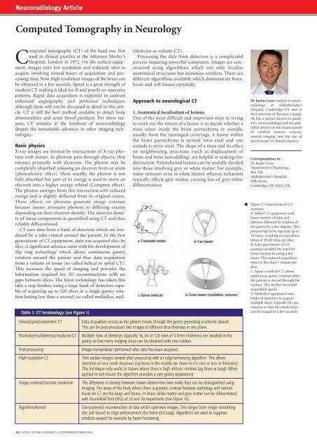
Figure 1: Generations of CT<br />
scanners.<br />
A. Initial CT equipment used<br />
linear motion of tube <strong>and</strong><br />
detector followed by rotation of<br />
the gantry by a few degrees. This<br />
process had to be repeated up to<br />
30 times, resulting in acquisition<br />
times of 30-60 mins per slice.<br />
B. Later generations of CT<br />
scanners avoided the need for<br />
linear motion by using a fan<br />
beam. This reduced acquisition<br />
times to less than 1 minute per<br />
slice.<br />
C. Spiral or helical CT allows<br />
continuous gantry rotation while<br />
the patient is moved through the<br />
scanner. This further increased<br />
acquisition speed.<br />
D. Multislice equipment uses<br />
banks of detectors to acquire<br />
multiple slices (typically 64) per<br />
rotation so that the whole brain<br />
can be imaged in a few seconds.<br />
Helical/spiral/volumetric CT<br />
Multislice/multidetector/multirow CT<br />
Post-processing<br />
High resolution CT<br />
Image contrast/Contrast resolution<br />
Algorithm/Kernel<br />
Data acquisition occurs as the patient moves through the gantry generating a volume dataset.<br />
This can be post-processed into images of different slice thickness in any plane.<br />
Multiple rows of detectors (typically 16, 64 or 128 rows of 0.5mm thickness) are installed in the<br />
gantry so that many imaging slices can be obtained with one rotation.<br />
Image manipulation performed after data has been acquired.<br />
Thin section images viewed after processing with an edge-enhancing algorithm. This allows<br />
detection of very small structures (eg bone in the middle ear down to 0.5 mm or less in thickness).<br />
This technique only works in tissues where there is high intrinsic contrast (eg bone or lung). When<br />
applied to soft tissues the algorithm provides a very grainy appearance.<br />
The difference in density between tissues determines how easily they can be distinguished using<br />
imaging. The areas of the body where there is greatest contrast between pathology <strong>and</strong> normal<br />
tissue on CT are the lungs <strong>and</strong> bones. In brain, white matter <strong>and</strong> grey matter can be differentiated<br />
with Hounsfield Unit (HU) of 20 <strong>and</strong> 30 respectively (see Figure 10).<br />
Computerised reconstruction of data which optimises images. This ranges from image smoothing<br />
(for soft tissue) to edge enhancement (for bone <strong>and</strong> lung). Algorithms are used to suppress<br />
artefacts caused for example by beam hardening.<br />
22 I <strong>ACNR</strong> • VOLUME 8 NUMBER 5 • NOVEMBER/DECEMBER 2008