Unn Ljøstad and Åse Mygland Jone Furlund Owe and Nils ... - ACNR
Unn Ljøstad and Åse Mygland Jone Furlund Owe and Nils ... - ACNR
Unn Ljøstad and Åse Mygland Jone Furlund Owe and Nils ... - ACNR

Create successful ePaper yourself
Turn your PDF publications into a flip-book with our unique Google optimized e-Paper software.
Neuropathology Article<br />
sclerosis in adulthood <strong>and</strong> also increases<br />
risk of febrile convulsions in the early years<br />
of life. Examination of resected material in<br />
HS patients shows evidence for the persistence<br />
of Cajal-Retzius cells in the superficial<br />
temporal cortex <strong>and</strong> alterations within the<br />
reelin signalling pathway, both of which<br />
may signify a disturbance of neuronal<br />
migration. 15 Further evidence to support<br />
this contention, derived from patients with<br />
pathologically-confirmed HS, includes: an<br />
increased incidence of subtle hippocampal<br />
malformations; excess ectopic white matter<br />
neurons in the mesial temporal lobe; <strong>and</strong><br />
association with other lesions that may<br />
have a malformative origin (including lowgrade<br />
glioneuronal tumours). 16 Further -<br />
more, subtle hippocampal malformations<br />
have been found in relatives of patients<br />
with HS (compared to age-matched controls)<br />
most of whom did not have a history<br />
of febrile convulsions or HS themselves. 17 A<br />
maldevelopmental origin would perhaps<br />
also help to explain why hippocampal sclerosis<br />
is often unilateral.<br />
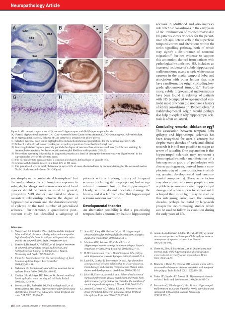
Figure 1: Microscopic appearances of (A) normal hippocampus <strong>and</strong> (B-I) hippocampal sclerosis.<br />
(A) Normal hippocampal anatomy. CA1-CA3=Ammon’s horn (Latin: cornu ammonis). DG=dentate gyrus. Sub=subiculum.<br />
(B) In hippocampal sclerosis, collapse of CA1 (arrows) is evident even at low power.<br />
(C) Selective neuronal drop-out is highlighted by immunohistochemical preparations for the neuronal marker NeuN.<br />
(D) Reduced width of CA1 is more striking on a myelin preparation (Luxol fast blue/cresyl violet).<br />
(E) Reactive gliosis/astrocytosis generally parallels the degree of neuronal loss, demonstrated here (dark brown staining) by<br />
immunohistochemistry for the astrocytic marker glial fibrillary acidic protein (GFAP).<br />
(F) Mossy fibre sprouting is identified in diagnostic practice as a b<strong>and</strong> of dynorphin-immunoreactivity (light brown) in the<br />
supragranular layer of the dentate gyrus.<br />
(G) The normal dentate gyrus contains a compact <strong>and</strong> sharply-defined layer of granule cells.<br />
(H)Granule cell dispersion is seen in at least 40% of HS cases.<br />
(I) The granule cell layer is focally bilaminar in up to 10% of cases, illustrated here by immunostaining for the neuronal marker<br />
NeuN. [Scale bar: A-F=2mm; G-I=200μm].<br />
sive atrophy in the contralateral hemisphere 12 but<br />
the confounding effects of long-term exposure to<br />
antiepileptic drugs <strong>and</strong> seizure-associated head<br />
injuries should be borne in mind. In general,<br />
prospective MRI studies have failed to show a<br />
consistent relationship between the degree of<br />
hippocampal sclerosis <strong>and</strong> the duration/severity<br />
of epilepsy or the total number of generalised<br />
seizures. 13 Furthermore, a quantitative postmortem<br />
study has identified a subgroup of<br />
patients with a life-long history of frequent<br />
seizures (including status epilepticus) but no significant<br />
neuronal loss in the hippocampus. 14<br />
Clearly, seizures do not inevitably damage the<br />
brain – <strong>and</strong> it is far from clear that hippocampal<br />
sclerosis worsens over time.<br />
Developmental theories<br />
An alternative possibility is that a pre-existing<br />
temporal lobe abnormality leads to hippocampal<br />
Concluding remarks: chicken or egg?<br />
The association between temporal lobe<br />
epilepsy <strong>and</strong> hippocampal sclerosis has<br />
been recognised for over a century, but<br />
despite many decades of basic <strong>and</strong> clinical<br />
research it is still not possible to assign an<br />
arrow of causality. One explanation is that<br />
hippocampal sclerosis may represent a<br />
phenotypically-similar manifestation of a<br />
heterogeneous group of pathologies with<br />
diverse pathogenesis, derived from a complex<br />
interplay of numerous factors (including<br />
genetic, developmental <strong>and</strong> environmental<br />
components). These same factors<br />
may also explain why some people are susceptible<br />
to seizure-associated hippocampal<br />
damage <strong>and</strong> others appear to be resistant. It<br />
is hoped that more light may be shed on<br />
this intriguing issue over the coming<br />
decades, perhaps facilitated by large-scale<br />
prospective neuroimaging studies which<br />
can be used to follow its evolution during<br />
the early years of life.<br />
References<br />
1. Margerison JH, Corsellis JAN. Epilepsy <strong>and</strong> the temporal<br />
lobes: a clinical, electroencephalographic <strong>and</strong> neuropathological<br />
study of the brain in epilepsy, with particular reference<br />
to the temporal lobes. Brain 1966;89:499-530.<br />
2. Zentner J, Hufnagel A, Wolf HK, et al. Surgical treatment<br />
of temporal lobe epilepsy: clinical, radiological, <strong>and</strong><br />
histopathological findings in 178 patients. J Neurol,<br />
Neurosurg <strong>and</strong> Psych 1995;58:666-73.<br />
3. Thom M. Recent advances in the neuropathology of focal<br />
lesions in epilepsy. Expert Rev Neurother<br />
2004;4(6):973-84.<br />
4. Meldrum BS. Excitotoxicity <strong>and</strong> selective neuronal loss in<br />
epilepsy. Brain Pathol 2008;3(4):405-12.<br />
5. Coulter DA, McIntyre DC, Löscher W. Animal models of<br />
limbic epilepsies: what can they tell us? Brain Pathol<br />
2002;12(2):240-56.<br />
6. Provenzale JM, Barboriak DP, VanL<strong>and</strong>ingham K, et al.<br />
Hippocampal MRI signal hyperintensity after febrile status<br />
epilepticus is predictive of subsequent mesial temporal sclerosis.<br />
AJR 2007;190:976-83.<br />
7. Scott RC, King MD, Gadian DG, et. Al. Hippocampal<br />
abnormalities after prolonged febrile convulsion: a longitudinal<br />
MRI study. Brain 2003;126:2551-7.<br />
8. Mathern GW, Adelson PD, Cahcal LD, et al.<br />
Hippocampal neuron damage in human epilepsy: Meyer’s<br />
hypothesis revisited. Prog Brain Res 2002;135:237-51.<br />
9. ILAE Commission report. Mesial temporal lobe epilepsy<br />
with hippocampal sclerosis. Epilepsia 2004;45(6):695-714.<br />
10. Lado FA, Dankar R, Lowenstein D, et al. Age-dependent<br />
consequences of seizures: relationship to seizure frequency,<br />
brain damage, <strong>and</strong> circuitry reorganisation. Mental retardation<br />
<strong>and</strong> developmental disabilities 2000;6:242-52.<br />
11. Jokeit H, Ebner A, Arnold S, et al. Bilateral reductions of<br />
hippocampal volume, glucose metabolism <strong>and</strong> Wada hemispheric<br />
memory performance are related to the duration of<br />
mesial temporal lobe epilepsy. J Neurol 1999;246:926-33.<br />
12. Araújo D, Santos AC, Velasco RT, et al. Volumetric evidence<br />
of bilateral damage in unilateral mesial temporal<br />
lobe epilepsy. Epilepsia 2006;47(8):1354-9.<br />
13. Cendes F, Andermann F, Gloor P, et al. Atrophy of mesial<br />
structures in patients with temporal lobe epilepsy: cause or<br />
consequence of repeated seizures. Ann Neurol<br />
1993;34:795-801.<br />
14. Thom M, Zhou J, Martinian L, et al. Quantitative postmortem<br />
study of the hippocampus in chronic epilepsy:<br />
seizures do not inevitably cause neuronal loss. Brain<br />
2005;128:1344-57.<br />
15. Blümcke I, Thom M, Wiestler OD. Ammon's horn sclerosis:<br />
a maldevelopmental disorder associated with temporal<br />
lobe epilepsy. Brain Pathol 2002;12(2):199-211.<br />
16. Fisher PD, Sperber EF, Moshé SL. Hippocampal sclerosis<br />
revisited. Brain <strong>and</strong> development 1998;20(8):563-73.<br />
17. Fern<strong>and</strong>ez G, Effenberger O, Vinz B, et al. Hippocampal<br />
malformation as a cause of familial febrile convulsions <strong>and</strong><br />
subsequent hippocampal sclerosis. Neurology<br />
1998;50:909-17.<br />
18 I <strong>ACNR</strong> • VOLUME 8 NUMBER 5 • NOVEMBER/DECEMBER 2008