Unn Ljøstad and Åse Mygland Jone Furlund Owe and Nils ... - ACNR
Unn Ljøstad and Åse Mygland Jone Furlund Owe and Nils ... - ACNR
Unn Ljøstad and Åse Mygland Jone Furlund Owe and Nils ... - ACNR

You also want an ePaper? Increase the reach of your titles
YUMPU automatically turns print PDFs into web optimized ePapers that Google loves.
Neurosurgery Article<br />
Management of Chronic Subdural Haematoma<br />
Chronic subdural haematoma (CSDH) is one of<br />
the most common clinical entities encountered<br />
in daily neurosurgical practice. It generally<br />
occurs in the elderly population in whom age related<br />
reductions in brain volume with a corresponding<br />
increase in the size of the subdural space increase the<br />
vulnerability to this disease. Cerebral atrophy is also<br />
important in increasing the risk of CSDH in patients<br />
with epilepsy, alcoholism, Huntington’s disease <strong>and</strong><br />
those with overdrainage from a ventriculo-peritoneal<br />
shunt. Patients with a coagulopathy, including<br />
antiplatelet <strong>and</strong> antithrombotic therapy (e.g. aspirin,<br />
dypyridamole, warfarin <strong>and</strong> heparin) are also at an<br />
increased risk of CSDH.<br />
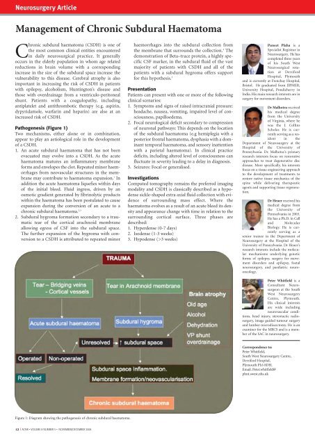
Pathogenesis (Figure 1)<br />
Two mechanisms, either alone or in combination,<br />
appear to play an aetiological role in the development<br />
of a CSDH.<br />
1. An acute subdural haematoma that has not been<br />
evacuated may evolve into a CSDH. As the acute<br />
haematoma matures an inflammatory membrane<br />
forms <strong>and</strong> envelopes the clot. Repeated minor haemorrhages<br />
from neovascular structures in the membrane<br />
may contribute to haematoma expansion. 1 In<br />
addition the acute haematoma liquefies within days<br />
of the initial bleed. Fluid ingress, driven by an<br />
osmotic gradient generated by fibrinolytic products<br />
within the haematoma has been postulated to cause<br />
expansion during the conversion of an acute to a<br />
chronic subdural haematoma. 2,3<br />
2. Subdural hygroma formation secondary to a traumatic<br />
tear of the cortical arachnoid membrane<br />
allowing egress of CSF into the subdural space.<br />
The further expansion of the hygroma with conversion<br />
to a CSDH is attributed to repeated minor<br />
haemorrhages into the subdural collection from<br />
the membrane that surrounds the collection. 4 The<br />
demonstration of Beta–trace protein, a highly specific<br />
CSF marker, in the subdural fluid of the vast<br />
majority of patients with CSDH <strong>and</strong> all of the<br />
patients with a subdural hygroma offers support<br />
for this hypothesis. 5<br />
Presentation<br />
Patients can present with one or more of the following<br />
clinical scenarios:<br />
1. Symptoms <strong>and</strong> signs of raised intracranial pressure:<br />
headache, nausea, vomiting, impaired level of consciousness,<br />
papilloedema.<br />
2. Focal neurological deficit secondary to compression<br />
of neuronal pathways: This depends on the location<br />
of the subdural haematoma (e.g hemiplegia with a<br />
posterior frontal haematoma, dysphasia with a dominant<br />
temporal haematoma, <strong>and</strong> sensory inattention<br />
with a parietal haematoma). In clinical practice<br />
deficits, including altered level of consciousness can<br />
fluctuate in severity leading to a delay in diagnosis.<br />
3. Seizures: Focal or generalised.<br />
Investigations<br />
Computed tomography remains the preferred imaging<br />
modality <strong>and</strong> CSDH is classically described as a hypodense<br />
sickle-shaped extra axial fluid collection with evidence<br />
of surrounding mass effect. Where the<br />
haematoma evolves as a result of an acute bleed its density<br />
<strong>and</strong> appearance change with time in relation to the<br />
surrounding cortical surface. Three phases are<br />
described:<br />
1. Hyperdense (0-7 days)<br />
2. Isodense (1-3 weeks)<br />
3. Hypodense (>3 weeks)<br />
Puneet Plaha is a<br />
Specialist Registrar in<br />
Neurosurgery. He has<br />
completed three years<br />
of his South West<br />
Neurosurgical rotation<br />
at Derriford<br />
Hospital, Plymouth<br />
<strong>and</strong> is currently at Frenchay Hospital,<br />
Bristol. He graduated from JIPMER,<br />
University Hospital, Pondicherry in<br />
India. His main research interests are in<br />
surgery for movement disorders.<br />
Dr Malhotra received<br />
his medical degree<br />
from the University<br />
of Virginia, where he<br />
was the J. Collins<br />
Scholar. He is currently<br />
serving as a resident<br />
in the<br />
Department of Neurosurgery at the<br />
Hospital of the University of<br />
Pennsylvania. Dr. Malhotra's primary<br />
research interests focus on restorative<br />
approaches to treat degenerative disc<br />
disease. More specifically, his interests<br />
focus on a tissue engineering approach<br />
to the development of treatments to<br />
restore native tissue mechanics of the<br />
spine while delivering therapeutic<br />
agents <strong>and</strong> supporting tissue regeneration.<br />
Dr Heuer received his<br />
medical degree from<br />
the University of<br />
Pennsylvania in 2003.<br />
He has a Ph.D. in Cell<br />
<strong>and</strong> Molecular<br />
Biology. He is currently<br />
serving as a<br />
senior trainee in the Department of<br />
Neurosurgery at the Hospital of the<br />
University of Pennsylvania. Dr Heuer’s<br />
research interests include the molecular<br />
mechanisms underlying genetic<br />
forms of epilepsy, surgery for movement<br />
disorders <strong>and</strong> epilepsy, foetal<br />
neurosurgery, <strong>and</strong> paediatric neurooncology.<br />
Peter Whitfield is a<br />
Consultant Neurosurgeon<br />
at the South<br />
West Neurosurgery<br />
Centre, Plymouth.<br />
His clinical interests<br />
are wide including<br />
neurovascular conditions,<br />
head injury, stereotactic radiosurgery,<br />
image guided tumour surgery<br />
<strong>and</strong> lumbar microdiscectomy. He is an<br />
examiner for the MRCS <strong>and</strong> is a member<br />
of the SAC in neurosurgery.<br />
Correspondence to:<br />
Peter Whitfield,<br />
South West Neurosurgery Centre,<br />
Derriford Hospital,<br />
Plymouth PL6 8DH.<br />
Email. Peter.whitfield@<br />
phnt.swest.nhs.uk<br />
Figure 1: Diagram showing the pathogenesis of chronic subdural haematoma.<br />
12 I <strong>ACNR</strong> • VOLUME 8 NUMBER 5 • NOVEMBER/DECEMBER 2008