Neonatal caffeine administration causes a permanent increase in ...
Neonatal caffeine administration causes a permanent increase in ...
Neonatal caffeine administration causes a permanent increase in ...

You also want an ePaper? Increase the reach of your titles
YUMPU automatically turns print PDFs into web optimized ePapers that Google loves.
SYNAPSE 60:450–455 (2006)<br />
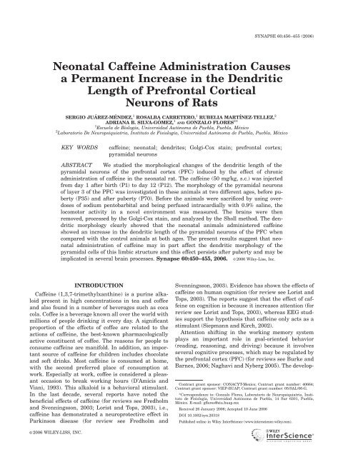
<strong>Neonatal</strong> Caffe<strong>in</strong>e Adm<strong>in</strong>istration Causes<br />
a Permanent Increase <strong>in</strong> the Dendritic<br />
Length of Prefrontal Cortical<br />
Neurons of Rats<br />
SERGIO JUÁREZ-MÉNDEZ, 1 ROSALBA CARRETERO, 1 RUBELIA MARTÍNEZ-TELLEZ, 2<br />
ADRIANA B. SILVA-GÓMEZ, 1 AND GONZALO FLORES 2 *<br />
1 Escuela de Biología, Universidad Autónoma de Puebla, Puebla, México<br />
2 Laboratorio De Neuropsiquiatria, Instituto de Fisiología, Universidad Autónoma de Puebla, Puebla, México<br />
KEY WORDS<br />
<strong>caffe<strong>in</strong>e</strong>; neonatal; dendrites; Golgi-Cox sta<strong>in</strong>; prefrontal cortex;<br />
pyramidal neurons<br />
ABSTRACT We studied the morphological changes of the dendritic length of the<br />
pyramidal neurons of the prefrontal cortex (PFC) <strong>in</strong>duced by the effect of chronic<br />
<strong>adm<strong>in</strong>istration</strong> of <strong>caffe<strong>in</strong>e</strong> <strong>in</strong> the neonatal rat. The <strong>caffe<strong>in</strong>e</strong> (50 mg/kg, s.c.) was <strong>in</strong>jected<br />
from day 1 after birth (P1) to day 12 (P12). The morphology of the pyramidal neurons<br />
of layer 3 of the PFC was <strong>in</strong>vestigated <strong>in</strong> these animals at two different ages, before puberty<br />
(P35) and after puberty (P70). Before the animals were sacrificed by us<strong>in</strong>g overdoses<br />
of sodium pentobarbital and be<strong>in</strong>g perfused <strong>in</strong>tracardially with 0.9% sal<strong>in</strong>e, the<br />
locomotor activity <strong>in</strong> a novel environment was measured. The bra<strong>in</strong>s were then<br />
removed, processed by the Golgi-Cox sta<strong>in</strong>, and analyzed by the Sholl method. The dendritic<br />
morphology clearly showed that the neonatal animals adm<strong>in</strong>istered <strong>caffe<strong>in</strong>e</strong><br />
showed an <strong><strong>in</strong>crease</strong> <strong>in</strong> the dendritic length of the pyramidal neurons of the PFC when<br />
compared with the control animals at both ages. The present results suggest that neonatal<br />
<strong>adm<strong>in</strong>istration</strong> of <strong>caffe<strong>in</strong>e</strong> may <strong>in</strong> part affect the dendritic morphology of the<br />
pyramidal cells of this limbic structure and this effect persists after puberty and may be<br />
implicated <strong>in</strong> several bra<strong>in</strong> processes. Synapse 60:450–455, 2006. VC 2006 Wiley-Liss, Inc.<br />
INTRODUCTION<br />
Caffe<strong>in</strong>e (1,3,7-trimethylxanth<strong>in</strong>e) is a pur<strong>in</strong>e alkaloid<br />
present <strong>in</strong> high concentrations <strong>in</strong> tea and coffee<br />
and also found <strong>in</strong> a number of beverages such as coca<br />
cola. Coffee is a beverage known all over the world with<br />
millions of people dr<strong>in</strong>k<strong>in</strong>g it every day. A significant<br />
proportion of the effects of coffee are related to the<br />
actions of <strong>caffe<strong>in</strong>e</strong>, the best-known pharmacologically<br />
active constituent of coffee. The reasons for people to<br />
consume <strong>caffe<strong>in</strong>e</strong> are manifold. In addition, an important<br />
source of <strong>caffe<strong>in</strong>e</strong> for children <strong>in</strong>cludes chocolate<br />
and soft dr<strong>in</strong>ks. Most <strong>caffe<strong>in</strong>e</strong> is consumed at home,<br />
with the second preferred place of consumption at<br />
work. Especially at work, coffee is considered a pleasant<br />
occasion to break work<strong>in</strong>g hours (D’Amicis and<br />
Viani, 1993). This alkaloid is a behavioral stimulant.<br />
In the last decade, several reports have noted the<br />
beneficial effects of <strong>caffe<strong>in</strong>e</strong> (for reviews see Fredholm<br />
and Svenn<strong>in</strong>gsson, 2003; Lorist and Tops, 2003), i.e.,<br />
<strong>caffe<strong>in</strong>e</strong> has demonstrated a neuroprotective effect <strong>in</strong><br />
Park<strong>in</strong>son disease (for review see Fredholm and<br />
Svenn<strong>in</strong>gsson, 2003). Evidence has shown the effects of<br />
<strong>caffe<strong>in</strong>e</strong> on human cognition (for review see Lorist and<br />
Tops, 2003). The reports suggest that the effect of <strong>caffe<strong>in</strong>e</strong><br />
on cognition is because it <strong><strong>in</strong>crease</strong>s attention (for<br />
review see Lorist and Tops, 2003), whereas EEG studies<br />
support the hypothesis that <strong>caffe<strong>in</strong>e</strong> only acts as a<br />
stimulant (Siepmann and Kirch, 2002).<br />
Attention shift<strong>in</strong>g <strong>in</strong> the work<strong>in</strong>g memory system<br />
plays an important role <strong>in</strong> goal-oriented behavior<br />
(read<strong>in</strong>g, reason<strong>in</strong>g, and driv<strong>in</strong>g) because it <strong>in</strong>volves<br />
several cognitive processes, which may be regulated by<br />
the prefrontal cortex (PFC) (for reviews see Burke and<br />
Barnes, 2006; Naghavi and Nyberg 2005). The develop-<br />
Contract grant sponsor: CONACYT-Mexico; Contract grant number: 40664;<br />
Contract grant sponsor: VIEP-BUAP; Contract grant number: 05/SAL/06-G.<br />
*Correspondence to: Gonzalo Flores, Laboratorio de Neuropsiquiatria, Instituto<br />
de Fisiología, Universidad Autónoma de Puebla, 14 Sur 6301, Puebla,<br />
México. E-mail: gflores@siu.buap.mx<br />
Received 26 January 2006; Accepted 10 June 2006<br />
DOI 10.1002/syn.20318<br />
Published onl<strong>in</strong>e <strong>in</strong> Wiley InterScience (www.<strong>in</strong>terscience.wiley.com).<br />
VC 2006 WILEY-LISS, INC.
ment of the PFC plays a critical role <strong>in</strong> the cognitive<br />
function, attention (Fuster, 2002). The role of the PFC<br />
<strong>in</strong> cognition is well-recognized when its connections<br />
are analyzed. The PFC is <strong>in</strong>terconnected via glutamatergic<br />
projections (Goldman-Rakic et al., 1984) with the<br />
hippocampus and with other limbic cortexes via <strong>in</strong>tracortical<br />
projections (Goldman-Rakic et al., 1984; Jay<br />
and Witter, 1991). Hippocampal neuronal activity<br />
exerts an important regulatory control on the layer-3<br />
pyramidal neurons of the PFC (F<strong>in</strong>ch et al., 1983; Jay<br />
and Witter, 1991; Lewis and Anderson, 1995; Sesack<br />
and Pickel, 1992; Sesack et al., 1989). The hippocampal<br />
to PFC synapses are modifiable synapses and may<br />
express different forms of plasticity <strong>in</strong> all cognitive<br />
processes (for review see Fuster, 2002).<br />
The effect of <strong>caffe<strong>in</strong>e</strong> consumption dur<strong>in</strong>g pregnancy<br />
is under discussion (for review see Signorello and<br />
McLaughl<strong>in</strong>, 2004). Some epidemiologic studies have<br />
not observed association between <strong>caffe<strong>in</strong>e</strong> <strong>in</strong>take dur<strong>in</strong>g<br />
pregnancy and a risk factor for the fetus (for review see<br />
Signorello and McLaughl<strong>in</strong>, 2004). The effects of <strong>caffe<strong>in</strong>e</strong><br />
<strong>in</strong> the neonatal human or animal bra<strong>in</strong> are<br />
unknown. We posed the question of whether <strong>caffe<strong>in</strong>e</strong><br />
exerts a positive or negative effect on the bra<strong>in</strong> dur<strong>in</strong>g<br />
development, especially on the pyramidal neurons of the<br />
PFC, a region related to the cognitive function. Our<br />
<strong>in</strong>vestigation was designed to assess whether the neonatal<br />
<strong>caffe<strong>in</strong>e</strong> <strong>adm<strong>in</strong>istration</strong> affects the dendritic length<br />
and sp<strong>in</strong>e density on pyramidal neurons of layer-3 of the<br />
PFC at both pre- and postpubertal ages. The results suggest<br />
an <strong>in</strong>terest<strong>in</strong>g and persistent effect of the neonatal<br />
<strong>adm<strong>in</strong>istration</strong> of <strong>caffe<strong>in</strong>e</strong> on these pyramidal cells.<br />
MATERIALS AND METHODS<br />
Pregnant Sprague–Dawley rats were obta<strong>in</strong>ed at<br />
gestational day 14–17 from our facilities (University of<br />
Puebla). Animals were <strong>in</strong>dividually housed <strong>in</strong> a temperature-<br />
and humidity-controlled environment on a<br />
12–12 h light–dark cycle, with free access to food and<br />
water. The day follow<strong>in</strong>g birth, litters with six male<br />
pups were grouped and each pup was assigned to either<br />
a <strong>caffe<strong>in</strong>e</strong> (dissolved <strong>in</strong> 0.9% NaCl solution)<br />
<strong>adm<strong>in</strong>istration</strong> (50 mg/kg s.c.) or sal<strong>in</strong>e <strong>adm<strong>in</strong>istration</strong><br />
(control). The <strong>caffe<strong>in</strong>e</strong> or sal<strong>in</strong>e solution was <strong>in</strong>jected<br />
every day <strong>in</strong> the morn<strong>in</strong>g (0800–0900) for 12 days. All<br />
experimental procedures were approved by the BUAP<br />
Animal Care Committee and met governmental guidel<strong>in</strong>es<br />
(Mexican Council for Animal Care, Norma Oficial<br />
Mexicana NOM-062-ZOO-1999). All efforts were made<br />
to m<strong>in</strong>imize animal suffer<strong>in</strong>g and to reduce the number<br />
of animals used. On P21, animals were weaned and<br />
grouped 2–3 animals per cage.<br />
Three weeks (P35, n ¼ 8–9 animals per group) and<br />
8 weeks (P70; n ¼ 8–9 animals per group) after f<strong>in</strong>ish<strong>in</strong>g<br />
the <strong>caffe<strong>in</strong>e</strong> <strong>in</strong>jections, spontaneous locomotor activity<br />
of sal<strong>in</strong>e-control and <strong>caffe<strong>in</strong>e</strong> <strong>adm<strong>in</strong>istration</strong> rats<br />
PREFRONTAL CORTEX AND CAFFEINE<br />
451<br />
was assessed for 60 m<strong>in</strong> <strong>in</strong> 8-photocell activity boxes<br />
(20 40 30 cm 3 ) connected to a computer to count<br />
beam crosses (Tecnologia Digital, Mexico). All animals<br />
were tested between 0800 and 1000. Immediately after<br />
the activity measurements, the rats were deeply anesthetized<br />
with sodium pentobarbital (60 mg/kg, ip) and<br />
perfused <strong>in</strong>tracardially with 0.9% sal<strong>in</strong>e. The bra<strong>in</strong>s<br />
were removed and were processed with modified Golgi-<br />
Cox sta<strong>in</strong><strong>in</strong>g us<strong>in</strong>g procedures described by Gibb and<br />
Kolb (1998). The bra<strong>in</strong>s were first stored <strong>in</strong> the dark<br />
for 14 days <strong>in</strong> Golgi-Cox solution, then 3 days <strong>in</strong> 30%<br />
sucrose. Sections of 200-mm thickness <strong>in</strong> the coronal<br />
plane at the level of the PFC were obta<strong>in</strong>ed us<strong>in</strong>g a<br />
vibratome (Camden Instrument, MA752, Leicester,<br />
England). Sections were collected on cleaned, gelat<strong>in</strong>coated<br />
microscope slides (four sections/slide) and sta<strong>in</strong>ed<br />
with ammonium hydroxide for 30 m<strong>in</strong>. They were immersed<br />
<strong>in</strong> Kodak Film Fixer for another 30 m<strong>in</strong>, and<br />
then washed with water, dehydrated, cleared, and<br />
mounted us<strong>in</strong>g a res<strong>in</strong>ous medium.<br />
The Golgi-impregnated pyramidal neurons of layer-3<br />
of the PFC (area Cg1 and 3, plate 7–9 of Pax<strong>in</strong>os and<br />
Watson, 1986, correspond<strong>in</strong>g to Cg1 and prelimbic cortex<br />
of Pax<strong>in</strong>os and Watson, 1998) were readily identified<br />
by their characteristic triangular soma-shape, apical<br />
dendrites extend<strong>in</strong>g toward the pial surface, and<br />
numerous dendritic sp<strong>in</strong>es. The criteria used to select<br />
neurons for reconstruction was essentially as described<br />
previously (Flores et al., 2005; Mart<strong>in</strong>ez-Tellez et al.,<br />
2005; Silva-Gómez et al., 2003; Vega et al., 2004). Five<br />
neurons <strong>in</strong> each hemisphere (10 neurons per animal)<br />
were drawn us<strong>in</strong>g camera lucida at a magnification of<br />
250 (DMLS, Leica Microscope) by a person bl<strong>in</strong>d to<br />
treatment conditions. Although the Golgi-Cox procedure<br />
has been extensively used to determ<strong>in</strong>e dendritic<br />
morphology, a caveat of the method is that only a small<br />
number of neurons are impregnated with the sta<strong>in</strong> and<br />
that some sp<strong>in</strong>es hidden beh<strong>in</strong>d the dendritic shafts<br />
may not be counted. Nevertheless, given the random<br />
nature of these events, we consider that the neurons<br />
studied here are representative of the total population<br />
and that sp<strong>in</strong>e density measurements are relative. For<br />
each neuron, the three-dimensional basal dendritic<br />
tree, <strong>in</strong>clud<strong>in</strong>g all branches, was reconstructed <strong>in</strong> a<br />
two-dimensional plane and the dendritic trac<strong>in</strong>g was<br />
quantified by Sholl analysis (Sholl, 1953). A transparent<br />
grid with concentric r<strong>in</strong>gs, equivalent to 10-mm<br />
spac<strong>in</strong>g, was placed over the dendritic draw<strong>in</strong>g and the<br />
number of branches <strong>in</strong>tersect<strong>in</strong>g each r<strong>in</strong>g was used to<br />
estimate the total dendritic length (TDL) (Flores et al.,<br />
2005). Us<strong>in</strong>g a branch<strong>in</strong>g analysis, the branch order of<br />
the dendritic arborization was estimated by count<strong>in</strong>g<br />
the number of r<strong>in</strong>g <strong>in</strong>tersections per every branch<br />
order for all the neurons. The density of dendritic<br />
sp<strong>in</strong>es was estimated by draw<strong>in</strong>g at least 10-mm long<br />
segments from the term<strong>in</strong>al tips at high power (1000)<br />
and count<strong>in</strong>g the number of sp<strong>in</strong>es.<br />
Synapse DOI 10.1002/syn
452 S. JUÁREZ-MÉNDEZ ET AL.<br />
Fig. 1. Photomicrograph show<strong>in</strong>g representative<br />
Golgi-Cox impregnated pyramidal<br />
neurons of the PFC from control<br />
animals (A) and animals with neonatal<br />
<strong>caffe<strong>in</strong>e</strong> <strong>adm<strong>in</strong>istration</strong> (B) at P35.<br />
Data from the Sholl analyses and the sp<strong>in</strong>e densities<br />
were analyzed us<strong>in</strong>g two-way ANOVA, then by<br />
Newman-Keuls tests for post hoc comparisons, with<br />
age and neonatal <strong>caffe<strong>in</strong>e</strong> <strong>adm<strong>in</strong>istration</strong> as <strong>in</strong>dependent<br />
factors (P < 0.05 was considered significant).<br />
RESULTS<br />
Locomotor activity<br />
The effect of the neonatal <strong>caffe<strong>in</strong>e</strong> <strong>adm<strong>in</strong>istration</strong><br />
on spontaneous locomotor activity <strong>in</strong> a novel environment<br />
was not different between rats. However, both<br />
control animals and animals with neonatal <strong>caffe<strong>in</strong>e</strong><br />
<strong>adm<strong>in</strong>istration</strong> <strong>in</strong>itially show an active exploratory<br />
behavior <strong>in</strong> a novel environment, without changes <strong>in</strong><br />
the locomotion when compared between groups at the<br />
same age, (P35: control: 1199 6 63, <strong>caffe<strong>in</strong>e</strong>: 1296 6<br />
122; P70: control: 930 6 51, <strong>caffe<strong>in</strong>e</strong>: 1043 6 96).<br />
Dendritic morphology<br />
The dendritic branch<strong>in</strong>g and density of dendritic sp<strong>in</strong>es<br />
on neurons of the PFC were measured by Golgi-Cox sta<strong>in</strong><br />
between neonatal <strong>caffe<strong>in</strong>e</strong> <strong>adm<strong>in</strong>istration</strong> and control<br />
rats. The maximum branch order, sp<strong>in</strong>e density, and TDL<br />
obta<strong>in</strong>ed were similar to our previous reports (Flores<br />
et al., 2005; Mart<strong>in</strong>ez-Tellez et al., 2005; Silva-Gomez<br />
et al., 2003; Vega et al., 2004). The Golgi-Cox impregnation<br />
procedure clearly filled the dendritic shafts and<br />
sp<strong>in</strong>es of layer-3 pyramidal neurons of the PFC (Fig. 1).<br />
As measured by Sholl analysis, a two-way ANOVA for the<br />
TDL showed there was a significant effect of the neonatal<br />
<strong>adm<strong>in</strong>istration</strong> of <strong>caffe<strong>in</strong>e</strong> (F 1, 30 ¼ 44.75, P < 0.001) with<br />
the <strong>in</strong>teraction neonatal <strong>caffe<strong>in</strong>e</strong> x age (F 1, 30 ¼ 11. 52,<br />
P ¼ 0.002) and without effect by the age (F 1, 30 ¼ 0.9, P ¼<br />
0.35). The post hoc test revealed that TDL was significantly<br />
longer <strong>in</strong> the pyramidal neurons from animals with<br />
neonatal <strong>caffe<strong>in</strong>e</strong> <strong>adm<strong>in</strong>istration</strong> at prepubertal (P <<br />
0.001) and at postpubertal (P < 0.05) age with significant<br />
differences between these two groups (P < 0.01) with neonatal<br />
<strong>caffe<strong>in</strong>e</strong> <strong>adm<strong>in</strong>istration</strong> but not between the control<br />
groups (Fig. 2A). A two-way ANOVA for the analysis of<br />
the density of the dendritic sp<strong>in</strong>es revealed that there was<br />
a significant effect of the neonatal <strong>adm<strong>in</strong>istration</strong> of <strong>caffe<strong>in</strong>e</strong><br />
(F 1, 30 ¼ 5.9, P ¼ 0.02) with effect by the age (F 1, 30 ¼<br />
8.88, P < 0.01) and not with the <strong>in</strong>teraction neonatal <strong>caffe<strong>in</strong>e</strong><br />
age (F 1, 30 ¼ 0.05, P ¼ 0.8). The post hoc test did<br />
Synapse DOI 10.1002/syn
PREFRONTAL CORTEX AND CAFFEINE<br />
453<br />
whereas that P60 rats with neonatal <strong>caffe<strong>in</strong>e</strong> <strong>adm<strong>in</strong>istration</strong><br />
show more arborization at the levels of the third<br />
order and higher (P < 0.05) (Figs. 2E and 2F).<br />
Fig. 2. Analysis of the pyramidal neurons <strong>in</strong> control animals<br />
and animals with neonatal <strong>caffe<strong>in</strong>e</strong> <strong>adm<strong>in</strong>istration</strong> (n ¼ 8–9 animals<br />
per group). A: Total dendritic length. Rats with neonatal <strong>caffe<strong>in</strong>e</strong><br />
<strong>adm<strong>in</strong>istration</strong> exhibit an <strong><strong>in</strong>crease</strong> <strong>in</strong> the total dendritic length. B:<br />
Sp<strong>in</strong>e density. C: Sholl analysis of <strong>in</strong>tersection per shell at P35. D:<br />
Sholl analysis of <strong>in</strong>tersection per shell at P70. The neonatal <strong>caffe<strong>in</strong>e</strong><br />
<strong>adm<strong>in</strong>istration</strong> produced an <strong><strong>in</strong>crease</strong> <strong>in</strong> the number of <strong>in</strong>tersections<br />
per concentric r<strong>in</strong>g at both ages. E: Branch order at P35. F: Branch<br />
order at P70. The <strong><strong>in</strong>crease</strong> <strong>in</strong> the branch order by the neonatal <strong>caffe<strong>in</strong>e</strong><br />
<strong>adm<strong>in</strong>istration</strong> was from the first to the fourth branch orders<br />
<strong>in</strong> the P35 animals, whereas at P70 the <strong><strong>in</strong>crease</strong> was from the third<br />
to sixth branch orders. a, P < 0.01 and b, P < 0.05.<br />
not show differences between neonatal <strong>caffe<strong>in</strong>e</strong> <strong>adm<strong>in</strong>istration</strong><br />
and control animals at both ages (Fig. 2B). However,<br />
the neonatal <strong>caffe<strong>in</strong>e</strong> <strong>adm<strong>in</strong>istration</strong> groups at both<br />
ages showed an apparent, but not statistically significant,<br />
trend towards an <strong><strong>in</strong>crease</strong>d density of the dendritic sp<strong>in</strong>es<br />
compared with the control animals (P35: P ¼ 0.09; P70:<br />
P ¼ 0.06) (Fig. 2B).<br />
Two other measured values obta<strong>in</strong>ed from the Sholl<br />
analysis, the <strong>in</strong>tersection per shell and the length per<br />
branch order, gave <strong>in</strong>formation about of the quality of<br />
the dendritic arborization. The analysis of <strong>in</strong>tersection<br />
per radius of shell also showed that the neonatal <strong>caffe<strong>in</strong>e</strong><br />
<strong>adm<strong>in</strong>istration</strong> rats showed significantly more<br />
dendritic arborization (P < 0.05)whencomparedtothe<br />
control animals at both ages (Figs 2C and 2D). The<br />
branch-order analysis suggests that P35 rats with neonatal<br />
<strong>caffe<strong>in</strong>e</strong> <strong>adm<strong>in</strong>istration</strong> exhibit more arborization<br />
at the levels of the first to fourth order compared to the<br />
controls (second order P < 0.01,restofordersP < 0.05),<br />
DISCUSSION<br />
Our major aim was to <strong>in</strong>vestigate the consequences<br />
of the neonatal <strong>caffe<strong>in</strong>e</strong> <strong>adm<strong>in</strong>istration</strong> on the development<br />
of the basilar dendritic structural morphology of<br />
layer-3 pyramidal cells of the PFC. We determ<strong>in</strong>ed that<br />
neonatal <strong>caffe<strong>in</strong>e</strong> <strong>adm<strong>in</strong>istration</strong> <strong>causes</strong> a persistent<br />
<strong><strong>in</strong>crease</strong> <strong>in</strong> the dendritic length <strong>in</strong> layer-3 pyramidal<br />
neurons of the PFC without significant changes <strong>in</strong> the<br />
density of the sp<strong>in</strong>es. Interest<strong>in</strong>gly, this <strong>permanent</strong><br />
change <strong>in</strong> the dendritic length did not alter the locomotor<br />
activity <strong>in</strong> response to a mild stress, such as a novel<br />
environment. Our f<strong>in</strong>d<strong>in</strong>gs contribute to the current<br />
knowledge of the effects of <strong>caffe<strong>in</strong>e</strong> on the limbic areas<br />
and cellular structures <strong>in</strong>volved <strong>in</strong> the underly<strong>in</strong>g basis<br />
of the cognitive function. Thus, these data suggest that<br />
changes <strong>in</strong> dendritic arborization caused by the neonatal<br />
<strong>caffe<strong>in</strong>e</strong> <strong>adm<strong>in</strong>istration</strong> may be l<strong>in</strong>ked <strong>in</strong> part to the<br />
<strong><strong>in</strong>crease</strong> of the cognitive function reported <strong>in</strong> children<br />
and rats (Bernste<strong>in</strong> et al., 1994; Elk<strong>in</strong>s et al., 1981;<br />
Prediger et al., 2005; Rapoport et al., 1981).<br />
Interest<strong>in</strong>gly, several studies have shown a variety of<br />
effects of <strong>caffe<strong>in</strong>e</strong> consumption <strong>in</strong> children and preadolescents,<br />
although it is surpris<strong>in</strong>g that so few studies<br />
have been specifically addressed to the effects on this<br />
population. At low doses, an <strong><strong>in</strong>crease</strong>d performance <strong>in</strong><br />
attention tests has been noted <strong>in</strong> children, whereas at<br />
high doses an <strong><strong>in</strong>crease</strong>d motor activity has been<br />
reported <strong>in</strong> children (for reviews see Castellanos and<br />
Rapoport, 2002; Nawrot et al., 2003). More <strong>caffe<strong>in</strong>e</strong> also<br />
caused an improvement <strong>in</strong> a manual dexterity test<br />
(Bernste<strong>in</strong> et al., 1994). In preadolescent boys it has<br />
been reported that a high dose of <strong>caffe<strong>in</strong>e</strong> produced an<br />
<strong><strong>in</strong>crease</strong> <strong>in</strong> motor activity with an <strong><strong>in</strong>crease</strong> <strong>in</strong> speech<br />
rate, whereas low doses produced a reduction <strong>in</strong> reaction<br />
time <strong>in</strong> a vigilance test, and a reduced number of errors<br />
<strong>in</strong> a susta<strong>in</strong>ed-attention-measure test (Elk<strong>in</strong>s et al.,<br />
1981; Rapoport et al., 1981). Other effects of <strong>caffe<strong>in</strong>e</strong> <strong>in</strong><br />
children caused their be<strong>in</strong>g nervous, anxious, fidgety, jittery,<br />
and restless and experienc<strong>in</strong>g hyperactivity and<br />
difficulty sleep<strong>in</strong>g (for reviews see Castellanos and<br />
Rapoport, 2002; Nawrot et al., 2003). However, all these<br />
studies only <strong>in</strong>cluded the effects of one dose of <strong>caffe<strong>in</strong>e</strong>,<br />
whereas for chronic <strong>caffe<strong>in</strong>e</strong> effects only attention to the<br />
symptoms associated with <strong>caffe<strong>in</strong>e</strong> withdrawal have<br />
been studied (for reviews see Castellanos and Rapoport,<br />
2002; Nawrot et al., 2003). Only one report suggests that<br />
<strong>caffe<strong>in</strong>e</strong> consumed for 2 months between ages 12 and 24<br />
months did not alter the cognitive development (Engle<br />
et al., 1999). There is not enough <strong>in</strong>formation about the<br />
effects of chronic <strong>caffe<strong>in</strong>e</strong> <strong>adm<strong>in</strong>istration</strong> dur<strong>in</strong>g early<br />
age on the morphological development of the cortex that<br />
may be present at puberty. However, mostly all children<br />
Synapse DOI 10.1002/syn
454 S. JUÁREZ-MÉNDEZ ET AL.<br />
at an early age start to consume chocolate and soft<br />
dr<strong>in</strong>ks, both important sources of <strong>caffe<strong>in</strong>e</strong>. Our results<br />
suggest that the early consumption of <strong>caffe<strong>in</strong>e</strong> plays a<br />
critical role <strong>in</strong> the development of the cortical neurons.<br />
In the rat, the first 2 weeks after birth is a critical<br />
period <strong>in</strong> the development and synaptogenesis because<br />
of the stimulation of the outgrowth and maturation of<br />
neurons (H<strong>in</strong>dley et al., 1997; Kalb and Agost<strong>in</strong>i, 1993;<br />
Roskams et al., 1994; Truman et al., 1996; Williams<br />
et al., 1994), <strong>in</strong> part correspond<strong>in</strong>g to the development of<br />
the human bra<strong>in</strong> <strong>in</strong> utero. For example, the <strong>in</strong>terconnection<br />
between the hippocampus and amygdala with the<br />
PFC <strong>in</strong> the rat is after P7 (Bouwmeester et al., 2002a,b;<br />
Lipska, 2004) and <strong>in</strong> the human this happens after the<br />
fifth gestational month (for reviews see Ulfig et al.,<br />
2003; We<strong>in</strong>berger and Lipska et al., 1995). Therefore,<br />
our data may <strong>in</strong> part have an implication about the consumption<br />
of <strong>caffe<strong>in</strong>e</strong> dur<strong>in</strong>g pregnancy.<br />
Dendritic growth and the density of sp<strong>in</strong>es on the dendritesarerelatedtothedegreeofconnectivityandafferent<br />
activity (McAllister, 2000). Further, changes <strong>in</strong> the<br />
cortical excitability dur<strong>in</strong>g early development produces<br />
alterations <strong>in</strong> dendritic sp<strong>in</strong>es (Friedlander et al., 1982;<br />
Globus and Scheibel, 1967; Guillery, 1973; Heumann and<br />
Rab<strong>in</strong>owicz, 1982; McMullen and Glaser, 1988; Wiesel<br />
and Hubel, 1963). Cortical excitability is ma<strong>in</strong>ly mediated<br />
by glutamate as the neurotransmitter and the glutamate<br />
by itself may control the sp<strong>in</strong>e creation and destruction<br />
at glutamatergic synapses (McAllister, 2000). In<br />
agreement with our dendritic sp<strong>in</strong>e density results, a<br />
recent report suggests that <strong>caffe<strong>in</strong>e</strong> did not alter the cortical<br />
excitability (Orth et al., 2005). By comparison with<br />
our results, coca<strong>in</strong>e-sensitized adult rats have an<br />
enhanced total basilar dendritic length with an <strong><strong>in</strong>crease</strong><br />
<strong>in</strong> the number of dendritic sp<strong>in</strong>es <strong>in</strong> the pyramidal neurons<br />
of the PFC (Rob<strong>in</strong>son and Kolb, 1999). However, <strong>in</strong><br />
these coca<strong>in</strong>e-sensitized animals, the morphological<br />
changes<strong>in</strong>thedendriteswerecorrelatedwithan<strong><strong>in</strong>crease</strong><br />
<strong>in</strong> the locomotor activity. The coca<strong>in</strong>e-sensitized effect<br />
may<strong>in</strong>partbecausedbyan<strong>in</strong>directeffectofthedopam<strong>in</strong>e<br />
(DA) that modulates the glutamate transmission<br />
(Rob<strong>in</strong>son and Kolb, 1999). In addition, a recent report<br />
demonstrated that <strong>in</strong> utero exposure to coca<strong>in</strong>e also produces<br />
an <strong><strong>in</strong>crease</strong> <strong>in</strong> the TDL of the PFC without changes<br />
<strong>in</strong> the somatosensory cortex (Lloyde et al., 2003).<br />
The mechanism by which <strong>caffe<strong>in</strong>e</strong> may cause an<br />
<strong><strong>in</strong>crease</strong> <strong>in</strong> the dendritic length of the pyramidal neurons<br />
of the PFC is not yet clear. However, consumption of <strong>caffe<strong>in</strong>e</strong>,<br />
at the dose size we have used, is primarily related<br />
to its actions to block adenos<strong>in</strong>e receptors (Fredholm<br />
et al., 1999; Phillis et al., 1991; Shi et al., 1993). The<br />
effects of <strong>caffe<strong>in</strong>e</strong> <strong>in</strong> the bra<strong>in</strong> are mediated particularly<br />
by its antagonistic actions at the A 1 and A 2A subtypes of<br />
the adenos<strong>in</strong>e receptors (for review see Lorist and Tops,<br />
2003). Adenos<strong>in</strong>e A 1 receptors are present <strong>in</strong> almost all<br />
bra<strong>in</strong> areas, but the cerebral cortex has the highest levels<br />
(Fastbom et al., 1987; Goodman and Snyder, 1982),<br />
whereas adenos<strong>in</strong>e A 2A receptors are ma<strong>in</strong>ly present <strong>in</strong><br />
the DA-rich regions of the bra<strong>in</strong>, such as the striatum<br />
and tuberculum olfactorium, but not <strong>in</strong> the cerebral cortex<br />
(for review see Lorist and Tops, 2003). In addition,<br />
theblockadeofA 2A receptors <strong>in</strong> striatopallidal neurons<br />
is crucial for the stimulatory action of <strong>caffe<strong>in</strong>e</strong>. There is<br />
ample evidence that an <strong>in</strong>tact dopam<strong>in</strong>ergic neurotransmission<br />
is necessary for <strong>caffe<strong>in</strong>e</strong> to be stimulatory (Ferre<br />
et al., 1992). Therefore, the effect of <strong>caffe<strong>in</strong>e</strong> on neonates<br />
maybe<strong>in</strong>partbecausedbyanadenos<strong>in</strong>eA 1 blockade or<br />
an <strong>in</strong>direct effect via DA <strong>in</strong>teraction. Other mechanisms<br />
of action of <strong>caffe<strong>in</strong>e</strong> are the <strong>in</strong>hibition of phosphodiesterase<br />
and mobilization of <strong>in</strong>tracellular calcium, but these<br />
effects require higher concentrations of <strong>caffe<strong>in</strong>e</strong> (for<br />
review see Lorist and Tops, 2003). It is well known that<br />
neural cells after embryonic day 18 (E18) are sensitive to<br />
<strong>caffe<strong>in</strong>e</strong>, and recent results suggest that after birth a differentiation<br />
of Ca 2+ release channels, which regulate the<br />
Ca 2+ <strong>in</strong>ternal (i) stores, are present <strong>in</strong> the cortical neurogenesis<br />
(Maric et al., 2000). Sp<strong>in</strong>al cord neurons express<br />
<strong>caffe<strong>in</strong>e</strong>-sensitive Ca 2+ i stores only <strong>in</strong> the more mature<br />
neurons. Perhaps, the block<strong>in</strong>g of the adenos<strong>in</strong>e receptors<br />
together with the <strong>in</strong>hibition of the mobilization of <strong>in</strong>tracellular<br />
calcium stores may <strong>in</strong> part be correlated with<br />
the morphological data reported here. Our results add to<br />
a grow<strong>in</strong>g body of literature show<strong>in</strong>g that changes <strong>in</strong> the<br />
dendritic arborization without changes of the dendrite<br />
sp<strong>in</strong>es may be caused by the effect of neonatal <strong>caffe<strong>in</strong>e</strong><br />
<strong>adm<strong>in</strong>istration</strong>. Our results are more evidence that neonatal<br />
<strong>caffe<strong>in</strong>e</strong> <strong>adm<strong>in</strong>istration</strong> produces changes <strong>in</strong> the<br />
bra<strong>in</strong>, and more studies are needed to understand what<br />
the physiological effect of these changes are.<br />
In summary, our results are the first evidence show<strong>in</strong>g<br />
significant dendritic morphological changes <strong>in</strong> the<br />
PFC at pre- and postpubertal animals with neonatal <strong>caffe<strong>in</strong>e</strong><br />
<strong>adm<strong>in</strong>istration</strong>. Our results add to a grow<strong>in</strong>g body<br />
of literature about the effect of <strong>caffe<strong>in</strong>e</strong> on the bra<strong>in</strong>. It<br />
is possible that dendritic changes <strong>in</strong> the pyramidal neurons<br />
of the PFC, by alter<strong>in</strong>g synaptic <strong>in</strong>puts and organization,<br />
may contribute to the cognitive changes reported<br />
<strong>in</strong> <strong>caffe<strong>in</strong>e</strong> consumers, such as attention and spatial<br />
learn<strong>in</strong>g (for reviews see Castellanos and Rapoport,<br />
2002; Nawrot et al., 2003).<br />
ACKNOWLEDGMENTS<br />
The authors are grateful to Dr. Carlos Escamilla for<br />
his help and suggestions related to keep<strong>in</strong>g of animals.<br />
S.J.M., R.C., and R.M.T. are students from<br />
BUAP. G.F. is a member of the Researcher National<br />
System of Mexico. The authors thank Dr. Ellis Glazier<br />
for edit<strong>in</strong>g the English-language text.<br />
REFERENCES<br />
Bernste<strong>in</strong> GA, Carroll ME, Crosby RD, Perwien AR, Go FS, Benowitz<br />
NL. 1994. Caffe<strong>in</strong>e effects on learn<strong>in</strong>g, performance, and anxiety<br />
<strong>in</strong> normal school-age children. J Am Acad Child Adolesc Psychiatry<br />
33:407–415.<br />
Synapse DOI 10.1002/syn
PREFRONTAL CORTEX AND CAFFEINE<br />
455<br />
Bouwmeester H, Wolter<strong>in</strong>k G, van Ree JM. 2002a. <strong>Neonatal</strong> development<br />
of projections from the basolateral amygdala to prefrontal,<br />
striatal, and thalamic structures <strong>in</strong> the rat. J Comp Neurol 442:<br />
239–249.<br />
Bouwmeester H, Smits K, Van Ree JM. 2002b. <strong>Neonatal</strong> development<br />
of projections to the basolateral amygdala from prefrontal<br />
and thalamic structures <strong>in</strong> rat. J Comp Neurol 450:241–255.<br />
Burke SN, Barnes CA. 2006. Neural plasticity <strong>in</strong> the age<strong>in</strong>g bra<strong>in</strong>.<br />
Nat Rev Neurosci 7:30–40.<br />
Castellanos FX, Rapoport JL. 2002. Effects of <strong>caffe<strong>in</strong>e</strong> on development<br />
and behavior <strong>in</strong> <strong>in</strong>fancy and childhood: A review of the published<br />
literature. Food Chem Toxicol 40:1235–1242.<br />
D’Amicis A, Viani R. 1993. The consumption of coffee. In: Garatt<strong>in</strong>i S,<br />
editor. Caffe<strong>in</strong>e, coffee and health. New York: Raven Press. p 1–16.<br />
Engle PL, VasDias T, Howard I, Romero-Abal ME, Quan de Serrano J,<br />
Bulux J, Solomons NW, Dewey KG. 1999. Effects of discont<strong>in</strong>u<strong>in</strong>g<br />
coffee <strong>in</strong>take on iron deficient Guatemalan toddlers’ cognitive. Early<br />
Hum Dev 53:251–269.<br />
Elk<strong>in</strong>s RN, Rapoport JL, Zahn TP, Buchsbaum MS, We<strong>in</strong>gartner H,<br />
Kop<strong>in</strong> IJ, Langer D, Johnson C. 1981. Acute effects of <strong>caffe<strong>in</strong>e</strong> <strong>in</strong><br />
normal prepubertal boys. Am J Psychiatry 138:178–183.<br />
Fastbom J, Pazos A, Palacios JM. 1987. The distribution of adenos<strong>in</strong>e<br />
A1 receptors and 5 0 -nucleotidase <strong>in</strong> the bra<strong>in</strong> of some commonly<br />
used experimental animals. Neuroscience 22:813–826.<br />
Ferre S, Fuxe K, vonEuler G, Johansson B, Fredholm BB. 1992. Adenos<strong>in</strong>e-dopam<strong>in</strong>e<br />
<strong>in</strong>teractions <strong>in</strong> the bra<strong>in</strong>. Neuroscience 51:501–512.<br />
F<strong>in</strong>ch DM, Nowl<strong>in</strong> NL, Babb TL. 1983. Demonstration of axonal<br />
projections of neurons <strong>in</strong> the rat hippocampus and subiculum by<br />
<strong>in</strong>tracellular <strong>in</strong>jection of HRP. Bra<strong>in</strong> Res 271:201–216.<br />
Flores G, Alquicer G, Silva-Gomez AB, Zaldivar G, Stewart J, Quirion<br />
R, Srivastava LK. 2005. Alterations <strong>in</strong> dendritic morphology<br />
of prefrontal cortical and nucleus accumbens neurons <strong>in</strong> postpubertal<br />
rats after neonatal excitotoxic lesions of the ventral hippocampus.<br />
Neuroscience 133:463–470.<br />
Fredholm BB, Svenn<strong>in</strong>gsson P. 2003. Adenos<strong>in</strong>e-dopam<strong>in</strong>e <strong>in</strong>teractions:<br />
Development of a concept and some comments on therapeutic<br />
possibilities. Neurology 61:S5–S9.<br />
Fredholm BB, Battig K, Holmén J, Nehlig A, Zvartau EE. 1999.<br />
Actions of <strong>caffe<strong>in</strong>e</strong> <strong>in</strong> the bra<strong>in</strong> with special reference. Pharmacol<br />
Rev 51:84–125.<br />
Friedlander MJ, Stanford LR, Sherman SM. 1982. Effects of monocular<br />
deprivation on the structure-function relationship of <strong>in</strong>dividual neurons<br />
<strong>in</strong> the cat’s lateral geniculate nucleus. J Neurosci 2:321–330.<br />
Fuster JM. 2002. Frontal lobe and cognitive development. J Neurocytol<br />
31:373–385.<br />
Gibb R, Kolb B. 1998. A method for vibrotome section<strong>in</strong>g of Golgi-<br />
Cox sta<strong>in</strong>ed whole bra<strong>in</strong>. J Neurosci Methods 79:1–4.<br />
Globus A, Scheibel AB. 1967. Synaptic loci on visual cortical neurons<br />
of the rabbit: The specific afferent radiation. Exp Neurol<br />
18:116–131.<br />
Goldman-Rakic PS, Selemon LD, Schwartz ML. 1984. Dual pathways<br />
connect<strong>in</strong>g the dorsolateral prefrontal cortex with the hippocampal<br />
formation and parahippocampal cortex <strong>in</strong> the rhesus monkey.<br />
Neuroscience 12:719–743.<br />
Goodman RR, Synder SH. 1982. Autoradiographic localization of<br />
adenos<strong>in</strong>e receptors <strong>in</strong> rat bra<strong>in</strong> us<strong>in</strong>g [3H]cyclohexyladenos<strong>in</strong>e.<br />
J Neurosci 2:1230–1241.<br />
Guillery RW. 1973. The effect of lid suture upon the growth of cells<br />
<strong>in</strong> the dorsal lateral geniculate nucleus of kittens. J Comp Neurol<br />
148:417–422.<br />
Heumann D, Rab<strong>in</strong>owicz T. 1982. Postnatal development of the visual<br />
cortex of the mouse after enucleation at birth. Exp Bra<strong>in</strong> Res<br />
46:99–106.<br />
H<strong>in</strong>dley S, Juurl<strong>in</strong>k BHJ, Gysbers JW, Middlemiss PJ, Herman<br />
MAR, Rathbone MP. 1997. Nitric oxide donors enhance neurotroph<strong>in</strong><br />
<strong>in</strong>duced neurite outgrowth through a cGMP dependent<br />
mechanism. J Neurosci Res 47:427–439.<br />
Jay TM, Witter MP. 1991. Distribution of hippocampal CA1 and<br />
subicular efferents <strong>in</strong> the prefrontal cortex of the rat studied by<br />
means of anterograde transport of Phaseolus vulgaris-leucoagglut<strong>in</strong><strong>in</strong>.<br />
J Comp Neurol 313:574–586.<br />
Kalb RG, Agost<strong>in</strong>i J. 1993. Molecular evidence for nitric oxide-mediated<br />
motor neuron development. Neuroscience 57:1–8.<br />
Lewis DA, Anderson SA. 1995. The functional architecture of the<br />
prefrontal cortex and schizophrenia. Psychol Med 25:887–894.<br />
Lipska BK. 2004. Us<strong>in</strong>g animal models to test a neurodevelopmental<br />
hypothesis of schizophrenia. J Psychiatry Neurosci 29:282–286.<br />
Lloyd SA, Wensley B, Faherty CJ, Smeyne RJ. 2003. Regional differences<br />
<strong>in</strong> cortical dendrite morphology follow<strong>in</strong>g <strong>in</strong> utero exposure<br />
to coca<strong>in</strong>e. Bra<strong>in</strong> Res Dev Bra<strong>in</strong> Res 147:59–66.<br />
Lorist MM, Tops M. 2003. Caffe<strong>in</strong>e, fatigue, and cognition. Bra<strong>in</strong><br />
Cogn 53:82–94.<br />
Maric D, Maric I, Barker JL. 2000. Developmental changes <strong>in</strong> cell<br />
calcium homeostasis dur<strong>in</strong>g neurogenesis of the embryonic rat<br />
cerebral cortex. Cereb Cortex 10:561–573.<br />
Mart<strong>in</strong>ez-Tellez R, Gomez-Villalobos Mde J, Flores G. 2005. Alteration<br />
<strong>in</strong> dendritic morphology of cortical neurons <strong>in</strong> rats with diabetes<br />
mellitus <strong>in</strong>duced by streptozotoc<strong>in</strong>. Bra<strong>in</strong> Res 1048:108–115.<br />
McAllister AK. 2000. Cellular and molecular mechanisms of dendrite<br />
growth. Cereb Cortex 10:963–973.<br />
McMullen NT, Glaser EM. 1988. Auditory cortical responses to neonatal<br />
deafen<strong>in</strong>g: Pyramidal neuron sp<strong>in</strong>e loss without changes <strong>in</strong><br />
growth or orientation. Exp Bra<strong>in</strong> Res 72:195–200.<br />
Naghavi HR, Nyberg L. 2005. Common fronto-parietal activity <strong>in</strong><br />
attention, memory and consciousness: Shared demands on <strong>in</strong>tegration?<br />
Conscious Cogn 14:390–425.<br />
Nawrot P, Jordan S, Eastwood J, Rotste<strong>in</strong> J, Hugenholtz A, Feeley M.<br />
2003. Effects of <strong>caffe<strong>in</strong>e</strong> on human health. Food Addit Contam 20:1–30.<br />
Orth M, Amann B, Ratnaraj N, Patsalos PN, Rothwell JC. 2005.<br />
Caffe<strong>in</strong>e has no effect on measures of cortical excitability. Cl<strong>in</strong><br />
Neurophysiol 116:308–314.<br />
Pax<strong>in</strong>os G, Watson C. 1986. The rat bra<strong>in</strong> <strong>in</strong> stereotaxic coord<strong>in</strong>ates,<br />
2nd ed. New York: Academic Press.<br />
Pax<strong>in</strong>os G, Watson C. 1998. The rat bra<strong>in</strong> <strong>in</strong> stereotaxic coord<strong>in</strong>ates,<br />
4th ed. New York: Academic Press.<br />
Phillis JW, Walter GA, Simpson RE. 1991. Bra<strong>in</strong> adenos<strong>in</strong>e and<br />
transmitter am<strong>in</strong>o acid release from the ischemic rat cerebral cortex:<br />
Effects of the adenos<strong>in</strong>e deam<strong>in</strong>ase <strong>in</strong>hibitor deoxycoformyc<strong>in</strong>.<br />
J Neurochem 56:644–650.<br />
Prediger RD, Pamplona FA, Fernandes D, Takahashi RN. 2005.<br />
Caffe<strong>in</strong>e improves spatial learn<strong>in</strong>g deficits <strong>in</strong> an animal model of<br />
attention deficit hyperactivity disorder (ADHD)–The spontaneously<br />
hypertensive rat (SHR). Int J Neuropsychopharmacol 8:583–594.<br />
Rapoport JL, Jensvold M, Elk<strong>in</strong>s R, Buchsbaum MS, We<strong>in</strong>gartner<br />
H, Ludlow C, Zahn TP, Berg CJ, Neims AH. 1981. Behavioral and<br />
cognitive effects of <strong>caffe<strong>in</strong>e</strong> <strong>in</strong> boys and adult males. J Nerv Ment<br />
Dis 169:726–732.<br />
Rob<strong>in</strong>son TE, Kolb B. 1999. Alterations <strong>in</strong> the morphology of dendrites<br />
and dendritic sp<strong>in</strong>es <strong>in</strong> the nucleus accumbens and prefrontal<br />
cortex follow<strong>in</strong>g repeated treatment with amphetam<strong>in</strong>e or coca<strong>in</strong>e.<br />
Eur J Neurosci 11:1598–1604.<br />
Roskams AJ, Bredt DS, Dawson TM, Ronnett GV. 1994. Nitric oxide<br />
mediates the formation of synaptic connections <strong>in</strong> develop<strong>in</strong>g and<br />
regenerat<strong>in</strong>g olfactory receptor neurons. Neuron 13:289–299.<br />
Sesack RS, Pickel VM. 1992. Prefrontal cortical efferents <strong>in</strong> the rat<br />
synapse on unlabeled neurons of catecholam<strong>in</strong>e term<strong>in</strong>als <strong>in</strong> the<br />
nucleus accumbens septi and on dopam<strong>in</strong>e neurons <strong>in</strong> the ventral<br />
tegmental area. J Comp Neurol 320:145–160.<br />
Sesack RS, Deutch AY, Roth RH, Bunney BS. 1989. Topographical<br />
organization of the efferent projections of the medial prefrontal<br />
cortex <strong>in</strong> the rat: An anterograde tract-trac<strong>in</strong>g study with phaseolus<br />
vulgaris leucoagglut<strong>in</strong><strong>in</strong>. J Comp Neurol 290:213–242.<br />
Shi D, Nikodijevic O, Jacobson KA, Daly JW. 1993. Chronic <strong>caffe<strong>in</strong>e</strong><br />
alters the density of adenos<strong>in</strong>e, adrenergic, chol<strong>in</strong>ergic, GABA,<br />
and seroton<strong>in</strong> receptors and calcium channels <strong>in</strong> mouse bra<strong>in</strong>.<br />
Cell Mol Neurobiol 13:247–261.<br />
Sholl DA. 1953. Dendritic organization <strong>in</strong> the neurons of the visual<br />
and motor cortices of the cat. J Anat 87:387–406.<br />
Siepmann M, Kirch W. 2002. Effects of <strong>caffe<strong>in</strong>e</strong> on topographic<br />
quantitative EEG. Neuropsychobiology 45:161–166.<br />
Signorello LB, McLaughl<strong>in</strong> JK. 2004. Maternal <strong>caffe<strong>in</strong>e</strong> consumption<br />
and spontaneous abortion: A review of the epidemiologic evidence.<br />
Epidemiology 15:229–239.<br />
Silva-Gomez AB, Rojas D, Juarez I, Flores G. 2003. Decreased dendritic<br />
sp<strong>in</strong>e density on prefrontal cortical and hippocampal pyramidal<br />
neurons <strong>in</strong> postwean<strong>in</strong>g social isolation rats. Bra<strong>in</strong> Res 5:128–136.<br />
Truman JW, De Vente J, Ball EE. 1996. Nitric oxide-sensitive<br />
guanylate cyclase activity is associated with the maturational<br />
phase of neuronal development <strong>in</strong> <strong>in</strong>sects. Development 122:<br />
3949–3958.<br />
Ulfig N, Setzer M, Bohl J. 2003. Ontogeny of the human amygdala.<br />
Ann N Y Acad Sci 985:22–33.<br />
Vega E, Gomez-Villalobos Mde J, Flores G. 2004. Alteration <strong>in</strong> dendritic<br />
morphology of pyramidal neurons from the prefrontal cortex<br />
of rats with renovascular hypertension. Bra<strong>in</strong> Res 1021:112–118.<br />
We<strong>in</strong>berger DR, Lipska BK. 1995. Cortical maldevelopment, antipsychotic<br />
drugs, and schizophrenia: A search for common ground.<br />
Schizophr Res 16:87–110.<br />
Wiesel TN, Hubel DH. 1963. Effects of visual deprivation on morphology<br />
and physiology of cells <strong>in</strong> the cats lateral geniculate body.<br />
J Neurophysiol 26:978–993.<br />
Williams CV, Nordquist D, McLoon SC. 1994. Correlation of nitric<br />
oxide synthase expression with chang<strong>in</strong>g patterns of axonal projections<br />
<strong>in</strong> the develop<strong>in</strong>g visual system. J Neurosci 14:1746–1755.<br />
Synapse DOI 10.1002/syn