ENZYMES & CELLULAR RESPIRATION
ENZYMES & CELLULAR RESPIRATION
ENZYMES & CELLULAR RESPIRATION

You also want an ePaper? Increase the reach of your titles
YUMPU automatically turns print PDFs into web optimized ePapers that Google loves.
Biology 20 Laboratory<br />
<strong>ENZYMES</strong> & <strong>CELLULAR</strong> <strong>RESPIRATION</strong><br />
OBJECTIVE<br />
• To be able to list the general characteristics of enzymes.<br />
• To study the effects of enzymes on the rate of chemical reactions.<br />
• To demonstrate the effect of some environmental conditions on enzymatic reactions.<br />
• To study anaerobic and aerobic respiration.<br />
• To study enzymes involved in the Krebs cycle of aerobic respiration.<br />
• To study and differentiate between oxidation and reduction.<br />
INTRODUCTION<br />
All living organisms require energy in order to sustain the many processes involved in life. The<br />
energy for these processes is provided by cellular respiration, a catabolic process that<br />
releases energy (exergonic), most often as ATP. It is essential that the chemical reactions<br />
involved in cellular respiration occur at a rapid rate and within optimum conditions. Enzymes<br />
are a critical in this process.<br />
Enzymes are biological catalysts that accelerate the multitude of anabolic and catabolic<br />
chemical reactions (movement, cellular respiration, digestion, growth, etc.), which occur in living<br />
organisms. Many of these reactions are not only accelerated by enzymes, but would not occur<br />
to any appreciable extent at body temperature without them.<br />
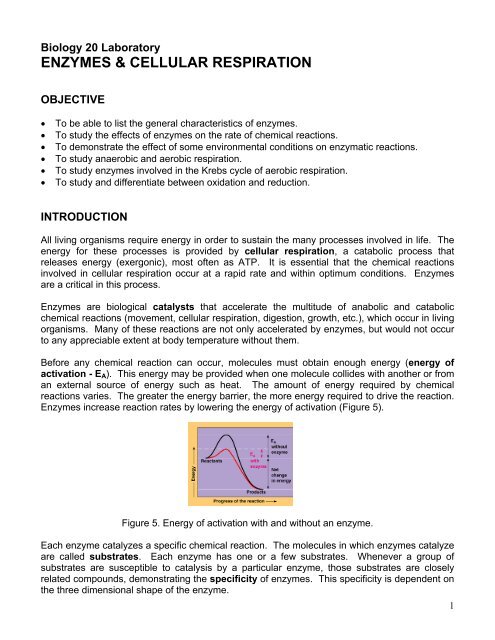
Before any chemical reaction can occur, molecules must obtain enough energy (energy of<br />
activation - E A ). This energy may be provided when one molecule collides with another or from<br />
an external source of energy such as heat. The amount of energy required by chemical<br />
reactions varies. The greater the energy barrier, the more energy required to drive the reaction.<br />
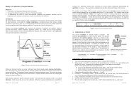
Enzymes increase reaction rates by lowering the energy of activation (Figure 5).<br />
Figure 5. Energy of activation with and without an enzyme.<br />
Each enzyme catalyzes a specific chemical reaction. The molecules in which enzymes catalyze<br />
are called substrates. Each enzyme has one or a few substrates. Whenever a group of<br />
substrates are susceptible to catalysis by a particular enzyme, those substrates are closely<br />
related compounds, demonstrating the specificity of enzymes. This specificity is dependent on<br />
the three dimensional shape of the enzyme.<br />
1
The catalytic cycle (Figure 5.1) for enzymatic reactions begins as the reactants (enzyme and<br />
substrate) collide and the substrate fits into the active site (Figure 5.2) of the enzyme. The<br />
substrate and enzyme forms the enzyme-substrate complex held together by temporary<br />
bonding (hydrophobic interactions, hydrogen and ionic bonds). It is during the complex<br />
formation that the chemical reaction(s) takes place resulting in the product(s). Notice that the<br />
enzyme also appears with the product(s) in the equation below (Figure 5.1). Enzymes emerge<br />
essentially unchanged upon completion of the chemical reaction and are capable of further<br />
catalysis (reusable).<br />
E S ES complex E P<br />
Enzyme + Substrate Enzyme-Substrate complex Enzyme + Product(s)<br />
Figure 5.1. Catalytic cycle for enzymatic reactions.<br />
Figure 5.2. Binding of the substrate<br />
in the active site of the enzyme.<br />
Figure 5.3. Interference of substrate binding<br />
by two types of inhibitors.<br />
Each enzymatic reaction has an optimum set of conditions, which produce the most efficient<br />
enzymatic activity (fastest reaction rate). Optimum conditions may vary with different enzymes<br />
and with the location of the reaction in the body. Many enzymes also require the presence of<br />
inorganic or organic enzyme helpers (cofactors and coenzymes) in order to function properly.<br />
Other factors that affect enzyme activity include: temperature, pH, the concentration of<br />
substrates, and the concentration of enzymes. Enzyme inhibitors (Figure 5.3) can also affect<br />
the binding of substrates by causing the active site to undergo a conformational change<br />
preventing substrate binding. When the three dimensional shape of the enzyme is disrupted,<br />
the protein is said to be denatured and the enzyme becomes inactivated.<br />
Let’s take a look at one example of an enzymatic reaction. Our cells utilize glucose for energy.<br />
To obtain energy from glucose, a number of reactions occur involving the oxidation/reduction<br />
of substrates, rearrangement of molecules, or removal of carbon dioxide. The initial use of<br />
glucose occurs in the cytoplasm of the cell. It involves oxidation/reduction reactions and<br />
molecular rearrangements, resulting in two molecules of pyruvic acid (pyruvate), two molecules<br />
of ATP and two molecules of reduced coenzyme, NADH. This series of reactions is known as<br />
glycolysis, an since the presence of oxygen has not been required, the process is considered<br />
anaerobic.<br />
The fate of the two pyruvic acids is dependent on the presence of oxygen. If oxygen is not<br />
present, the two pyruvic acids will remain in the cytosol and undergo the anaerobic process<br />
called fermentation. There is no “extra” energy yield from fermentation. If oxygen is present,<br />
2
the two pyruvic acids will be shuttled to mitochondria, altered and enter into a series of reactions<br />
involving the Krebs Cycle and the Electron Transport Chain (ETC). Both are dependent on<br />
oxygen and are aerobic in nature. The Krebs cycle only produces 1 ATP molecule directly per<br />
cycle. However, it is indirectly responsible for the greatest ATP production by generating<br />
coenzymes, both NADH and FADH 2 . When these coenzymes are reoxidized in the Electron<br />
Transport Chain, many molecules of ATP are generated (a theoretical 36 ATP per 1 glucose).<br />
A. AEROBIC <strong>RESPIRATION</strong><br />
SUCCINIC ACID DEHYDROGENASE ACTIVITY IN THE KREBS CYCLE<br />
Succinic acid dehydrogenase (SDH) is an oxidative enzyme that catalyzes the removal of<br />
hydrogen atoms from succinic acid, the substrate, according to Figure 5. This reaction is a vital<br />
step in the Krebs Cycle, which is a sequence of biochemical reactions occurring in the<br />
mitochondria of cells.<br />
Succinic acid loses two hydrogen atoms and is transformed into fumaric acid, which has a<br />
double bond between the middle two carbon atoms (Figure 5.4). Since two hydrogen atoms are<br />
given up in the reaction, there must be some molecule, which accepts them. Flavin adenine<br />
dinucleotide (FAD) is a molecule called a “coenzyme” which works along with SDH and<br />
accepts the hydrogen atoms released during the oxidation of the substrate, succinic acid, and<br />
passes them to an acceptor molecule.<br />
Figure 5.4: The conversion of succinic acid to fumaric acid.<br />
COOH-CH 2 -CH 2 -COOH<br />
Succinic acid (substrate – reduced)<br />
SDH<br />
2 H +<br />
COOH-CH=CH-COOH<br />
Fumaric acid (product – oxidized)<br />
FAD FADH 2<br />
(oxidized) (reduced)<br />
The FAD molecule with its hydrogen atoms, FADH 2 , passes its hydrogens on through a series<br />
of biochemical steps located in the inner mitochondrial membrane involving cytochrome<br />
enzymes. The hydrogens and their electrons (one electron/hydrogen atom) are eventually<br />
accepted by oxygen to form water (Figure 5.5). This stepwise sequence of events, involving<br />
electron transfer and cytochrome enzymes, is known as the electron transport chain (ETC) or<br />
the respiratory chain. Because the final reaction involves oxygen, this process is known as<br />
oxidation, the loss/removal of electrons from a molecule. SDH facilitates the oxidation of<br />
succinic acid to fumaric acid, removing hydrogen atoms and their electrons. Meanwhile FAD<br />
becomes reduced as it picks up the two hydrogens. Reduction is the gain of electrons (follow<br />
the hydrogens in Figure 5.5).<br />
Figure 5.5: The oxidation of FADH 2 and the reduction of O 2 .<br />
FADH 2 + ½ O 2 FAD + H 2 O<br />
3
The activity of enzymes can be inhibited in many ways. Some inhibitors “poison” the system by<br />
irreversibly binding to the enzymes active site or altering their structure. Other inhibitors,<br />
disguised as the normal substrate, “compete” for the active site on enzymes (Figure 5.2). An<br />
enzyme molecule is very large compared to its substrate and may contain several active sites.<br />
When inhibitors or “false” substrates bind to the active site, the substrate cannot undergo the<br />
desired reaction at the rate ordinarily observed.<br />
Malonic acid has the formula: COOH-CH 2 -COOH. Its structure is very similar to that of succinic<br />
acid (COOH-CH 2 -CH 2 -COOH), lacking only a second CH 2 group. Since it strongly resembles<br />
the normal substrate of SDH, it can “trick” the enzyme into binding with it. Hydrogens cannot be<br />
removed from the malonic acid, however, and the enzyme is temporarily inactive. When<br />
succinic acid and malonic acid are mixed together, succinic acid must compete with malonic<br />
acid for an active site on the enzymes therefore hydrogens are not passed on to methylene blue<br />
(Figure 5.6) as readily. If malonic acid is removed, the active site can be cleared and the SDH<br />
enzyme will return to its original active form. Figure 5.4 shows the relationship of the structure<br />
of fumaric acid to succinic acid.<br />
Experiment:<br />
In this experiment, a hydrogen acceptor that does not occur in nature, the dye methylene blue,<br />
is used. When oxidized it is blue, but when it accepts hydrogen and is thus reduced, it becomes<br />
colorless (Figure 5.6). Therefore, it can be used as an indicator to show when the reaction<br />
mixture has undergone some oxidative activity.<br />
Figure 5.6: The reduction of methylene blue.<br />
FADH 2 + methylene blue FAD + methylene blue-H 2 .<br />
(blue colored)<br />
(colorless)<br />
Since muscle is an excellent source of mitochondria, hamburger meat will serve as the source<br />
of the succinic acid dehydrogenase (SDH) enzyme of the Krebs Cycle. Succinic acid and<br />
malonic acid will both be available in dropper bottles.<br />
Materials:<br />
Homogenized hamburger, succinic acid, malonic acid, methylene blue, test tubes, stir rods,<br />
incubator (37C), DI water.<br />
Procedure:<br />
1. Obtain 4 test tubes and set them up as listed below.<br />
2. Test tube #1:<br />
a. Place approximately 2 cm (depth) of homogenized hamburger in the test tube.<br />
b. Add 10 drops of succinic acid.<br />
c. Add 3 drops of methylene blue.<br />
d. Mix thoroughly with a clean stir rod.<br />
e. NOTE the time the tube was setup and how long it takes for any color change to<br />
occur. This will indicate reaction rate.<br />
3. Test Tube #2:<br />
a. Place approximately 2 cm of homogenized hamburger in the test tube.<br />
b. Add 10 drops of DI water acid.<br />
4
c. Add 3 drops of methylene blue.<br />
d. Mix thoroughly with a clean stir rod.<br />
e. NOTE the time the tube was setup and how long it takes for any color change to<br />
occur. This will indicate reaction rate.<br />
4. Test Tube #3:<br />
a. Place approximately 2 cm of homogenized hamburger in the test tube.<br />
b. Add 10 drops of malonic acid.<br />
c. Add 3 drops of methylene blue.<br />
d. Mix thoroughly with a clean stir rod.<br />
e. NOTE the time the tube was setup and how long it takes for any color change to<br />
occur. This will indicate reaction rate.<br />
5. Test Tube #4:<br />
a. Place 5 ml of DI water in the test tube – NO meat.<br />
b. Add 10 drops of succinic acid.<br />
c. Add 3 drops of methylene blue.<br />
d. Mix thoroughly with a clean stir rod.<br />
e. NOTE the time the tube was setup and how long it takes for any color change to<br />
occur. This will indicate reaction rate.<br />
6. DO NOT SHAKE OR STIR THE TUBES, it will change the results.<br />
7. Make some predications regarding this experiment as the experiment is proceeding. Do<br />
not forget to check on your tubes.<br />
8. Do not allow the experiment to exceed 45 minutes.<br />
9. Carefully remove the test tubes and note any change on your worksheet.<br />
10. Have your instructor confirm your results and then answer question #11 on page 7.<br />
B. ANAEROBIC FERMENTATION<br />
Fermentation involves the oxidation of NADH by the removal of electrons (or hydrogens) from<br />
the NADH + H + and their acceptance by pyruvic acid, forming either lactic acid or ethyl<br />
alcohol. The products, which result from the reduction of pyruvic acid, depend upon the<br />
presence of the specific enzymes of the organisms involved. Many cells are capable of<br />
fermentation, but animal cells can produce only lactic acid. Bacterial cells can produce not only<br />
lactic acid, but also many other products, including ethyl alcohol. Yeast and certain other fungi<br />
are known for their fermentation abilities, producing ethyl alcohol and CO 2 in the process.<br />
Materials:<br />
3 fermentation tubes, water, molasses solution, yeast, metric ruler, cork<br />
Procedure:<br />
1. Three fermentation tubes will be used by the entire lab table and labeled #1 - #3.<br />
2. Test tube #1: fill the entire tube with DI water.<br />
3. Test tube #2: fill with the molasses solution.<br />
4. Test tube #3: fill with the molasses and activated yeast (that has been heated slightly).<br />
5. Develop a hypothesis.<br />
6. Make some predications regarding this experiment on your worksheet.<br />
7. After 20 – 30 minutes, measure any gas production with a millimeter ruler and note the<br />
appearance of the tubes.<br />
5
Biology 20<br />
Cellular Respiration & Enzymes Worksheet<br />
Name:<br />
Lecture Day & Time:<br />
A. SUCCINIC ACID DEHYDROGENASE ACTIVITY IN THE KREBS CYCLE<br />
Data Table 1: Tube contents and time required for color change.<br />
Tube # Tube Contents Time Required for<br />
Color Change (minutes)<br />
1<br />
2<br />
3<br />
4<br />
1. What is the hypothesis that is being tested?<br />
2. Make 2 – 3 predictions regarding this experiment:<br />
3. Was your hypothesis validated by your experiment? Explain why or why not.<br />
4. What is the substrate in this experimental setup?<br />
5. What enzyme is changing the substrate?<br />
6. Where, in the hamburger, are the enzymes-substrate complexes located?<br />
7. What is the role of methylene blue in this experiment?<br />
8. Although the process that is occurring is called “oxidation,” no oxygen is used. Explain.<br />
6
9. Did a color change occur in Tube #2? Since no succinic acid was added, should there<br />
have been a color change? Is there a difference in what you observed and what you<br />
expected? If so, explain.<br />
10. Why did the methylene blue become colorless at the bottom of the tube, yet remain<br />
colored (blue) at the surface of the hamburger?<br />
11. Would shaking or stirring the tube affect the color of the tube? Answer this question<br />
after your instructor has confirmed your results. If a change occurred, what was the<br />
cause?<br />
12. Graph the reaction rate results for this experiment. Do not forget to include all the<br />
components of a graph.<br />
7
B. ANAEROBIC <strong>RESPIRATION</strong><br />
Data Table 2: Anaerobic respiration.<br />
Tube # Gas Amount (mm) Appearance<br />
1<br />
2<br />
3<br />
13. Develop a hypothesis:<br />
14. Make 2 – 3 predictions regarding this experiment:<br />
15. Was your hypothesis validated by your experiment? Explain why or why not.<br />
16. What is the purpose of Tubes #1 and #2?<br />
17. What is occurring in Tube #3 to make it look differently?<br />
18. Do similar reactions occur in your body? If so, where?<br />
19. What was the role of the yeast in the experimental setup?<br />
8