Volume 6, Spring 2008 - Saddleback College
Volume 6, Spring 2008 - Saddleback College
Volume 6, Spring 2008 - Saddleback College

Create successful ePaper yourself
Turn your PDF publications into a flip-book with our unique Google optimized e-Paper software.
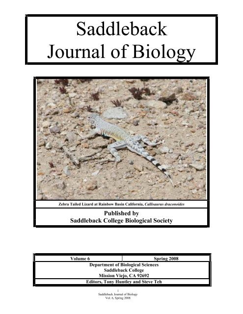
<strong>Saddleback</strong><br />
Journal of Biology<br />
Zebra Tailed Lizard at Rainbow Basin California, Callisaurus draconoides<br />
Published by<br />
<strong>Saddleback</strong> <strong>College</strong> Biological Society<br />
<strong>Volume</strong> 6 <strong>Spring</strong> <strong>2008</strong><br />
Department of Biological Sciences<br />
<strong>Saddleback</strong> <strong>College</strong><br />
Mission Viejo, CA 92692<br />
Editors, Tony Huntley and Steve Teh<br />
i<br />
<strong>Saddleback</strong> Journal of Biology<br />
Vol. 6, <strong>Spring</strong> <strong>2008</strong>
TABLE OF CONTENTS<br />
Peer Reviewed Manuscripts from the Biology 3B Class<br />
<strong>Spring</strong> <strong>2008</strong><br />
Author(s) Title Page<br />
Merielle Ebol Growth Difference in the Goldfish, Carassius auratus, 1<br />
&<br />
Shanon Carney<br />
Exposed to Different Diets with Varying Levels of Proteins<br />
and Carbohydrates<br />
Kyle Lutz & The Effect of a Lactate Supplement on Maximal Cycling 4<br />
John I. Miller<br />
Performance in Man<br />
Stephanie Anstadt & The Effect of Silver Nitrate on the Inhibiting Growth of 7<br />
Teo Fernandez<br />
Escherichia coli<br />
Nathaly Leal- Arteaga & Effect of Tide Level on Nitrate and Phosphate Concentration 10<br />
Saori Shimamoto<br />
in Marine Water<br />
Lancelot Beier and Effect of Differing Color on the Timed Length of Aggressive 14<br />
Harrison Pham<br />
Response of Betta splenens<br />
Crystine Gill & Intestinal Candida albicans Overgrowth in Autistic Children 17<br />
Samantha Lopez<br />
with Food Allergies<br />
Ryan C. Clark & Comparison of Chlorophyll Content in Shade and Sun 20<br />
Josue J. Mandujano Leaves of the Lemonade Berry Plant (Rhus integrifolia)<br />
Thao Nguyen & The Effects of Ethinyl Estradiol on Aggressive Behavior in 22<br />
IxChel Cruz-Gonzalez<br />
Siamese Fighting Fish (Betta splendens)<br />
Kevin Murray & Antibiotics (Tobramycin and Polymyxin) Resistance in 26<br />
David Stapleton Staphylococcus aureus & Effectiveness of These Antibiotics<br />
Yuriko Kayama The Effects of pH on Escherichia coli Fermentation 29<br />
Kaung Ko & Antibiotics (Tobramycin and Polymyxin) Resistance in 33<br />
Spencer Roberts Staphylococcus aureus & Effectiveness of These Antibiotics<br />
Dorothy Chang &<br />
Effects of temperature on metabolic rate in<br />
37<br />
Grant T. Huttar<br />
Gromphadorhina portentosa<br />
Dayana Vera & The Effect of Creatine Monohydrate on the Run Time of 41<br />
Michael Moeller<br />
Sceloporus occidentalis<br />
Ryan G. White & The Metabolic Cost of Digestion in the Ball Python, 44<br />
Michael M. Hadley<br />
Python regius<br />
Nicole Baumgartner & Recreational Water Safety Following Rain at T-street Beach, 47<br />
Karl Neil<br />
San Clemente, California<br />
Takahiro Ueno &<br />
Arash Moghaddam<br />
The Effect of Salinity on the Photosynthetic Rate of<br />
Pickleweed, Salicornia virginica<br />
49<br />
Matt Apke &<br />
Zachary Beam<br />
The effects of various light sources on the fruiting bodies of<br />
Citrus limonium<br />
52<br />
ii<br />
<strong>Saddleback</strong> Journal of Biology<br />
Vol. 6, <strong>Spring</strong> <strong>2008</strong>
TABLE OF CONTENTS<br />
Biology 20 Honors Manuscripts<br />
Fall 2007 & <strong>Spring</strong> 2007<br />
Author(s) Title Page<br />
Michelle Huynh and Is MiracleGro the Best Fertilizer for Impatiens wallerana? 55<br />
Jiwon Park<br />
Cynthia Tran and<br />
Camille Barlow<br />
The Effects of Organic Fertilizer vs. Inorganic Fertilizer<br />
(Miracle Gro) on Growth of Tomato Plants<br />
57<br />
TABLE OF CONTENTS<br />
Peer Reviewed Manuscripts from the Biology 3B Class<br />
Fall 2007<br />
Author(s) Title Page<br />
Lauren Ferris<br />
The Effects of fire on the Southern California Plant 60<br />
Succession and the Prevalence of Artichoke Thistle, Cynara<br />
cardunculus<br />
Gregory Nelson and The Effect of Mycorrhizae on the Growth and Development 63<br />
Elena Novak<br />
of Bush Beans Phaseolus vulgaris<br />
Thomas Caldwell Exhaustion due to Mental Stress and Metabolism of Sugar 66<br />
and Caffeine in Energy Drinks<br />
Heather Rufino and<br />
Alexa Milman<br />
Serial Lactate Levels Increase over Time under Anesthesia<br />
on Healthy Canine Patients Undergoing Elective, Minimally<br />
70<br />
Jasmine Mitchell and<br />
Tony Schofer<br />
Invasive, Lower Abdominal Surgery<br />
The Correlation Between the Vertical Jump Height and Calf<br />
Length in Athletes and Non-athletes<br />
74<br />
Chris Yang and<br />
Josue Mandujano<br />
Milad Danesh<br />
Catherine Pearson<br />
Shengchieh Chang<br />
Tarick Sheikh and Chris<br />
Glendinning<br />
Comparison of Chlorophyll Content of Leaves in a Green<br />
House and their normal environment of a Cyclamen Plant<br />
(Cyclamen Persicum)<br />
The Effects of Ginkgo Biloba on the Cognitive Thinking of<br />
Mus musculus<br />
Increased performance level due to carbohydrate vs.<br />
carbohydrate-protein composition in sports drinks<br />
Comparison of Drinks and Their Effects on Exercise<br />
Performance<br />
Correlation between attractiveness of male body odor and<br />
facial feature of Homo. sapiens<br />
77<br />
79<br />
83<br />
86<br />
90<br />
iii<br />
<strong>Saddleback</strong> Journal of Biology<br />
Vol. 6, <strong>Spring</strong> <strong>2008</strong>
Biology 3A abstracts for papers presented at the 6 th Annual Biology 3A/3B<br />
Scientific Meeting (<strong>Spring</strong> <strong>2008</strong>)<br />
The meeting organizers do not assume responsibility for any inconsistencies in quality or errors<br />
in abstract information. Abstracts are in numerical order according to the abstract number<br />
assigned to each presentation. Authorless abstracts appear at the end of all the abstracts,<br />
including non-submitted abstracts, if any. Abstracts begin on page 94.<br />
TABLE OF CONTENTS<br />
Biology 3A Abstracts<br />
<strong>Spring</strong> <strong>2008</strong><br />
Author(s) Title Page<br />
Alexandra M. Franco THE EFFECT OF SODIUM CHLORIDE CONCENTRATION ON 94<br />
and<br />
THE GROWTH OF THE COMMON BEAN (Phaseolus vulgaris)<br />
Earl-Eugene E. Ringpis<br />
Chris J. LaCroix and PREFERENCE OF DOGS (Canis familiaris) FOR HIGH FAT DOG<br />
Jocelyn A. Finley<br />
FOOD VERSUS LOW FAT DOG FOOD<br />
94<br />
Raiff Josey and TESTING FOR INTELLIGENCE IN THE SLIME MOLD Physarum<br />
Christopher Luna<br />
polycephalum<br />
94<br />
Jason R. Riggio EFFECT OF pH ON RATES OF CLOSURE IN VENUS FLY TRAPS<br />
(Dionaea Muscipula)<br />
95<br />
Aubrey Michi and EFFECT OF TEMPERATURE ON PHOTOSYNTHETIC RATE OF<br />
Jeremy Ward<br />
Elodea canadensis<br />
95<br />
Anne Kathreane Ebol THE EFFECT OF GUAVA Psidium guajava AGAINST Escherichia<br />
and Hannah Rae Manuel<br />
coli<br />
95<br />
Anoush A. Garakani and THE EFFECT OF TEMPERATURE ON BACTERIAL COLONY<br />
Cassra Minai.<br />
FORMATION IN Escherichia coli<br />
96<br />
Brooke A. Hargis and CYNIPIDAE GALL DISTRIBUTION ON YOUNGER AND OLDER<br />
Candice B. Archer<br />
COAST LIVE OAK TREES, QUERCUS AGRIFOLIA<br />
96<br />
Claudia M. Shuldberg and<br />
Ximena M. Horne<br />
EXTRACTION OF IRON FROM BREAKFAST CEREALS 96<br />
Andrew J. Shires and OPERCULAR PUMPING RATES OF GOLDFISH (Carassius<br />
Christopher J. Walsh auratus) IN DIFFERENT CONCENTRATIONS OF GLUCOSE<br />
97<br />
Shayda Haghgoo and EFFECTS OF TABLE SUGAR AND STEVIA SWEETNER ON<br />
Erin Kang<br />
BLOOD GLUCOSE LEVELS IN HUMANS<br />
97<br />
Angel R. Vargas GROWTH INHIBITON OF HYDROGEN PEROXIDE ON THE<br />
PATHOGENIC BACTERIA, PSEUDOMONAS AERUGINOSA<br />
97<br />
Robert Powers COMPARISON OF LACTATE DEHYDROGENASE ACTIVITY<br />
BETWEEN THE THRESHER SHARK (Alopias vulpinus) AND THE<br />
98<br />
PIKE MACKEREL (Cololabis saira)<br />
Hanwool Park<br />
THE EFFECT OF A HAIR GEL ON THE GROWTH OF<br />
MALASSEZIA FURFUR<br />
98<br />
David Guzman<br />
THERE IS NO SIGNIFICANT OXYGEN PRODUCTION OF<br />
SPINACIA OLERACEA PLACED INTO CALCIUM CARBONATE<br />
98<br />
SOLUTION<br />
Yoko Kamei and THE EFFECT OF MODERN ROLLER COASTER RIDE ON HUMAN<br />
Diana Nguyen<br />
HEART RATE<br />
99<br />
Robert E. Maloney and THE AFFECT OF ROUNDUP ® ON OXYGEN PRODUCTION OF<br />
Bianca Christensen<br />
RED ALGAE (RHODOPHYTA)<br />
99<br />
Lindy A. Ackerman and<br />
Brittany N. Lincoln<br />
THE EFFECT OF SOIL pH ON THE GROWTH OF PHASEOLUS<br />
VULGARIS AND RAPHANUS SATIVUS<br />
99<br />
iv<br />
<strong>Saddleback</strong> Journal of Biology<br />
Vol. 6, <strong>Spring</strong> <strong>2008</strong>
TABLE OF CONTENTS<br />
Biology 3A Abstracts<br />
<strong>Spring</strong> <strong>2008</strong><br />
Author(s) Title Page<br />
Nicholas Schmidt and IMPACT OF RED AND GREEN LIGHT ON GROWTH RATE OF<br />
Bobby Stangl<br />
SNAPDRAGONS (Antirrhinum majus)<br />
100<br />
Grady S. Counts and EFFECTS OF ENVIRONMENTAL TEMPERATURE ON THE<br />
Eric T. Rueda<br />
SPEED OF COMMON GARDEN SNAILS (Helix aspersa)<br />
100<br />
Hilda Gonzalez and THE EFFECT OF PH ON THE RESPIRATION RATE OF<br />
Natsumi Iwata<br />
GOLDFISH (Carassius auratus)<br />
100<br />
Tommy L. Roberts and<br />
Emmanuel Romero<br />
DETERRENT EFFECTS OF PEPPERMINT OIL IN MICE 101<br />
Samantha T. Dinh and THE EFFECT OF ROLLER COASTERS ON HEART RATE IN<br />
Cole P. Lyou<br />
RELATION TO GENDER<br />
101<br />
Harris M. Elhan and THE COMPARATIVE COST OF LOCAMOTION IN FEEDER MICE<br />
Tracy L. Kubas (Mus musculus) AND ROBOROVSKI HAMSTERS (Phodopus<br />
101<br />
roborovskii)<br />
Bobby Fujimoto EFFECT OF CHLORINE ON ALGAL GROWTH 102<br />
Grady S. Counts and EFFECTS OF ENVIRONMENTAL TEMPERATURE ON THE<br />
Eric T. Rueda<br />
SPEED OF COMMON GARDEN SNAILS (Helix aspersa)<br />
102<br />
Scott Mitchell and THE EFFECT OF TEMPERATURE ON THE METABOLIC RATE OF<br />
Amir Zand<br />
ANOLE LIZARDS (Anolis Carolinensis)<br />
102<br />
Roky Coria<br />
THE EFFICACY OF GARLIC (ALLIUM SATIVUM ) AS A<br />
BIOLOGICAL CONTROL OF MOSQUITO LARVA<br />
103<br />
John Lowd and<br />
EFFECTS OF AUDITORY AND CHEMICAL STIMULI IN<br />
Charles Steinfeld<br />
MAMMALIAN MEMORY (Mus musculus)<br />
103<br />
Sally A. Hutson A COMPARISON OF SUCROSE AND STEVIA EXTRACT AS A<br />
SUBSTRATE FOR YEAST METABOLISM<br />
104<br />
Natasha V. Polanski EFFECTS OF PESTICIDE ON OXYGEN PRODUCTION RATES OF<br />
RED ALGEA (Aharghiella tenera)<br />
104<br />
Anonymous POST FIRE PLANT FREQUENCY IN NORTH AND SOUTH SIDED<br />
HILLS IN SANTIAGO CANYON<br />
104<br />
Biology 3A abstracts for papers presented at the Annual Biology 3A<br />
Poster Presentation Scientific Meeting (Fall 2007)<br />
The meeting organizers do not assume responsibility for any inconsistencies in quality or errors<br />
in abstract information. Abstracts are in numerical order according to the abstract number<br />
assigned to each presentation. Authorless abstracts appear at the end of all the abstracts,<br />
including non-submitted abstracts, if any. Abstracts begin on page 105.<br />
TABLE OF CONTENTS<br />
Biology 3A Abstracts<br />
Fall 2007<br />
Author(s) Title Page<br />
Thao Nguyen and EFFECTS OF SPIRULINA ALGAE VERSUS REGULAR FISH<br />
Nelson Huang FLAKES ON METABOLISM IN BETTA FISH (Betta splendens)<br />
105<br />
Janelle Reed and<br />
Irina Alexandrova<br />
ASCORBIC ACID CONCENTRATION OF ORGANIC AND<br />
CONVENTIONALLY GROWN PRODUCE<br />
105<br />
v<br />
<strong>Saddleback</strong> Journal of Biology<br />
Vol. 6, <strong>Spring</strong> <strong>2008</strong>
TABLE OF CONTENTS<br />
Biology 3A Abstracts<br />
Fall 2007<br />
Author(s) Title Page<br />
Kevin Murray and ANTIBACTERIAL EFFECTS OF RED WINE ON ORAL BACTERIA<br />
Arash Moghaddam<br />
105<br />
Izaak Miller and THE EFFECT OF PYRUVATE ON OXYGEN CONSUMPTION OF<br />
Lancelot Beier<br />
MUS MUSCULUS<br />
106<br />
Michael G. Moeller EFFECT OF LIGHT ON CHLOROPHYHLL CONCENTRATION IN<br />
ENGLISH IVY (Hedera helix)<br />
106<br />
Anne Merielle Ebol and SENSITIVITY OF PATHOGENIC BACTERIA STAPHYLOCOCCUS<br />
Shanon Carney<br />
AUREUS TO HONEY<br />
106<br />
Kaung Ko and THE EFFECT OF TEMPERATURE ON PHOTOSYNTHETIC RATE<br />
Spencer Roberts<br />
OF ELODEA CANADENSIS<br />
107<br />
Y Leho and<br />
THE EFFECT OF WEIGHT LOAD ON THE OXYGEN<br />
Christopher Triana CONSUMPTION OF PARAKEETS DURING TERRESTRIAL<br />
107<br />
LOCOMOTION<br />
Valerie Bowen and EFFECT OF SUCRALOSE AND SUGAR ON BLOOD GLUCOSE<br />
Samantha Lopez<br />
IN HOMO SAPIENS<br />
107<br />
Sherri Burnett and<br />
IxChel Cruz- Gonzalez<br />
Krystle Salazar and<br />
Matt Apke<br />
Saori Shimamoto and<br />
Yuriko Kayama<br />
Kyle Lutz and<br />
Yohsuke Kobayashi<br />
Deepa A. Thaker and<br />
Stanley Lin<br />
Nathaly Leal-Arteaga<br />
Aaron Echols and<br />
Crystine Gill<br />
Greg M. Fitzgerald and<br />
Michael B. Zilly<br />
Monica Mehran and<br />
Paris Aliyazdi<br />
Ryan G. White and<br />
Michael Hadley<br />
Micaela K. Dora<br />
Alvin P. Jogasuria and<br />
Takahiro Ueno<br />
John K. Davis and<br />
Jaclyn R. Kuluris<br />
Dorothy Chang and<br />
Grant T. Huttar<br />
COMPARISON OF LACTATE DEHYDROGENASE ACTIVITY IN 108<br />
SKELETAL MUSCLE OF BLUE ANCHOVY (ENCRASICHOLINA<br />
DEVISI) AND COW (BOS TAURUS)<br />
EFFECT OF PH ON GERMINATION RATE IN LIMA BEANS 108<br />
OXYGEN PRODUCTION OF BROWN ALGAE (Egregia laevigata) 108<br />
AND RED ALGAE (Gelidium robustum) IN DIFFERENT COLORS<br />
OF LIGHT<br />
THE EFFECT OF SODIUM BICARBONATE ON TIME TO 109<br />
EXHAUSTION IN THE WESTERN FENCE LIZARD (Sceloporus<br />
occidentalis)<br />
EFFECT OF CAFFEINE ON MEMORY IN MICE (Mus musculus) 109<br />
THE EFFECT OF PH ON THE GROWTH OF BACTERIA<br />
(Escherichia coli)<br />
THE EFFECTS OF OZONE ON ESCHERICHIA COLI ON SPINACH<br />
LEAVES<br />
THE EFFECT OF WAVELENGTH OF LIGHT ON THE<br />
DISCOLORATION OF WINE.<br />
THE EFFECT OF APPLES ON THE RIPENING OF VALENCIA<br />
ORANGES (Citrus aurantium)<br />
A COMPARISON OF THE WATER UPTAKE RESPONSES TO<br />
INTRAPERITONEAL BOLUS INJECTIONS OF ARGININE<br />
VASOTOCIN IN THE TOAD, BUFO AMERICANUS AND THE<br />
LEOPARD FROG, RANA PIPIENS<br />
EFFECT OF AMBIENT TEMPERATURE ON THE FORCE<br />
EXERTED BY CARIBBEAN HERMIT CRABS (Coenobita clypeatus)<br />
THE EFFECT OF HYDROGEN PEROXIDE ON THE<br />
GERMINATION RATE AND GROWTH OF STRING BEANS<br />
(Phaseolus vulgaris)<br />
EFFECT OF CAFFEINE ON BLOOD LACTATE IN EXERCISING<br />
HUMANS<br />
INHIBITORY EFFECTS OF CAM PLANT FLUID ON<br />
SATPHYLOCOCCUS AUREUS<br />
109<br />
110<br />
110<br />
110<br />
111<br />
111<br />
111<br />
112<br />
112<br />
vi<br />
<strong>Saddleback</strong> Journal of Biology<br />
Vol. 6, <strong>Spring</strong> <strong>2008</strong>
TABLE OF CONTENTS<br />
Biology 3A Abstracts<br />
Fall 2007<br />
Author(s) Title Page<br />
Reza Ghassemi and DIABETES MELLITUS CONTRIBUTES TO THE WEIGHT<br />
Jonathan Willner<br />
DIFFERENCE IN FELIS DOMESTICUS<br />
112<br />
Madina Ali and<br />
THE EFFECTS OF VITAMINS A, C, AND E ON THE<br />
Rhonda Cheikh<br />
GERMINATION OF RADISH SEEDS<br />
113<br />
Nicole K. Baumgartner THE EFFECTS OF ACID RAIN ON O 2 PRODUCTION IN ELODEA<br />
and Karl M. Neil<br />
(Elodea camadensis)<br />
113<br />
Larry T. Lam EFFECT OF NORMAL SALINE AND CONTACT LENS SOLUTION<br />
ON THE EPITHELIUM OF THE CORNEA<br />
113<br />
Kasra Abolhosseini and<br />
Harrison Pham<br />
THE EFFECT OF JADE PLANT ON ESCHERICHIA COLI 113<br />
vii<br />
<strong>Saddleback</strong> Journal of Biology<br />
Vol. 6, <strong>Spring</strong> <strong>2008</strong>
Fall 2007 Biology 3A Abstracts<br />
Growth Difference in the Goldfish, Carassius auratus, Exposed to Different Diets with<br />
Varying Levels of Proteins and Carbohydrates<br />
Anne Merielle Ebol and Shanon Carney<br />
Department of Biological Sciences<br />
<strong>Saddleback</strong> <strong>College</strong><br />
Mission Viejo, California USA<br />
Good quality food is necessary to achieve adequate health and nutrition for all living<br />
organisms. Carbohydrates and proteins are two main components found in food, which<br />
are required for proper metabolism and growth. Several studies have been performed on<br />
goldfish to determine whether maximum growth based on weight and body size was<br />
achieved with a diet higher in protein or with a diet higher in carbohydrate. In the current<br />
study, fifteen goldfish were weighed before and after exposure to different diets consisting<br />
of various ratios of proteins and carbohydrates. While both food components play a role in<br />
growth, a difference in body mass was observed in the goldfish while exposed to the<br />
different food contents. Group A was given a diet higher in protein, Group B was given a<br />
diet higher in carbohydrate and Group C was given a diet containing both elements. The<br />
diets were administered for a period of one month and changes in body mass were<br />
evaluated every week. The average final weight measurements were: 1.76 ± 0.02 g (±se) for<br />
Group A, 1.61 ± 0.03 g (±se) for Group B, and 1.44 ± 0.03 g (±se) for Group C. The<br />
percentage weight gained for Group A was 15.03% from the high protein diet, Group B<br />
was 8.78% from the high carbohydrate diet, and Group C was 2.13% from the combo diet.<br />
Results indicated that there is a difference in growth in the goldfish, Carassius auratus,<br />
when given different diets with varying components (p
Fall 2007 Biology 3A Abstracts<br />
carbohydrates improved growth and feed utilization<br />
of the fish (Tan et al, 2006).<br />
Although both proteins and carbohydrates<br />
contribute to the development of the organism, the<br />
influence of each nutrient is not essentially equal to<br />
the other (Lovell, 1991). Since each component has<br />
various effects on the development of the fish, its<br />
effect on growth was evaluated. In the current study,<br />
growth of fifteen juvenile goldfish was observed<br />
before and after exposure to three different diets: a<br />
high protein, a high carbohydrate and a combination<br />
diet comprising of both elements. Growth was<br />
measured as an increase in body size, change in body<br />
condition and overall weight gain. As mentioned<br />
earlier, each nutrient has variable influence on<br />
growth; therefore, there should be a difference in<br />
growth or in the net weight gain of the goldfish<br />
between the different diets.<br />
Materials and Methods<br />
Experimental Diets<br />
Fifteen juvenile goldfish obtained from PetSmart<br />
at Mission Viejo, California were used in the study<br />
and were maintained in aerated, filtered water in<br />
three separate glass aquarium tanks. The goldfish<br />
were divided evenly into the three separate tanks and<br />
each tank was designated a different diet. Group A<br />
were given Micro Pellets (Kyorin Food Industry,<br />
Japan) with high protein content, Group B were given<br />
Baby Brine Shrimp Cubes (San Francisco Bay<br />
Manufacturing; Newark, California) with high<br />
carbohydrate content, and Group C were given<br />
TopFin Flakes (Pacific Coast Distributing, Phoenix),<br />
the control treatment, which contains both<br />
carbohydrates and proteins.<br />
When coupled with organic wastes discharged by<br />
the fish, excess feed in the tanks decreases dissolved<br />
oxygen in the water, which also decreases appetite<br />
and growth rate; hence, tanks were cleaned everyday<br />
before administering new sets of food, to ensure<br />
suitable environment for survival (Cacho et al, 1991).<br />
Weight Measurements and<br />
Calculations<br />
Each fish was weighed before the trial. Fish was<br />
removed from the tank using a net and was placed<br />
into a bucket containing tank water to carry out the<br />
measurements. Each fish was placed in a tared glass<br />
container of tank water on an electronic balance<br />
(Biology Department, <strong>Saddleback</strong> <strong>College</strong>) for body<br />
weight measurements, before being returned to their<br />
original tanks.<br />
Environmental changes, such as changes in<br />
temperature or pH levels, can cause stress to the<br />
normal physiology of the fish (Smith, 1966). In order<br />
to minimize stress, the groups were allowed to<br />
acclimatize to their new environment for one week<br />
prior to exposure to the different diets. After<br />
acclimatization for one week, each group was<br />
introduced to its new diet and was fed 1 gram of the<br />
food every 12 hours. Each group was given the same<br />
type of food for one week and subsequent weight<br />
measurements were made every week using an<br />
electronic balance. The study was conducted at room<br />
temperature (25–30 ºC) for four weeks in Mission<br />
Viejo, California starting February 24, <strong>2008</strong> to March<br />
12, <strong>2008</strong> and changes in growth were evaluated.<br />
Percentage of weight gained was obtained by using<br />
the nutritional index (Eq.1) (Bandyopadhyay et al,<br />
2005):<br />
Equation 1.<br />
Net Weight Gain = Final weight – Initial weight<br />
Weight Gain (%) = Net Weight Gain<br />
Food Analysis<br />
Initial weight<br />
X 100<br />
Percentage %<br />
Protein Carbos Fat Fiber Phosph<br />
orus<br />
(A) 49.0 20.0 4.0 0.8 2.0<br />
(B) 3.0 42.0 1.5 0.2 0.1<br />
(C) 39.0 43.0 9.0 2.0 1.0<br />
Table 1. Percent composition of diets administered<br />
to fish during trial. Percentage of vitamins and other<br />
minerals are not included in the overall composition<br />
of the food. Values acquired from each food<br />
container.<br />
Results<br />
Average initial weight measurements were 1.53 ±<br />
0.24 g for Group A, 1.48 ± 0.12 g for Group B and<br />
1.41 ± 0.07 g for Group C. Growth difference<br />
between the three experimental diet groups was<br />
demonstrated by the average final weight<br />
measurements obtained from the fish in comparison<br />
to its initial weight (Table 2). The results of the<br />
study indicate that there was a significant difference<br />
in growth observed between the groups of fish when<br />
9<br />
<strong>Saddleback</strong> Journal of Biology<br />
<strong>Spring</strong> <strong>2008</strong>
Fall 2007 Biology 3A Abstracts<br />
given different types of diet, (p
Fall 2007 Biology 3A Abstracts<br />
Food preference and required nutrition<br />
change over the course of fish development and<br />
the types of food that are beneficial for the<br />
growing juvenile fish may not essentially be<br />
beneficial for the adult (Lovell, 1991). Aside<br />
from the purpose of determining the best food<br />
source, future experiments might include<br />
studying the difference in nutritional<br />
requirements of juvenile and adult goldfish to<br />
determine whether assimilation rates or<br />
absorption efficiencies vary between the two<br />
groups.<br />
Literature Cited<br />
Bandyopadhyay, P, Swain, S, and Mishra, S (2005).<br />
Growth and dietary utilization in goldfish fed<br />
diets formulated with various local agro-produces.<br />
BioResource Technology. 96: 731-740.<br />
Cacho, O, Kinnucan, H, and Hatch, U (1991).<br />
Optimal control of fish growth. American Journal<br />
of Agricultural Economics. 73(1): 174-183.<br />
Garling, D, and Wilson, R (1977). Optimum<br />
dietary protein to energy ratio for channel catfish<br />
fingerlings, Ictalurus punctatus. Journal of<br />
Nutrition. 106(9): 1368-1375.<br />
Kaiser, H, Endemann, F, and Paulet, T (2003). A<br />
comparison of artificial and natural foods and<br />
their combinations in the rearing of goldfish.<br />
Aquaculture Research. 34: 943-950.<br />
Lopez, R (2006). Exploring the assimilation rate of<br />
goldfish: a comparison of protein and<br />
carbohydrate diets. Biology Journal. 1: 273-282.<br />
Lovell, R (1991). Nutrition of aquaculture species.<br />
Journal of Animal Science. 69(10): 4193-4200.<br />
Lovell, T (1998). Nutrition and feeding of fish.<br />
Massachusetts, US: Kluwer Academic Publishers.<br />
266p.<br />
Priestley, S, Stevenson, A, and Alexander, A<br />
(2006). Growth rate and body condition in<br />
relation to group size in black widow Tetras<br />
(Gymnocorymbus ternetzi) and common Goldfish<br />
(Carrasius auratus). Journal of Nutrition. 136(7):<br />
2078S-2080S.<br />
Smith, M.W (1966). Influence of temperature<br />
acclimatization on sodium-glucose interactions in the<br />
goldfish intestine. Journal of Physiology. 182: 574-<br />
590.<br />
Tan, Q, Xie, S, Zhu, X, Lei, W and Yang, Y<br />
(2007). Effect of dietary carbohydrate-to-lipid<br />
ratios on growth and feed utilization in Chinese<br />
longsnout catfish (eiocassis longirostris). J. Appl.<br />
Ichthyol. 23: 605-610<br />
The Effect of a Lactate Supplement on Maximal Cycling Performance in Man<br />
Kyle Lutz and John I. Miller<br />
Department of Biological Sciences<br />
<strong>Saddleback</strong> <strong>College</strong><br />
Mission Viejo, CA 92692<br />
High lactate concentrations have traditionally been considered a causative factor in<br />
muscle fatigue during strenuous exercise in man. Recent research has challenged this view.<br />
A lactate supplement (SportLegs®) claims to increase time until fatigue in strenuous<br />
activity. Our study seeks to determine if supplementation with lactate before strenuous<br />
activity will increase time until fatigue in cyclists. Cycle power and time to failure were<br />
measured in two trials: the first unsupplemented and the second supplemented. Mean time<br />
to fatigue in trial 1 was 14.1 minutes and in trial 2 was 14.0 minutes. The supplement did<br />
however increase time to peak lactate production. Other studies have shown that high<br />
lactate concentrations have an insignificant effect on muscle fatigue and that other factors<br />
may be more important (Nielson 2003, Bangsbo 1992). We found that there was no<br />
significant difference in time to fatigue or maximum power output between cyclists taking<br />
the lactate supplement and those that did not. Our results strengthen the notion that<br />
lactate has a minimal effect on muscle fatigue.<br />
11<br />
<strong>Saddleback</strong> Journal of Biology<br />
<strong>Spring</strong> <strong>2008</strong>
Fall 2007 Biology 3A Abstracts<br />
Introduction<br />
It has widely been accepted that lactate build<br />
up contributes to fatigue during exercise. More<br />
recently, however, that notion has been increasingly<br />
challenged. During exercise, initially lactate<br />
concentrations rise rapidly. But with continued,<br />
submaximal exercise, there is a net usage of lactate.<br />
Lactate plays a role in gluconeogenesis and is<br />
shuttled from exercising and non-exercising muscles<br />
to splanchnic regions where it may be converted to<br />
glucose for use by the exercising muscles (Ahlborg,<br />
1982).<br />
Lactate is a by-product of glycolysis<br />
produced when oxygen levels are insufficient to<br />
metabolize pyruvate via the Citric Acid Cycle.<br />
Gluconeogenesis may consume lactate in splanchnic<br />
tissues to fuel active muscles during exercise.<br />
During prolonged (120 minutes), moderate intensity<br />
(30% and 58% maximal oxygen consumption,<br />
VO 2 max) exercise, a rise in splanchnic glucose<br />
output relative to work load has been associated with<br />
a 60 – 100% increase in the splanchnic uptake of<br />
gluconeogenic precursors, including lactate (Alhborg,<br />
1986; Ahlborg, 1982). In fact, after periods of<br />
moderate intensity exercise, previously active<br />
muscles become net consumers of lactate (Gladden,<br />
2004).<br />
During short bouts of intense activity (>60%<br />
VO 2 max) arterial lactate concentrations increase<br />
rapidly (Ahlborg, 1982). This results in an increased<br />
intramuscular lactate concentration ([La - ]) and net<br />
output of lactate into the blood. Whether this intense,<br />
short-term increase in [La - ] has a negative affect on<br />
muscle contractility or not is still a topic of debate.<br />
Research has shown that when [La - ] is kept at ~ 4<br />
mMolar by exogenous La - infusion, during moderate<br />
intensity exercise, lactate competes effectively with<br />
glucose as a carbohydrate fuel source, sparing<br />
glucose (Gladden, 2004).<br />
In addition to the effects of [La - ] on muscle<br />
performance, other factors such as concentration of<br />
hydronium ion ([H + ]) (pH), and potassium ion [K + ]<br />
have been proposed as potential mechanisms of<br />
fatigue. [K + ] at the onset of fatigue and ultimately<br />
muscle failure may be constant in trained and<br />
untrained human subjects. A delayed onset of this<br />
[K + ], and fatigue, has been associated with trained<br />
subjects (Nielsen, 2003). Studies have reported that<br />
lactic acidosis can protect against negative effects of<br />
altered [K + ] on muscle excitability and force<br />
(Gladden, 2004).<br />
While H + transport seems to be coupled with<br />
La - transport in inactive muscles (Bangsbo, 1995), H +<br />
transport (efflux out of the muscle cell) seems to<br />
exceed and be somewhat independent of La - transport<br />
in intensely active muscles (Bangsbo, 1993). Where<br />
some research has shown that high [H + ] may increase<br />
fatigue, other research illustrates that under normal<br />
physiological conditions the negative effects of<br />
elevated [H + ] are absent (Gladden, 2004). If a<br />
correlation exists between H + and La - co-transport,<br />
then a decreased [H+] by buffers in inactive muscle<br />
(Bangsbo, 1995) might impact [La - ], and thus impact<br />
energy production via gluconeogenesis. The<br />
mechanism for shuttling lactate between cells and<br />
tissues is still undetermined (Gladden, 2004).<br />
The SportLegs® supplement’s proposed<br />
mechanism states that by increasing lactate<br />
production prior to exercise, the body is stimulated to<br />
begin consuming lactate, therefore delaying the onset<br />
of fatigue from high [La - ]. While the net consumption<br />
of lactate by exercising muscle has been well<br />
documented with moderate intensity (
Fall 2007 Biology 3A Abstracts<br />
blood lactate was measured (see following<br />
paragraph) with the Lactate Scout using blood taken<br />
from the finger. They continued this until exhaustion.<br />
Blood lactate concentration was measured again<br />
every minute for the first five minutes during<br />
recovery.<br />
Each subject was required to draw blood<br />
using a lancet from one of the fingers. The Lactate<br />
Scout uses testing strips that require a 0.5 uL whole<br />
blood sample. The Lactate Scout automatically<br />
analyzes blood and displays a lactate concentration<br />
that is accurate between 0.5 mM and 25.0 mM [La - ]<br />
and gives results within 15 seconds.<br />
All readings for heart rate and power output<br />
are from the Saris Powertap and were obtained with<br />
the Saris Poweragent (Saris Company, Madison,<br />
Wisconsin) software. The Poweragent software<br />
converted data to comma-separated-values (*.csv)<br />
and all analyses were performed using Microsoft<br />
Excel (Microsoft Corporation, Redmond,<br />
Washington). A paired T-test was used to evaluate<br />
the differences between the two trials.<br />
Results<br />
Time until failure with and without the<br />
supplement had little variance, the largest change in<br />
an individual participant was an improved<br />
performance of 1.9 minutes. The mean time to failure<br />
without the supplement was 14.1 minutes and with<br />
the supplement was 14.0 minutes (Figure 1). Overall<br />
there was no statistically significant increase in time<br />
to failure between participants riding with the<br />
supplement and without the supplement. The p-value<br />
is 0.419 (One-tailed T-Test).<br />
Time Until Failure (m)<br />
18<br />
16<br />
14<br />
12<br />
10<br />
8<br />
6<br />
4<br />
2<br />
0<br />
1 2 3 4 5<br />
Participant<br />
Without Supplement<br />
With Supplement<br />
Figure 1. Time to failure for each participant with<br />
and without the SportLegs lactate supplement.<br />
Use of the supplement increased the blood<br />
lactate levels in the participants. The mean lactate<br />
level without the supplement was 8.08 mM (Figure 2)<br />
and with the supplement was 9.89 mM (Figure 3).<br />
Time to peak lactate was significantly<br />
greater with supplementation (One-tailed Students T-<br />
test, p < 0.05). Exogenous lactate did delay muscle<br />
lactate saturation in the cyclists (Figure 2 and Figure<br />
3).<br />
16.0<br />
14.0<br />
12.0<br />
10.0<br />
8.0<br />
6.0<br />
4.0<br />
2.0<br />
0.0<br />
0 5 10 15 20<br />
Time (min)<br />
Figure 2. Blood lactate concentration<br />
during exercise in each cyclist without the lactate<br />
supplement. Mean lactate without the supplement<br />
was 8.07 mM. Cyclists in this group rode for a mean<br />
time of 14.1 minutes. A unique shape denotes each<br />
individual participant.<br />
30<br />
25<br />
20<br />
15<br />
10<br />
5<br />
0<br />
0 5 10 15 20 25<br />
Time (min)<br />
Figure 3. Blood lactate concentration in<br />
each cyclist during exercise with the lactate<br />
supplement. Mean lactate with the supplement was<br />
9.89 mM. Cyclists in this group rode for a mean time<br />
of 14.0 minutes. A unique shape denotes each<br />
individual participant.<br />
Discussion<br />
This study looked at the effects of<br />
exogenous lactate on muscle fatigue in cyclists.<br />
While supplementation (Trial 2) did increase arterial<br />
lactate concentration ([La - ]), there was no significant<br />
change in the time to failure during short bouts of<br />
near maximal intensity cycling between trials with<br />
supplementation (Trial 1) and without<br />
supplementation (Trial 2).<br />
Recent research indicates that during longterm,<br />
moderate intensity exercise lactate may be an<br />
important intermediate metabolite. Although short<br />
13<br />
<strong>Saddleback</strong> Journal of Biology<br />
<strong>Spring</strong> <strong>2008</strong>
Fall 2007 Biology 3A Abstracts<br />
bouts of exercise result in an accumulation of lactate,<br />
that accumulation has not been directly related to<br />
fatigue or performance.<br />
Traditionally, lactate was considered to be a<br />
waste product resulting in fatigue. Previous studies<br />
have shown that the detrimental effects of elevated<br />
[La - ] may be due more to [H + ] and [K + ]. Although<br />
[H + ]’s role in the onset of fatigue is highly debatable,<br />
the role of the [K + ] on fatigue seems to be more well<br />
documented. Nielsen, 2003, determined that muscle<br />
fatigue during exercise is caused, at least in part, by<br />
high interstitial potassium levels. Here again, [La - ]<br />
and lactic acid may help to maintain a proper [K + ]<br />
balance, reducing fatigue (Gladden, 2004).<br />
Although performance was not improved by<br />
La - supplementation, negative effects of a high [La - ]<br />
were also absent. During short-bouts of near maximal<br />
intensity exercise, blood [La - ] has no effect on<br />
performance. The findings in this research are<br />
consistent with others in that the contribution of [La-]<br />
to fatigue is insignificant (Alhborg, 1986; Ahlborg,<br />
1982; Gladden, 2004; Bangsbo, 1993). High [La-]<br />
associated with fatigue in other studies may have to<br />
do with other factors such as decreased ability of the<br />
body to undergo gluconeogenesis effectively.<br />
Ahlborg, G., Felig, P. (1982). Lactate and glucose<br />
exchange across the forearm, legs, and splanchnic<br />
bed during and after prolonged leg exercise. Journal<br />
of Clinical Investigation, 69, 45-54.<br />
Gladden, L. B. (2004). Lactate metabolism: A new<br />
paradigm for the third millennium. Journal of<br />
Physiology, 558.1, 5-30.<br />
Bangsbo, J., Johansen, L., Graham, T., Saltin, B.<br />
(1993). Lactate and H + effluxes from human skeletal<br />
muscles during intense, dynamic exercise. Journal of<br />
Physiology, 462, 115-133.<br />
Bangsbo, J., Aagaard, T., Olsen, M., Kiens, B.,<br />
Turcotte, L. P., Richter, E. A. (1995). Lactate and H +<br />
uptake in inactive muscles during intense exercise in<br />
man. Journal of Physiology, 488.1, 219-229.<br />
Nielsen, J. J., Mohr, M., Klarkov, C., Kristensen, M.,<br />
Krustrup, P., Juel, C., Bangsbo, J. (2003). Effects of<br />
high-intensity intermitten training on potassium<br />
kinetics and performance in human skeletal muscle.<br />
Journal of Physiology, 554.3, 857-870.<br />
Literature Cited<br />
Alhborg, G., Wahren, J., Felig, P. (1986). Splanchnic<br />
and peripheral glucose and lactate metabolism during<br />
and after prolonged arm exercise. Journal of Clinical<br />
Investigation, 77, 690-699.<br />
The Effect of Silver Nitrate on the Inhibiting Growth of Escherichia coli<br />
Stephanie Anstadt and Teo Fernandez<br />
Department of Biological Science<br />
<strong>Saddleback</strong> <strong>College</strong><br />
Mission Viejo, CA 92692<br />
During this experiment the effect of silver nitrate on the inhibition growth of<br />
Escherichia coli was studied. An agar solution was made and placed into a total of fifteen<br />
petri dishes. For this experiment a 0.5% silver nitrate solution was made from a 1.0%<br />
solution and were both used for this study. A controlled group with no silver nitrate<br />
solution was used. 0.5mL of E. coli were pipetted with a P1000 micro pipette and<br />
incorporated into the agar from which a lawn was made. The corresponding silver nitrate<br />
solution was incorporated into each correctly labeled petri dish by using chads. All dishes<br />
were placed into a 37°C incubator and results were read every 48 hour period. The<br />
hypothesis being tested in this experiment was supported by the results obtained during the<br />
second 48 hour period in which p= 0.008.<br />
Introduction By conducting this experiment the<br />
investigators were able to achieve a better<br />
14<br />
<strong>Saddleback</strong> Journal of Biology<br />
<strong>Spring</strong> <strong>2008</strong>
Fall 2007 Biology 3A Abstracts<br />
understanding of using silver nitrate (AgNO 3 ) to<br />
inhibit the growth of the bacteria Escherichia coli.<br />
An article published by Nataro and Kaper (1998) in<br />
the Clinical Microbiology Reviews, Escherichia coli<br />
is the predominant nonpathogenic facultative flora of<br />
the human intestine. Some E. coli strains, however,<br />
have developed the ability to cause disease of the<br />
gastrointestinal, urinary, or central nervous system in<br />
even the most robust human hosts. This makes quite a<br />
challenge in the inhibiting treatment of E. coli.<br />
According to Feng and Wu (2000) on the<br />
antibacterial effect of silver ions on E. coli and<br />
Staphylococcus aureus after the use of silver ions<br />
there were morphological changes in both bacteria.<br />
Silver is an agent known to have antibacterial<br />
properties and its inhibiting effect on E. coli would<br />
be a very cost effective way to treat outbreaks of this<br />
bacteria. Investigators Sondi and Sondi (2004) at the<br />
Center for Marine and Environmental Research<br />
found that nanosized silver particles damaged E. coli<br />
cells, showing formation of “pits” in the cell wall of<br />
the bacteria, while the silver nanoparticles were<br />
found to accumulate in the bacterial membrane. A<br />
membrane with such morphology exhibits a<br />
significant increase in permeability, resulting in the<br />
death of the cell. Schreurs and Rosenberg (1982)<br />
concluded that silver ions inhibit the respiratory chain<br />
of E. coli possibly at two sites of different<br />
sensitivities, and were also reported, in the journal of<br />
bacteriology, to exert an uncoupler-like action. It<br />
seems that silver will affect the E. coli in some way<br />
most often damaging the replication process and<br />
contributing to the death of the cell. Rosenkranz and<br />
Carr found that silver sulfadiazine blocked<br />
macromolecular synthesis in treated bacteria. Silver<br />
has been a known antibacterial agent against gram<br />
negative bacteria and could also have an effect on the<br />
gram positive bacteria E. coli. Gram positive bacteria<br />
is very resistant to antibiotic treatment. According to<br />
Jack and Tagg (1995) the journal of microbiology<br />
and molecular biology reviews published an article<br />
on the bacteriocins of gram positive bacteria and<br />
stated that the antibacterial action against a sensitive<br />
cell of a gram-positive strain is produced principally<br />
by destabilization of membrane functions. Silver<br />
nitrate is a chemical agent with properties that may<br />
demobilize the growth of E. coli. The hypothesis for<br />
this experiment is that the agar with the highest<br />
concentration of Silver Nitrate will have the greatest<br />
effects on inhibiting the growth of Escherichia coli.<br />
We anticipate the inability for the E. coli eukaryotic<br />
cells to replicate with the treatment of silver nitrate.<br />
department were used to prepare an agar solution<br />
within 500mL of water to determine the inhibition<br />
growth of Escherichia coli. The agar solution was<br />
introduced to fifteen petri dishes all correctly labeled<br />
to the amount of silver nitrate they were introduced to<br />
(0%, 0.5%, and 1.0%). After the agar solution<br />
hardened, 0.5mL of Escherichia coli were pipetted<br />
with a P1000 micro pipette onto each plate and<br />
spread with a sterile glass rod to form a lawn. The<br />
glass rod was dipped into ethanol, flamed, and cooled<br />
in between each plate. Following the incorporation of<br />
E. coli silver nitrate was placed in both the 0.5% and<br />
1.0% labeled plates. The 0% sample was used as a<br />
control group and left without silver nitrate.<br />
A 1.0% silver nitrate solution was obtained<br />
and used to make a 0.5% silver nitrate solution by<br />
diluting 1.0mL of it into 1.0mL of water. Chads were<br />
used to incorporate the corresponding silver nitrate<br />
solution into the correctly labeled petri dish. There<br />
were a total of four chads placed across from each<br />
other into each dish. The same procedure was<br />
followed for the remaining concentrations. Before<br />
placing a chad in each plate forceps were sterilized<br />
by dipping them into ethanol, placing them over a<br />
flame, and letting them cooled in between each plate.<br />
This process was strictly followed in order to avoid<br />
any possible contamination that could interfere with<br />
the case being studied. All petri dishes were placed<br />
into a 37°C incubator and results were interpreted<br />
within a 48 hour period. After each 48 hour period<br />
silver nitrate formed a perimeter of inhibition around<br />
each chad. The perimeter formed was measured in<br />
millimeters and used to determine the inhibition<br />
growth of E. coli.<br />
Results<br />
Materials and Methods<br />
Ten grams of nutrient agar obtained from<br />
the <strong>Saddleback</strong> <strong>College</strong> Biological Science<br />
15<br />
<strong>Saddleback</strong> Journal of Biology<br />
<strong>Spring</strong> <strong>2008</strong>
Fall 2007 Biology 3A Abstracts<br />
Figure 1. Average inhibition growth of E. coli after the<br />
first 48 hour period. Error bars indicate SE. Average<br />
inhibited growth of E. coli in 1.0% AgNO3 was 0.52 ± 0.11<br />
(SE). The 0.5% AgNO3 sample obtained an average<br />
inhibition of 0.26 ± 0.02 (SE). A two tailed t- test was<br />
calculated to have a p value of 0.08.<br />
The inhition growth of E. coli under the two<br />
types of silver nitrate solutions appeared to increase<br />
within the 1.0% sample over a 96 hour period as<br />
shown in Figure 2, but started to remain constant<br />
within the 0.5% silver nitrate samples. Although the<br />
inhibition growth increased in both concentrations<br />
there was no net significance in the first trial, P value<br />
was more than 0.05 (p = 0.08). The experiment was<br />
run for 48 hours longer to further determine if there<br />
was a significance in inhibition. After the second trial<br />
p= 0.008, which indicated a statistical significance in<br />
the inhibition growth of E. coli under the 1.0%<br />
solution. The final results confirm our initial<br />
hypothesis and conclude that there is a statistical<br />
difference in the inhibition growth of E. coli under<br />
both silver nitrate solutions.<br />
concentration. According to an article by Wong and<br />
Jelacic (2000) in the New England Journal of<br />
Medicine, individuals that were treated with<br />
antibiotics for E. coli infection experienced<br />
symptoms of hemolytic uremic syndrome. Factors<br />
significantly associated with the hemolytic–uremic<br />
syndrome were a higher initial white-cell count<br />
(relative risk, 1.3; 95 percent confidence interval, 1.1<br />
to 1.5), evaluation with stool culture soon after the<br />
onset of illness (relative risk, 0.3; 95 percent<br />
confidence interval, 0.2 to 0.8), and treatment with<br />
antibiotics (relative risk, 14.3; 95 percent confidence<br />
interval, 2.9 to 70.7). Through trial and error<br />
investigators can uncover the most effective<br />
antibacterial agent to ward off E. coli. The 1.0%<br />
silver nitrate concentration did exhibit growth<br />
inhibition, as well as the 0.5% silver nitrate<br />
concentration and the results concluded that there is a<br />
significant difference with the use of silver nitrate.<br />
Previous investigators had similar findings in with<br />
the use of silver derivatives such as Feng and Wu,<br />
who concluded that morphological changes took<br />
place in bacteria treated with silver ions. Jack and<br />
Tagg also found that gram positive strains of bacteria,<br />
such as E. coli, can only by inhibited by<br />
destabilization of membrane functions. A function<br />
known of silver nitrate is to cause destruction of the<br />
cell according to Sondi and Sondi. This knowledge is<br />
a positive indication that silver nitrate is effective in<br />
the inhibition of E. coli, and if it could be derived in<br />
antibiotic form to treat infected humans, it would be a<br />
turning point in the field of medicine and bacterial<br />
control.<br />
Literature Cited<br />
Feng, Q.L. and Wu, J. (2000). A mechanic study of<br />
the antibacterial effect of silver ions on Escherichia<br />
coli and Staphylococcus aureus. Journal of<br />
Biomedical Materials Research. 52: 662-668.<br />
Figure 2. Average inhibition growth of E. coli after the<br />
second 48 hour period. Error bars indicate SE. Average<br />
inhibited growth of E. coli in 1.0% AgNO3 was 0.57 ± 0.06<br />
(SE). The 0.5% AgNO3 sample obtained an average<br />
inhibition of 0.26 ± 0.01 (SE). A two tailed t- test was<br />
calculated to have a p value of 0.008.<br />
Discussion<br />
Growth inhibition of E. coli and many other<br />
bacteria has been an issue at large in the medical field<br />
and investigators continue to pursue new research<br />
methods to improve the modes of treatment. The<br />
results of this trial concluded that there is a<br />
statistically significant difference in the inhibiting<br />
growth of E. coli between the 0.5% silver nitrate<br />
concentration and the 1.0% silver nitrate<br />
Jack, R.W. and Tagg J.R. (1995). Bacteriocins of<br />
gram positive bacteria. Microbiology and Molecular<br />
Biology Reviews. 171-200, Vol 59, No. 2<br />
Nataro, J and Kaper, J. (1998) Diarrheagenic<br />
Escherichia coli. Clinical Microbiology Reviews. p.<br />
142-201, Vol. 11, No. 1<br />
Schreurs, W.J. and Rosenberg, H. (1982) Effect of<br />
silver ions on transport and retention of phosphate by<br />
Escherichia coli. Journal of Bacteriology. 152: 7-13<br />
Sondi, I. and Sondi, B. (2004) Silver nanoparticles as<br />
antimicrobial agent: a case study on E. coli as a<br />
model for Gram-negative bacteria. Journal of Colloid<br />
and Interface Science. 275: 177-182<br />
16<br />
<strong>Saddleback</strong> Journal of Biology<br />
<strong>Spring</strong> <strong>2008</strong>
Fall 2007 Biology 3A Abstracts<br />
Wong, C. and Jelacic, S. (2000). The Risk of the<br />
Hemolytic–Uremic Syndrome after Antibiotic<br />
Treatment of Escherichia coli O157:H7 The New<br />
England Journal of Medicine. <strong>Volume</strong> 342:1930-<br />
1936<br />
Effect of Tide Level on Nitrate and Phosphate Concentration in Marine Water<br />
Nathaly Leal- Arteaga and Saori Shimamoto<br />
Department of Biological Science<br />
<strong>Saddleback</strong> <strong>College</strong><br />
Mission Viejo, CA 92692<br />
It is known that several factors such as tide level, temperature, water nutrient level,<br />
seasons and salinity affect phytoplankton activity. The objective of this study was to see<br />
the relationship between the tide level and the concentration of nitrogen gas and<br />
phosphorous ions, both of which affect the phytoplankton level in marine environments.<br />
The sea water was collected at Dana Point Harbor off the California coast on April 17,<br />
<strong>2008</strong>. A DR/850 Colorimeter was used to measure the concentration of two ions. Three<br />
10mL water sample were observed and the average value was analyzed. The nitrate ion<br />
concentration was 1.07 ± 0.3 ppm (± se) at low tide and 1.5 ± 0.1 ppm (± se) at high tide.<br />
The mean phosphate acid concentration resulted 0.28 ± 0.02 ppm (± se) (N=3) at low tide<br />
and 0.77 ± 0.09 ppm (± se) (N=3) at high tide. Both nitrate ions and phosphate acid levels<br />
increased as the sea level increased; however, there was no significant difference between<br />
low tide and high tide in the nitrate ion concentration (p>0.05).<br />
Introduction<br />
Combined inorganic nitrogen gas and<br />
phosphorus acid is the major limiting nutrient in<br />
many aquatic ecosystems (Small et al, 1989).<br />
Nitrogen fixation is the major way for blue-green<br />
algae and phytoplankton to precede the nitrogen<br />
metabolism by reducing the atmospheric nitrogen to<br />
ammonia. Nitrogen fixation is related to blue-green<br />
algal blooms, nitrogen compounds in lakes, and the<br />
role of the heterocyst (Horne et al, 1972). Heterocyst<br />
is the section where the cells in a filament carried out<br />
only by nitrogen fixation. Horne et al (1972)<br />
represented the role of the transparent heterocyst cell<br />
giving good measurement rates of nitrogen fixation<br />
by Anabaena; a freshwater algae that contaminate the<br />
water with a fishy odor and taste.<br />
Nitrogen gas ends up in the environment<br />
mainly through agricultural processes, and thereby<br />
also ends up in the ocean. The most widely applied<br />
nitrogen fertilizers is sodium nitrate; these fertilizers<br />
mainly contain nitrate, ammonia, urea, ammonium<br />
ions and amines are adding to the abundance of<br />
nitrogen compounds found in water masses, such as<br />
lakes, oceans and rivers. After fertilization, crops use<br />
a relatively small amount of added nitrogen<br />
compounds (Horne et al, 1972), therefore leaving the<br />
rest to run off into the water.<br />
Not only do the nutrients such as nitrogen gas<br />
and phosphorus gas control the phytoplankton<br />
population but other marine systems affect the water.<br />
May et al (2003) sustained observations and<br />
experimentations in South San Francisco Bay with<br />
numerical modeling analyses to search for general<br />
principles that define phytoplankton population<br />
responding to physical dynamics. Characteristics of<br />
shallow nutrient-rich coastal waters, tides, wind and<br />
the flow of water influence the phytoplankton<br />
concentrations. May et al (2003) indicated in their<br />
study that the sensitivity of estuarine phytoplankton<br />
dynamics to spatial and temporal variations in<br />
17<br />
<strong>Saddleback</strong> Journal of Biology<br />
<strong>Spring</strong> <strong>2008</strong>
Fall 2007 Biology 3A Abstracts<br />
turbidity depends on available light, rather than<br />
nutrients limiting phytoplankton growth.<br />
Large amounts of nitrate ions and phosphate<br />
gases may cause eutrophication, an excess of<br />
nutrients resulting in the abundance of photosynthetic<br />
algae and plankton causing a serious effect on the<br />
marine organisms’ life. Domoic acid (C 15 H 21 NO 6 ) is<br />
a naturally occurring amino acid phycotoxin<br />
produced by algae and plankton in all marine coasts<br />
(Pan et al, <strong>2008</strong>). This extreme chemical<br />
proliferation is triggered by temperature in seasonal<br />
algae blooms during the months of March and June<br />
contaminating phytoplankton, shellfish and sardines<br />
which later poison lipid rich sea mammals. California<br />
sea lions are primarily affected by domoic acid; this<br />
neurotoxin attacks the hippocampus part of their<br />
brain causing memory loss, blindness and seizures<br />
that will lead to death. Borchelt (1997) has a handful<br />
of possible explanations for the increase of toxic<br />
tides: increase in the amount of nitrogen gas,<br />
phosphorus gas and other nutrients are disposed from<br />
land fertilizers and animal waste; sewage and effluent<br />
pollution in oceans.<br />
Temperature, salinity, wave conditions, sea<br />
levels caused by high tide and low tide influence<br />
phytoplankton activity- Nitrogen gas and Phosphate<br />
ion levels are well known to increase phytoplankton<br />
concentrations in ocean waters (Lehman, 2006).<br />
Therefore, in this study, nitrogen gas and phosphate<br />
ion levels are being tested to determine the<br />
relationship between tides and the abundance of<br />
phytoplankton which lead to severe toxin<br />
concentrations.<br />
Materials and Methods<br />
This experiment was conducted over a period<br />
of three days in April of <strong>2008</strong>. Water samples were<br />
collected behind the Ocean Institute at the Dana Point<br />
Harbor in Southern California and the analysis were<br />
performed in the laboratory at <strong>Saddleback</strong> <strong>College</strong>,<br />
Mission Viejo, CA. On Sunday April 6, <strong>2008</strong>, three<br />
sterilized bottles were used to collect sea water<br />
during high tide. According to <strong>2008</strong> Dana Point<br />
Harbors Tide Calendar, high tide was at 9:30 am with<br />
a 4.7 MSL (mean sea level) which are recorded by<br />
stationed tide clocks and gauges. The bottles were<br />
then placed in a refrigerator to prevent bacterial<br />
growth. Later that day at 3:50 pm three other<br />
sterilized bottles were used to collect water samples<br />
during low tide, 0.6 MSL. The samples were also<br />
placed refrigerator to obtain accurate results. Samples<br />
were taken to laboratory to examine on April 7,<br />
<strong>2008</strong>.<br />
A DR/850 Colorimeter (Hach Company, CO.<br />
U.S.A) was used to measure the concentration of<br />
dissolved nitrate ions and phosphate ions in the water<br />
samples. This devise had several settings and packets<br />
that contain compounds that create a reaction; a blank<br />
test tube with sample water was always needed to<br />
calibrate the calorimeter between readings. Nitrogen<br />
was first tested, with a Nitra Ver 5 Nitrate packet<br />
mainly containing: Cadmium, Gentisic Acid,<br />
Magnesium Sulfate, Potassium Phosphate,<br />
Monobasic and Sulfanilic Acid. Using the low tide<br />
samples ten mL of sea water was placed into three<br />
special glass bottles with the Ver 5 Nitrate packets,<br />
they were shaken vigorously for one minute and were<br />
set aside for five minutes while the Colorimeter read<br />
the blank sample which was then zeroed out (0.0<br />
mg/L No3-N). The water samples were also tested for<br />
Phosphorus; a 3 Phosphate Reagent packet was used<br />
that contains: Ascorbic Acid, Potassium Pyrosulfate<br />
and Sodium Molybdate. Using low tide samples, 10<br />
mL of each bottle was placed in three special bottles<br />
that the colorimeter could read. Phos Ver 3 powder<br />
was added to each test tube, shaken for 15 seconds,<br />
were then left to stand for two minutes to allow the<br />
reaction occur. The blank tube was placed in the<br />
colorimeter to zero out (0.0 mg/L P04). Each water<br />
sample was placed in the device to be read.<br />
For both nitrogen gas and phosphorus gas, the<br />
average concentrations of the three measurements<br />
were recorded by summing all the three values and<br />
dividing by three. Those average values were<br />
demonstrated on bar graphs using Microsoft Excel<br />
2007 to compare the concentration differences at low<br />
tide and high tide.<br />
Results<br />
The nitrate ion concentration measurements were<br />
taken from the three water samples from low tide and<br />
were placed into the digital colorimeter that measures<br />
in gram per liter (mg/L) which is also known as parts<br />
per million (ppm). The samples displayed 1.6 mg/L,<br />
0.8mg/L and 0.8mg/L with an average concentration<br />
of 1.07 ± 0.3 ppm (± se) (N=3). For high tide, the<br />
nitrate concentration was measured to be 1.6mg/L,<br />
1.3 mg/L and 1.6mg/L with a mean of 1.5 ± 0.1 ppm<br />
(± se) (N=3). T-test was performed to see whether<br />
the difference is significant or not; resulting with a p-<br />
value of 0.1, the concentration of nitrate ions at low<br />
tide and high tide was not significantly different<br />
(Figure 1).<br />
Phosphate ion concentration was measured in<br />
the same manner for both low and high tide. The<br />
samples displayed 0.29 mg/L, 0.32 mg/L and 0.24<br />
mg/L and that average concentration was 0.28 ± 0.02<br />
ppm (± se). For high tide, the phosphate ion<br />
concentration sample was 0.57 mg/L, 0.69 mg/L, and<br />
0.94 mg/L. Due to the high concentration of<br />
phosphate ions between low and high tide, the water<br />
18<br />
<strong>Saddleback</strong> Journal of Biology<br />
<strong>Spring</strong> <strong>2008</strong>
Fall 2007 Biology 3A Abstracts<br />
turned to a bluish clear color with blue color<br />
precipitate floating in the water.<br />
Conc of N (ppm)<br />
1.8<br />
1.6<br />
1.4<br />
1.2<br />
1<br />
0.8<br />
0.6<br />
0.4<br />
0.2<br />
0<br />
low<br />
high<br />
Figure 1. There was no difference in nitrate concentration<br />
between low tide (1.07 ± 0.3 ppm (± se) and high tide (1.5<br />
± 0.1 ppm (± se), (p = 0.1, one tailed t- test). Error bars<br />
show the standard error.<br />
1<br />
Conc of P (ppm)<br />
0.8<br />
0.6<br />
0.4<br />
0.2<br />
0<br />
low<br />
high<br />
Figure 2. Phosphate concentration was greater during<br />
high tide (0.77 ± 0.09 ppm (± se) than low tide ( 0.28 ±<br />
0.02 ppm (± se,) (p= 0.02, one-tailed t-test). Error bars<br />
indicate the standard error.<br />
Discussion<br />
For nitrate ion concentration was 1.07 ± 0.3<br />
ppm (± se) at low tide and 1.5 ± 0.1 ppm (± se) at<br />
high tide. Although hypothesis stated that nitrate ion<br />
concentration and the tide level were corresponding,<br />
the difference of nitrogen gas concentration at low<br />
tide and high tide were not significant. The<br />
concentration of nitrogen did not depend on the level<br />
of tide in this study. The result may suggest that<br />
dissolved nitrogen gas concentration varies<br />
depending on the season of the year or water<br />
conditions; for example salinity, temperature and<br />
tides.<br />
In this study, the phosphate concentration<br />
showed the relatively high value. For phosphate<br />
concentration, it was 0.3 ± 0.02 ppm (± se) at low<br />
tide and was 0.77 ± 0.09 ppm (± se) at high tide.<br />
Increased rate of phosphate supply led to the growth<br />
of two species of photosynthetic alga, O. agardhii<br />
and A. Formosa (Tilman et al, 1982). Tilman et al<br />
(1982) proved that nitrogen and phosphate limitation<br />
was the factor which led to a succession of algal<br />
growth.<br />
Horne et al, (1972) measured dissolved<br />
nutrients in the lake. In Oaks arm, nitrogen fixation<br />
was very low, but much higher in the two basins<br />
where high nitrogen fixation was occurring. This<br />
observation suggests that the possibility of a lasting<br />
nitrogen gas and phosphate gas limitation in Oaks<br />
arm following the peak of the bloom at the end of<br />
August. In contrast, organic dissolved nitrogen<br />
showed no significant variation. Horne et al (1972)<br />
provide a reasonable answer as to why low nitrogen<br />
concentration was found at certain times and only in<br />
non oligotrophic lakes. In oligotrophic lakes, these<br />
conditions of some combined inorganic nitrogen and<br />
sufficient dissolved organic nitrogen may never<br />
occur. In stratified non-oligotrophic lakes, the<br />
condition is normally only provided toward the end<br />
of a spring bloom and during autumn overturn. In<br />
non-stratified, non-oligotrophic lakes, the condition<br />
may occur at irregular intervals throughout the year<br />
were dependent on nitrogen turnover rates. Carbon,<br />
Nitrogen and Phosphorus are the chemical building<br />
blocks for living organism, therefore nitrogen<br />
fixation is a natural process that is needed in order for<br />
all organisms to survive, (Tzortziou, 2007 ) but the<br />
over use of nitrogen gas becomes a toxin that may<br />
also harm marine life. As water was collected and<br />
nitrogen gas and phosphorous gas levels were<br />
calculated with a DR/850 Calorimeter (Hach<br />
Company, CO. U.S.A) from Dana Point Harbor in<br />
Southern California at both low and high tide; Nitrate<br />
ions and Phosphorous gas levels did in fact increase<br />
as sea levels increased. This DR/850 Calorimeter<br />
devise has several settings and packets that contain<br />
compounds that create reactions obtaining accurate<br />
results. The mean of phosphate gas concentration was<br />
0.28 ± 0.02 ppm (± se) (N=3) at low tide and 0.77 ±<br />
0.09 ppm (± se) (N=3) at high tide. Nitrate ion<br />
concentrations were 1.07 ± 0.27 ppm (± se) at low<br />
tide and 1.5 ± 0.1 ppm (± se) at high tide; although<br />
there was no significant difference of nitrate ion<br />
levels between low and high tide. Nitrogen gas that<br />
ends up in the ocean waters by run offs of farmlands<br />
are contributing to the outbreak of Domoic acid (Pan<br />
et al, 1996) ; as the fish eat the polluted plankton, the<br />
toxins become more potent, then marine mammals<br />
consume this contaminated food source, affecting<br />
them tremendously. As a result their bodies can no<br />
longer digest the toxins and their brain’s<br />
hippocampus becomes paralyzed resulting in seizures<br />
19<br />
<strong>Saddleback</strong> Journal of Biology<br />
<strong>Spring</strong> <strong>2008</strong>
Fall 2007 Biology 3A Abstracts<br />
that make them too weak to defend themselves from<br />
other predators or they drown (Lefebvre et al, 1999).<br />
The ocean and the environment are being<br />
impacted by the amounts of chemicals and other<br />
pollutants around our neighborhoods carried via<br />
storm drains to surface waters which result in beach<br />
closings, polluted drinking water and endangered<br />
wildlife (Sixeas, 2000). The public is largely unaware<br />
of the problem because its effects are gradual and<br />
mostly hidden. Unlike most pollutants, nitrogen gas<br />
in reasonable levels is essential nutrient for marine<br />
life; too little nitrogen is a bad thing, but so is too<br />
much resulting in a decrease of biodiversity. Every<br />
individual can take small steps to decrease the use of<br />
toxic household chemicals and fertilizers. Septic<br />
systems should be inspected annually and the volume<br />
of wastewater can be reduced as well. The only way<br />
that there will be a great impact is if everyone<br />
educates themselves and those around them into<br />
taking care of our planet. Overall, this study supports<br />
the concept that Nitrogen levels and Phosphorous<br />
levels are affected by both low and high tides in<br />
Southern California.<br />
Literature Cited<br />
Borchelt, N. (1997). A Blooming Presence.<br />
Environment, 39 (10), 22-24.<br />
Horne, A.J, Dillard J.E, Fujita, D.K. and Goldman,<br />
C.R. (1972). Nitrogen Fixation in Clear Lake,<br />
California. Synoptic Studies on the Autumn<br />
Anabaena Bloom. The American Society of<br />
Limnology and Oceanography, 17 (5), 693-704.<br />
Lefebvre, K.A, Powell, C.L, Busman, M., Doucette,<br />
G. J., Moeller, P.D., Silver, J.B., Miller, P.E.,<br />
Hughes, M.P., Singaram, S., Silver, M.W. and<br />
Tjeerdema, R.S. (1999). Detection of Domoic Acid in<br />
Northern Anchovies and California Sea Lions<br />
Associated with an Unusual Mortality Event. Natural<br />
Toxins, 7(3), 85-92.<br />
Lehman, P.W. (2006). The Influence of<br />
Phytoplankton Community Composition on Primary<br />
Productivity Along the Riverine to Freshwater Tidal<br />
Continuum in the San Joaquin River, California.<br />
Estuaries and Coasts, 30(1), 82-93<br />
May, C.L., Koseff, J. R., Lucas, L.V., Cloern, J. E.<br />
and Schoellhamer, D.H. (2003). Effects of Spatial<br />
and Temporal Variability of Turbidity on<br />
Phytoplankton Blooms. Marine Ecology Progress<br />
Series, 254, 111-128.<br />
Pan, Y., Rao, D.S., Mann, K. H., Brown, R.G., and<br />
Pocklington, R. (1996). Effect of Silicate Limitation<br />
on Production of Domoic Acid, A Neurotoxin, by a<br />
Diatom Pseudonitzschia multiseries. I. Batch Culture<br />
Studies. Marine Ecology Progress Series, 131, 225-<br />
233.<br />
Sixeas, V. (2000). Deteriorating Coasts.<br />
Environment, 42 (6), 6-7.<br />
Small, L.F, Landry, M.R, Eppley, R.W, Azam, F. and<br />
Carlucci, A. F. (1989). Role of Plankton in the<br />
Carbon and Nitrogen Budgets of Santa Monica<br />
Basin, California. Marine Ecology Progress Series,<br />
56, 57-74.<br />
Tilman, D., Kilham, S.S. and Kilham, P. (1982).<br />
Phytoplankton Community Ecology: The Role of<br />
Limiting Nutrients. Annual Review of Ecology and<br />
Systematics, 13, 349-372.<br />
Tzortziou, M., Neale, P.J., Osburn, C.L., Megonigal,<br />
J.P., Maie, N and Jaffe, R. (<strong>2008</strong>). Tidal Marshes as a<br />
Source of Optically and Chemically Distinctive<br />
Colored Dissolved Organiz Matter in the Chesapeake<br />
Bay. The American Society of Limnology and<br />
Oceanography, 53(1), 148-159.<br />
20<br />
<strong>Saddleback</strong> Journal of Biology<br />
<strong>Spring</strong> <strong>2008</strong>
Fall 2007 Biology 3A Abstracts<br />
Effect of Differing Color on the Timed Length of Aggressive Response of Betta splenens<br />
Lancelot Beier and Harrison Pham<br />
Department of Biological Sciences<br />
<strong>Saddleback</strong> <strong>College</strong><br />
Mission Viejo, CA 92692<br />
Siamese Fighting Fish are famous for their aggressive behavior towards other male<br />
fighting fish. It is known that fin size has a major effect on the evoked response of the<br />
opposing fish but few studies have been preformed to test whether or not color has an effect<br />
on the aggressive response of bettas. This study introduced betta fish to images of other<br />
male bettas in aggressive display that varied in colors. The aggressive response was<br />
measured by calculating the mean timed display length per aggressive show for each<br />
different color stimuli. The mean timed length of aggressive display for green, red, yellow<br />
and blue stimuli were 7.24 seconds (± 0.833 s.e.), 6.59 seconds (± 0.957 s.e.), 6.95 seconds<br />
(± 0.678 s.e.) and 7.69 seconds (± 1.306 s.e.) respectively. When compared, statistical<br />
analyses showed that there was no significant difference between the average lengths of<br />
aggressive displays when presented with differing colors of stimuli.<br />
Introduction<br />
Male Betta splendens, or Siamese Fighting<br />
Fish are well known for their aggressive behavior,<br />
especially towards other male betta fish. They typically<br />
exhibit an unlearned, or instinctual, aggressive display<br />
prior to attacking an opposing fish; this aggressive<br />
response consists of alternating displays (Thompson<br />
1963). The first method involves the displaying fish to<br />
be facing the opponent head-on with its opercular<br />
covers flared to increase its apparent size. The second<br />
method involves the displaying fish to be profile to the<br />
opponent fish while fanning out its dorsal, caudal and<br />
pelvic fins also to increase its apparent size (Bando<br />
2004). Betta fish respond to an opponent with an<br />
aggressive response that consists of numerous shows.<br />
They typically flare their fins and opercula for a short<br />
period of time per show. This will continue until the<br />
fish attack one another usually ending when one<br />
retreats or dies (Bronstein 1981a).<br />
Domesticated Betta fish, which can be easily<br />
found at almost all pet and aquarium supply stores,<br />
have been selectively bred to have larger fins and<br />
greater varieties of color. In addition to fin size and<br />
color, domesticated Betta fish are much more<br />
aggressive than their wildtype counterparts (Simpson<br />
1968). Fin size has already been shown to be a major<br />
contributor to the aggressive response seen in male to<br />
male Betta interaction (Allan and Nicoletto 1997), but<br />
this study looks to test whether or not color of fish<br />
plays an important role as well. It is hypothesized that<br />
the new variety of colors will evoke different degrees<br />
of aggressive response, which will be monitored as the<br />
length of display per show.<br />
Materials and Methods<br />
Five male Betta fish were purchased from<br />
Petco pet stores (Capo Beach, Ca). The fish were kept<br />
in a quiet, sun-lit room at a residential home in Dana<br />
Point in individual containers that were visually<br />
separated from each other. In this way, they were in<br />
complete isolation for five days before testing was to<br />
begin, as to get them acclimated to being alone, and<br />
more importantly, not seeing other male Betta fish<br />
(they were kept in visual contact before being<br />
purchased and their aggressive response had been<br />
diminished due to over exposure to other males). After<br />
isolation and the return of their aggressive response the<br />
first of the trials was preformed in which the aggressive<br />
display was monitored in response to visual stimuli.<br />
This study looked to test whether or not color of visual<br />
stimuli affected the average time of aggressive display<br />
per show.<br />
Betta fish were in isolation when not in trial,<br />
kept in dechlorinated water that was changed twice a<br />
week, and fed once every three days. When trials began<br />
the fish were introduced into a larger container (8”x5”)<br />
with flat surfaces so that they could see outside the<br />
container clearly with no distortion for even slight<br />
distortion of a curved container could weaken the<br />
aggressive response of Bettas (Bronstein 1983). Three<br />
of the four sides were covered with white paper so as to<br />
keep any other outside stimuli from distracting the fish.<br />
21<br />
<strong>Saddleback</strong> Journal of Biology<br />
<strong>Spring</strong> <strong>2008</strong>
Fall 2007 Biology 3A Abstracts<br />
On the fourth side was placed the experimental visual<br />
stimuli. The visual stimuli consisted of a profile image<br />
of a betta fish in an aggressive show (Figure 1). Image<br />
stimuli has been shown to elicit an aggressive response<br />
comparable to live stimuli (Thompson 1966). The<br />
stimulus was kept at a constant distance roughly 4cm<br />
away from the container. This was done for it has been<br />
shown that the aggressive response of bettas is<br />
inversely proportional to the distance between the<br />
subject and stimulus (Bronstein 1981b). The image was<br />
edited using Adobe Photoshop to exhibit the four<br />
colors that were used in experimentation: blue, red,<br />
yellow and green.<br />
Figure 1. Profile view of Male Betta splendens in<br />
aggressive display as was used as the variant visual<br />
stimuli (edited to be Green, Red, yellow & blue).<br />
As each fish was introduced into the testing<br />
container they would be given five minutes to become<br />
accustomed to the new environment. Soon after, the<br />
stimulus was introduced to the fish by placing the<br />
image in visual contact with the fish from outside the<br />
container. Once the first aggressive display was<br />
observed a five minute timer was started. At the same<br />
time a stopwatch would be used to record the length of<br />
time the fish showed aggressive display out of the five<br />
minutes. In addition, a number counter would also<br />
count the number of times an aggressive show<br />
occurred. At the end of the five minutes all timing and<br />
counting would cease and the data recorded. (The trial<br />
length was chosen to be five minutes because during<br />
initial testing the fish showed that they would lose<br />
interest after this amount of time). More trials would<br />
follow in the same fashion using the remaining four<br />
fish. After each fish was introduced to the visual<br />
stimulus a thirty minute time period was allowed to<br />
pass before they were to be introduced to the next<br />
stimuli. This same process was repeated for each color<br />
of the visual stimuli for each fish. This was the<br />
conclusion of trial set no. 1. Five more days were<br />
allowed to pass before trial set no. 2 was to be<br />
preformed so as to give the fish time to be acclimated<br />
once more to being isolated from other fish and visual<br />
stimuli. As betta fish are exposed to other male betta<br />
for extensive amounts of time, their aggressive<br />
response diminishes. A recovery period is required in<br />
order to enable their full response (Clayton and Hinde<br />
1968). Trial set no. 2 was carried out in the same way<br />
as the first trial.<br />
Calculations<br />
The data collected consisted of total amount<br />
of time the fish showed aggressive display (t r ) out of<br />
the five minutes as well as number of times an<br />
aggressive show was observed during each trial (N f ). t r<br />
was then divided by N f allowing the average length of<br />
aggressive response per show to be obtained (t ave ).<br />
t r / N f = t ave<br />
Each color of visual stimuli had two sets (one<br />
for each trial set) of t ave for each of the five fish. This<br />
was then averaged together to give the final data points<br />
that were to be inserted into an Analysis of Variance<br />
(ANOVA) test between groups test to test for<br />
significance (Table 1). Significance was determined at<br />
P < 0.05.<br />
Results<br />
The mean length of display per show for the<br />
green stimuli was 7.24 seconds (± 0.833 s.e.), for the<br />
red stimuli it was 6.59 seconds (± 0.957 s.e.), for<br />
yellow it was 6.95 (± 0.678 s.e.) and for blue it was<br />
7.69 seconds (± 1.306 s.e.) Although there are<br />
differences in the mean lengths of displays per show<br />
there is not an apparent contrast between them (Figure<br />
2).<br />
Mean Length of Display per Show;<br />
tave (sec)<br />
10<br />
9<br />
8<br />
7<br />
6<br />
5<br />
4<br />
3<br />
2<br />
1<br />
0<br />
Green Red Yellow Blue<br />
Color of Stimuli<br />
Figure 2. Mean length of Display t ave per show for each color<br />
of visual Stimuli. Y-error bars indicate Standard Error<br />
An Analysis of Variance between Groups test<br />
confirms this observation and does not support this<br />
experiments hypothesis for there is not a significant<br />
difference of mean length of display per show between<br />
the four variant stimuli. (P = 0.878).<br />
22<br />
<strong>Saddleback</strong> Journal of Biology<br />
<strong>Spring</strong> <strong>2008</strong>
Fall 2007 Biology 3A Abstracts<br />
fish# Green Red Yellow Blue<br />
#1 5.19 5.48 5.92 3.69<br />
#2 7.28 6.6 8 7.87<br />
#3 6.29 10 4.77 11.5<br />
#4 10.2 6.6 8.12 6.38<br />
#5 7.26 4.25 7.91 8.96<br />
Ave. 7.24 5.59 6.94 7.68<br />
Table 1. Mean length of display (t ave ) for all<br />
five fish for each color in seconds.<br />
Discussion<br />
Color of stimuli had no significant effect on<br />
the mean length of aggressive display per show. This<br />
could be due to a number of factors. Betta fish are<br />
territorial animals that usually spend their time in the<br />
still waters of rice patties (Bronstein 1980).<br />
Throughout experimentation the fish were constantly<br />
being transferred from one container to another leaving<br />
little chance for the fish to ever claim their territory.<br />
Because the fish were introduced into a foreign<br />
container during the trials their aggressive response<br />
may have been limited on account that they were no<br />
longer protecting their territory, but invading that of<br />
another fish. Although they still evoked an aggressive<br />
display it may be more out of necessity so that it can<br />
try to intimidate the perceived threat, the stimuli<br />
(Bronstein 1983).<br />
Size of the opposing betta has been shown to<br />
be a major contributor to the aggressive response of the<br />
test betta (Allen and Nicoletto 1997). In this study the<br />
visual stimuli was a constant size but the test betta fish<br />
varied in sizes. Some fish were smaller than that of the<br />
stimuli and some were larger. Because of this it may be<br />
possible that the smaller fish would not produce such a<br />
long aggressive display due to intimidation. The larger<br />
fish would in turn not see the stimuli as a threat, thus<br />
be more inclined to show a longer display. The size of<br />
the stimulus remained the same throughout hoping that<br />
that the results would be consistent and therefore, still<br />
comparable.<br />
The resulting ANOVA test values showed that<br />
there was no significant difference between any of the<br />
four colors of stimuli. Furthermore, showing no<br />
indication that the fish would show an increased<br />
aggressive display towards a particular color. Color<br />
therefore, in the case of B. splendens, is not a visual<br />
trigger for aggressive displays. Color variation may<br />
exist in Betta fish not for male-male dominant behavior<br />
contact but instead for sexual selection.<br />
Literature Cited<br />
Allen, Joseph., Paul Nicoletto. (1997). Response of<br />
Betta splendens to Computer Animations of<br />
Males with Fins of Different Length. Copeia<br />
Vol. 1, 195-99<br />
Bando, Toshirhiro. (2004). Visual Perception of<br />
Texture in Aggressive Behavior of Betta<br />
splendens. Journal of Comparative Physiology<br />
A: Neuroethology, Sensory, Neural, and<br />
Behavioral Physiology. Vol. 169, 51-58<br />
Bronstein, Paul. (1980). Betta splendens: a Territorial<br />
Note. Bulletin of the Psychonomic Society.<br />
Vol. 16, 484-485<br />
Bronstein, Paul. (1981). Commitments to aggression<br />
and to nest sites in male Betta splendens.<br />
Journal of Comparative and Physiological<br />
Psychology. Vol. 95, 436-449 (a)<br />
Bronstein, Paul. (1981). Social Reinforcment in Betta<br />
splendens: A Reconsideration. Journal of<br />
Comparitive and physiological Physchology.<br />
Vol. 95, 943-950 (b)<br />
Bronstein, Paul. (1983). Agonistic Sequences and the<br />
Assessment of Opponents in Male Betta<br />
splendens. The American Journal of<br />
Psychology. Vol. 96:2, 163-173<br />
Clayton, Frances., Robert Hinde. (1968). The<br />
Habituation and Recovery of Aggressive<br />
Display in Betta splendens. Behaviour. Vol.<br />
30:1, 96-105<br />
Simpson, M.J.A. (1968). The Threat Display of the<br />
Siamese Fighting Fish, Betta splendens.<br />
Animal Behavior Vol. 1, 1-73<br />
Thompson, Travis I. (1963). Visual reinforcement in<br />
Siamese Fighting Fish. Science, New Series.<br />
Vol. 141:3575, 55-57<br />
Thompson, Travis I. (1966). Operant and Classically-<br />
Conditioned Aggressive Behavior in Siamese<br />
Fighting Fish. American Zoologist. Vol. 6:4<br />
,629-641<br />
23<br />
<strong>Saddleback</strong> Journal of Biology<br />
<strong>Spring</strong> <strong>2008</strong>
Fall 2007 Biology 3A Abstracts<br />
Intestinal Candida albicans Overgrowth in Autistic Children with Food Allergies<br />
Crystine Gill and Samantha Lopez<br />
Department of Biological Science<br />
<strong>Saddleback</strong> <strong>College</strong><br />
Mission Viejo, Ca 92692<br />
Candida albicans overgrowth in the intestines has been proposed as a causative factor in<br />
a portion of autism cases. An overgrowth of C. albicans in the intestine can cause<br />
perforations in the intestinal walls that permit undigested substances to enter directly into<br />
the bloodstream, which can affect the central nervous system, and/or result in food<br />
sensitivities. Urine samples were collected from autistic and non-autistic children<br />
Homosapiens between the ages of 4-14 years old. Samples were plated on Bacto Candida<br />
BCG formula agar, for specific identification of Candida species. The plates were examined<br />
for fungal growth after 72 hours of incubation at 37° C. Variations in species of Candida<br />
were found, including Candida albicans, Candida stellatoidea, and/or Candida<br />
guilliermondii. Significant differences were not found in the frequency of Candida growth<br />
between specimens from autistic children and non-autistic children (p=0.234, one-tailed t-<br />
test). A correlation between children with food allergies and Candida growth was noted,<br />
but a significant difference was not supported (p=0.267, one tailed t-test).<br />
Introduction<br />
Autism is a condition, which can vary in degree of<br />
symptoms, characterized by impaired communication<br />
(delayed or lack of language), repetitive behaviors<br />
(preoccupations, motor mannerisms) and impaired<br />
social interaction (lack of eye gaze, peer relationships,<br />
sharing interests with others, reciprocity) (Vig and<br />
Jedrysek 1999). The diagnosis of Autism spectrum<br />
disorders has become an increasing syndrome among<br />
children within the United States. About one per 150<br />
children is diagnosed with Autism Spectrum Disorders<br />
(CDC). Presently, the specific etiology of autism is<br />
unknown. Genetics and environmental combinative<br />
factors are lead potential causes of the disorder<br />
(Szatmari 2000).<br />
A higher frequency of gastrointestinal dysfunction<br />
is found in among people diagnosed with autism and<br />
other developmental delays (Erickson and others<br />
2005). Studies have found evidence of immune<br />
response to intestinal fungus (Torrente and others<br />
2002, Edelson 2000). Further studies have addressed<br />
the alleviation of the overproduction of immune<br />
responses that manifest as GI dysfunctions (Torrente<br />
and others 2004). The presence of epithelial IgG and<br />
other immunoglobulins in foveolar and glandular<br />
epithelium suggests the body is responding to an<br />
infection within the digestive tract.<br />
The focus of this study is the causative factor of GI<br />
dysfunction in autistic children, proposed as Candida<br />
albicans. C. albicans is known to cause diseases<br />
varying from simple mucocutaneous infections to fatal<br />
candidiasis. An overgrowth of C. albicans can invade<br />
intestinal tissue causing Spontaneous Intestinal<br />
Perforation (Robertson and others 2003). Undigested<br />
proteins or substances may enter directly into the<br />
bloodstream, possibly causing toxic effects possibly<br />
attributing to characteristics of autism, or causing food<br />
sensitivities (Andrutis 2000).<br />
A symptom of a Candida can be food allergy or<br />
food sensitivity. Those diagnosed with autism show<br />
cognitive dysfunctions, which may be a secondary<br />
result of by the toxic effects to a fungal infection.<br />
The presence of C. albicans in urine is expected to<br />
be found at a greater frequency in children diagnosed<br />
with autism and having food allergies than in nonautistic<br />
children having food allergies<br />
Materials and Methods<br />
Twenty-one children between the ages of four and<br />
fourteen years old participated in this study (mean age:<br />
7); ten males and eleven females. Twelve, of the<br />
twenty-one participants, are diagnosed with autism,<br />
ranging from severe to high functioning. Food allergies<br />
are reported in six of the autistic children and two of<br />
the non-autistic children. All participants reside within<br />
Orange County, California.<br />
Urine samples were collected in sterile specimen<br />
cups between April 1-9, <strong>2008</strong>. Urine was plated, via<br />
aseptic techniques, within 48 hours from collection<br />
24<br />
<strong>Saddleback</strong> Journal of Biology<br />
<strong>Spring</strong> <strong>2008</strong>
Fall 2007 Biology 3A Abstracts<br />
onto Agar plates, prepared with bromcresol green (pH<br />
6.1 ± 0.1; at 25 °C) according to a Bacto Candida BCG<br />
agar formula (Difco, NJ). Urine not plated within 1<br />
hour was refrigerated at 10°C until plating.<br />
Inoculated plates were incubated at 37 °C.<br />
Examination for Candida albicans growth on the<br />
plates, was performed after 24 hours and after 72 hours<br />
of incubation. Difco Manual was used as a reference<br />
for morphology and pH indications to identify various<br />
Candida species.<br />
The morphology of the fungal colonies present was<br />
interpreted as Candida albicans, Candida stellatoidea<br />
and/or Candida guilliermondii (Difco reference<br />
manual). An unpaired, t-test and A-Nova test was<br />
performed in MS Excel (Microsoft Corporation,<br />
Sylmar, Ca) to assess the statistical differences<br />
between the quantity of specimens positive for<br />
Candida fungus in children diagnosed with Autism<br />
compared to children not diagnosed with Autism.<br />
Results<br />
After three days of incubation, inoculated agar plates<br />
were observed for growth and morphological<br />
identification. Twelve of the twenty-one plates had<br />
positive Candida growth (table 1). Fifty percent of the<br />
plates with specimen from children diagnosed with<br />
autism and sixty seven percent of the plates with<br />
specimen from non-autistic children were positive for<br />
Candida growth.<br />
Table 1. Results of urine samples plated on pH<br />
indicated agar, to indicate intestinal Candida growth in<br />
children with and without autism and food allergies.<br />
+Growth<br />
+ Food<br />
allergies<br />
-Growth<br />
+ Food<br />
allergies<br />
+ Growth<br />
-Food<br />
allergies<br />
-Growth<br />
-Food<br />
allergies<br />
Autistic<br />
children 4 2 2 4<br />
Non-autistic<br />
children 2 0 4 3<br />
Plates that were positive for Candida growth varied in<br />
morphology most commonly found configuration was<br />
round with raised margin, smooth margins, and<br />
convexed elevation. Few of the colonies showed<br />
variations in morphology including one concentric<br />
configuration, undulate or irregular margins, and<br />
elevations ranging from raised to drop-like. All<br />
growths were characteristically deep green to blue with<br />
margins fading to a pale yellow (figure 1). A decrease<br />
in pH was also observed as the blue in the agar change<br />
d to green or yellow around colonial growths.<br />
Figure 1. Candida growth showing characteristic<br />
morphology round configuration with smooth (some<br />
undulated in this figure), raised margin, and convexed<br />
elevation. Deep green to blue coloring, with<br />
diminishing color to pale yellow toward margins.<br />
Interpretation of identification was ambiguous, due<br />
to less than 100% agreement between defined<br />
morphological characteristics and morphology<br />
characteristics observed on the plates. However, each<br />
colony was deduced to be one of two types of Candida.<br />
Eight of twelve growths were identified as either<br />
Candida albicans or Candida stelloidea. Three were<br />
identified as Candida albicans or Candida<br />
guilliermondi. One was deduced to be Candida<br />
albicans.<br />
There was not a significant difference in the number<br />
of positive plates of Candida growth between autistic<br />
and non-autistic children (p= 0.234, one-tailed t-test;<br />
n=21).<br />
Sixty-seven percent of the plates from children<br />
having food allergies were positive for Candida<br />
growth. There was not a significant difference in the<br />
number of plates positive for Candida growth between<br />
children with and without food allergies (p= 0.267,<br />
one-tailed t-test; n=6).<br />
Discussion<br />
This study did not support a significant difference in<br />
Candida growth between autistic children and any<br />
other group including autistic children without food<br />
allergies, non-autistic children with and without food<br />
allergies.<br />
The small number of participants yielded a limited<br />
result with narrow statistics in this study.<br />
False positives may be a variable, as independent<br />
collection of specimens may have been exposed to<br />
non-aseptic environments. Additionally, females are<br />
25<br />
<strong>Saddleback</strong> Journal of Biology<br />
<strong>Spring</strong> <strong>2008</strong>
Fall 2007 Biology 3A Abstracts<br />
more prone to Candida contamination in their samples<br />
due to exposure to naturally occurring Candida of the<br />
external genitalia.<br />
One autistic subject having reported food allergies<br />
regularly takes an anti-fungal medication. Those results<br />
were negative for fungal growth; wherein he has had<br />
positive results in independent studies.<br />
Ambiguity in interpretation of morphology could be<br />
reduced in future studies with an alternative agar<br />
medium to further distinguish species of Candida.<br />
While the results of this test supported the null<br />
hypothesis, reports of GI dysfunction in autistic<br />
children remain prevalent. The presence of epithelial<br />
IgG and other immunoglobulins in foveolar and<br />
glandular epithelium in biopsies (Torrente and others<br />
2004), suggests the body is responding to an infection<br />
within the digestive tract. This study may not support<br />
the causative factor to be Candida overgrowth leading<br />
to undigested substances in the blood stream; further<br />
investigation is needed to determine the cause of<br />
elevation in the immune response system.<br />
A further occurrence of interest, with regard to<br />
intestinal Candida overgrowth in autistic children, is<br />
the perpetuation of Candida growth due to diet. Many<br />
autistic children are on gluten-free and casein-free<br />
diets. The fermentation of carbohydrates provides<br />
lactic acid, which inhibits the Candida from an<br />
overgrowth (Matsen 2004). Although, this reason gives<br />
cause to speculation, as GI dysfunction is frequent in<br />
those without special diets as well.<br />
There is not a current standard of measurement of<br />
dysfunction level of the gastrointestinal system<br />
(Yamaguchi N 2006). As such, a hypothesis was not<br />
made based on subjective data. It remains to be an area<br />
of further study to determine the correlation between<br />
GI dysfunction and autism. As further research<br />
methods are introduced and additional studies<br />
performed the cause of GI dysfunction can be<br />
determined and symptoms then alleviated.<br />
Literature Cited<br />
Andrutis K.A. 2000. Intestinal Lesion Associated<br />
with<br />
Disseminated Candidiasis in an Experimental<br />
Animal Model. Journal of Clinical Microbiology.<br />
38(6), 2317-233.<br />
Edelson S, Carter D. 2000. The neurotoxic<br />
etiology of<br />
the autistic spectrum disorders: a replication<br />
study. 2000. Toxicology & Industrial Health.<br />
16(6):239-247.<br />
Eichler, Evan E., Zimmerrman, Andrew W. (<strong>2008</strong>). A<br />
Hot Spot of Genetic Instability in Autism. The New<br />
England Journal of Medicine.<br />
Ericson C, Stigler K, Corkins M, Posey D, Fitzgerald J,<br />
McDougle C. 2005. Gasterointestinal Factors in<br />
Autistic Disorder: A Critical review. Journal of<br />
Autism and Developmental Disorders. 35 (6): 713-<br />
726.<br />
Matsen J. 2004. Intestinal Yeast and bacterial<br />
overgrowth. Better Nutrition. 66(9)29-31.<br />
Office of Communications and Public Liaison. (CDC)<br />
2006.Autism Fact Sheet. National Institute of<br />
Nerological Disorders and Stroke.<br />
Roberts N, Kuna J, Cox P, Lakhoo K. 2003.<br />
Spontaneous intestinal perforation and Candida<br />
peritonis presenting as extensive necrotizing<br />
enterocolitlis. Acta Paediatrica. 92(2): 258.<br />
Szatmari P. (2003) The causes of autism spectrum<br />
disorders. British Medical Journal. 326(1):732.<br />
Torrente F, Anthony A, Path M, Heuschkel R,<br />
Thomson M, Ashwood P, Murch S. 2004. Focal-<br />
Enhanced Gastritis in Regressive Autism with<br />
Features Distinct from Crohn's and Helicobacter<br />
Pylori Gastritis. The American Journal of<br />
Gastroenterology 99 (4): 598–605.<br />
Torrente F, Ashwood P, Day R, Machado N, Furlano<br />
R, Anthony A, Davies S, Wakefield A, Thomson<br />
M, Walker-Smith J, Murch S. 2002. Small<br />
intestinal enteropathy with epithelial IgG and<br />
complement deposition in children with regressive<br />
autism. Moleculary Psychiatry 7(4): 375.<br />
Vig, S, Jedrysek, E. 1999. Autistic features in young<br />
children with significant cognitive impairment:<br />
Autism or mental retardation? Journal of Autism<br />
& Developmental Disorders. 29(3):235-248.<br />
Yamaguchi N, Sugita R, Miki A, Takemura N,<br />
Kawabata J, Watanabe J, Sonoyama K. 2006.<br />
Gastrointestinal Candida colonisation promotes<br />
sensitisation against food antigens by affecting the<br />
mucosal barrier in mice. Gut 55:954-960<br />
26<br />
<strong>Saddleback</strong> Journal of Biology<br />
<strong>Spring</strong> <strong>2008</strong>
Fall 2007 Biology 3A Abstracts<br />
Comparison of Chlorophyll Content in Shade and Sun Leaves of the Lemonade<br />
Berry Plant (Rhus integrifolia).<br />
Ryan C. Clark and Josue J. Mandujano<br />
Department of Biological Sciences<br />
<strong>Saddleback</strong> <strong>College</strong><br />
Mission Viejo, CA 92692<br />
Chlorophyll is a green, photosynthetic pigment that absorbs sunlight, and<br />
uses this energy to produce ATP and NADPH. It is, therefore, the foundation for<br />
the life functions of all plants. Chlorophyll content varies with different plants but<br />
can vary in different leaf types of a certain plant. The amount of chlorophyll in a<br />
leaf depends on the quantity of sunlight the leaf in question receives. Given that<br />
photosynthesis occurs with more efficiency if it has more sunlight, it was predicted<br />
that the sun leaves, which receive more direct sunlight than the shade leaves would<br />
contain higher chlorophyll content than shade leaves. A spectrophotometer and<br />
chlorophyll extraction were used to determine whether sun or shade leaves, would<br />
contain more chlorophyll. Five mL of 80% concentrated acetone were mixed with<br />
two 6 mm leaf chads in scintillation vials. A 1 mL solution was inserted via styrene<br />
cuvette into the spectrophotometer for analysis. It was discovered that in the<br />
samples taken, half of the shade leaves of the lemonade berry contained more<br />
chlorophyll than the sun leaves and half of the sun leaves contained more<br />
chlorophyll than the shade leaves. However, the total combined average of the<br />
samples taken show that the sun leaves have a higher chlorophyll content than<br />
shade leaves. The results of the experiments therefore, supported the hypothesis<br />
that the sun leaves would contain a higher concentration of chlorophyll than the<br />
shade leaves.<br />
Introduction<br />
Pigments are chemical compounds which<br />
reflect only certain wavelengths of visible light (Speer,<br />
1995). Chlorophyll is a green pigment that contains a<br />
porphyrin ring and it is located in the thylakoids. It is<br />
the utilization of this porphyrin ring with its freemoving<br />
electrons that is the basic pathway by which<br />
chlorophyll captures sunlight’s energy. Of the several<br />
different kinds of chlorophyll, chlorophyll “a”, which<br />
is found in all plants and algae that photosynthesize, is<br />
the most important type of chlorophyll (Speer, 1995).<br />
Chlorophyll a is the type of chlorophyll that<br />
makes photosynthesis possible. It does this by passing<br />
on its energized electrons to molecules which will<br />
manufacture sugars (Speer, 1995). A second type of<br />
chlorophyll, chlorophyll b only occurs in plants and<br />
green algae that transfers energy to chlorophyll a.<br />
Photosynthesis is divided into two different and distinct<br />
stages – the Light Reaction, and the Calvin Cycle<br />
(Farabee, 2001). In the Light Reaction, which occurs<br />
continuously during the process, in the grana of the<br />
thylakoid membrane contained in the chloroplast in<br />
Photosystem II, photophosphorylation occurs. This is<br />
due to light energy causing the removal of an electron<br />
from P680 in Photosystem II (Campbell and Reece,<br />
2005). The P680 replaces the electron by taking it<br />
from a water molecule, which is split into its H + ions<br />
and O 2− ions. The O 2− ions then combine to form<br />
diatomic oxygen, which is released. The electron is<br />
captured by the primary electron acceptor and passed<br />
from Photosystem II to Photosystem I via an electron<br />
transport chain. While the electron moves through the<br />
electron transport chain to Photosystem I, it moves to a<br />
lower energy level, and it, along with other electrons<br />
moving along the chain, provides energy for the<br />
synthesis of ATP. Light energy excites an electron in<br />
P700 reaction center of Photosystem I, the electron is<br />
boosted to higher energy potential, and the electron is<br />
captured by Photosystem I’s primary electron acceptor.<br />
The electron that moved down the transport chain from<br />
27<br />
<strong>Saddleback</strong> Journal of Biology<br />
<strong>Spring</strong> <strong>2008</strong>
Fall 2007 Biology 3A Abstracts<br />
Photosystem II replaces the electron excited in<br />
Photosystem I, and as this excited electron is passed<br />
down a second electron transport chain through the<br />
protein ferrodoxin, the enzyme NADP + reductase<br />
transfers electrons from ferrodoxin to NADP + to form<br />
NADPH (Campbell and Reece, 2005).<br />
The Calvin Cycle of photosynthesis, which<br />
occurs in the stroma of the chloroplast, takes place<br />
three times (Farabee, 2001). Once every revolution of<br />
the cycle, a carbon dioxide molecule is attached to<br />
RuBP, which reacts with rubisco to form a product that<br />
immediately breaks down into two molecules of 3-<br />
phosphoglycerate. Each of the 3-phosphoglycerates<br />
obtains another phosphate group from ATP and<br />
becomes 1, 3-biphosphoglycerate. That is reduced by<br />
NADPH to from G3P. One G3P is released, the<br />
remaining molecules of G3P are turned into RuBP with<br />
the assistance of ATP, and the cycle repeats (Campbell<br />
and Reece, 2005). Chlorophyll content varies with<br />
different environmental factors. A plant that is<br />
healthier will have a higher amount of chlorophyll<br />
overall, not just in one type of leaf it possesses, but<br />
both sun and shade leaves. The amount of chlorophyll<br />
in a leaf is directly related to the amount of direct<br />
sunlight it receives. It is the main purpose of this<br />
experiment to determine which leaves, either sun or<br />
shade, will contain a higher chlorophyll content. It is<br />
predicted that the sun leaves will contain more<br />
chlorophyll than the shade leaves.<br />
Materials and Methods<br />
Leaf samples were acquired for the analysis of<br />
chlorophyll content in shade and sun leaves of the<br />
lemonade berry plant (Rhus integrifolia). The samples<br />
were collected on 7 April <strong>2008</strong>. The area from which<br />
the samples were obtained was near the entrance to the<br />
trail of Seaview Park in Laguna Niguel, CA. One<br />
hundred leaves were collected: fifty sun leaves, and<br />
fifty shade leaves. From each of 10 different mature<br />
lemonade berry plants, five sun leaves and five shade<br />
leaves were taken. The leaf samples were placed into<br />
labeled plastic bags which differentiated the sun from<br />
the shade leaves, and corresponded to the plant from<br />
which they had been chosen.<br />
The leaves were prepared for chlorophyll<br />
analysis on 8 April <strong>2008</strong> from 12:00 PM to 5:30 PM at<br />
<strong>Saddleback</strong> <strong>College</strong>. One hundred scintillation vials<br />
were filled with 5 mL of 80% acetone, measured using<br />
a Labnet International Inc. Labmax Dispenser. Two<br />
leaf chads, made with a standard hole-punch (diameter<br />
of 6 mm), were added to each vial, which were then<br />
labeled according to specimen number and condition,<br />
whether shade or sun. All vials were placed in a 4°C<br />
environment for a period of 48 hours.<br />
Chlorophyll measurements were taken on 10<br />
April <strong>2008</strong> using a Beckman DU 730<br />
spectrophotometer, calibrated for measurement of<br />
chlorophyll content in acetone at two wavelengths in<br />
nm. One milliliter of an 80% acetone solution was<br />
measured into a Plastibrand styrene cuvette, for the<br />
purpose of zeroing-out the spectrophotometer. Then,<br />
one milliliter of each of the sample mixtures was<br />
measured into a Plastibrand styrene cuvette and the<br />
readings of chlorophyll content were taken for each<br />
leaf. For each of the categorized groups of samples,<br />
the average amount of chlorophyll content was<br />
calculated in mg/L.<br />
Results<br />
The sun leaves of the lemonade berry plant<br />
contained more chlorophyll than the shade leaves. The<br />
combined averages of the groups of samples showed<br />
that half of sun-leaf groups had a higher chlorophyll<br />
concentration and half of the shade-leaf groups had a<br />
higher chlorophyll concentration. However, the total<br />
combined average of all sun leaf and shade leaf<br />
samples showed that sun leaves contain more<br />
chlorophyll (Figure 1). The total average measurement<br />
of milligrams of chlorophyll per liter of 80%<br />
concentrated acetone in the sun leaves of the lemonade<br />
berry plant were 4.03 mg/L (+ se, N=50). The total<br />
average measurement of milligrams of chlorophyll per<br />
liter of 80% concentrated acetone in the shade leaves of<br />
the lemonade berry plant were 3.73 mg/L (+ se, N=50).<br />
The difference between the average amounts of<br />
chlorophyll in sun leaves from the average amounts of<br />
chlorophyll in shade was not very significant (Figure<br />
1). A two-tailed, paired t-test revealed that the sun<br />
leaves contained more chlorophyll than the sun leaves<br />
(p= 1.52 ×10 -6 ).<br />
Avg. Chlorophyll Concentration<br />
(mg/L)<br />
5.00<br />
4.50<br />
4.00<br />
3.50<br />
3.00<br />
2.50<br />
2.00<br />
1.50<br />
1.00<br />
0.50<br />
0.00<br />
Shade Leaves<br />
Sun Leaves<br />
Lemonade Berry (Rhus integrifolia)<br />
Figure 1. Bar graph showing the mean + SE values<br />
for chlorophyll concentration.<br />
Discussion<br />
In measuring and comparing the chlorophyll<br />
content in sun and shade leaves of the lemonade berry<br />
plant, the results showed that the difference in amount<br />
of chlorophyll in shade leaves from sun leaves varies<br />
28<br />
<strong>Saddleback</strong> Journal of Biology<br />
<strong>Spring</strong> <strong>2008</strong>
Fall 2007 Biology 3A Abstracts<br />
from plant to plant. It was also demonstrated that same<br />
sample types can themselves vary significantly from<br />
one plant to another.<br />
The experiment attests that the major factors<br />
contributing to chlorophyll concentration in the sun and<br />
shade leaves is overall location of the plant, and how<br />
much direct sunlight the leaf type receives. In the<br />
experiment concerning the monitoring of chlorophyll<br />
in sugar maples tree leaves (Cate and Perkins, 2003), it<br />
was stated that angle of incidence and PAR irradiance<br />
affect chloroplast distribution and angle (Haupt 1982)<br />
and chlorophyll content index values are significantly<br />
affected by the incident irradiance, typically giving<br />
lower values at higher irradiances (Hoel and Solhaug,<br />
1998). In addition, in an experiment to determine the<br />
absorption of light by chlorophyll solutions (G.<br />
MacKinney, 1941), it was found that solvents,<br />
including 80% anhydrous acetone, have an affect on<br />
the absorption coefficients of chlorophyll a and b,<br />
altering them.<br />
Given that fact that the overall average of<br />
chlorophyll content was higher in sun leaves versus<br />
shade leaves, it would seem that sun leaves of the<br />
lemonade berry plants typically contain more<br />
chlorophyll than the shade leaves. This may be due to<br />
the fact that since the sun leaves receive much more<br />
sunlight to undergo photosynthesis than the shade<br />
leaves, they have more chlorophyll and, therefore,<br />
more effectively undergo the process that sustains the<br />
plant. Since the sun leaves have a larger amount of<br />
chlorophyll spread out over their surface area, it<br />
increases the leaves’ ability of going through<br />
photosynthesis more efficiently and producing the<br />
necessary amount of nutrients the plant needs to be and<br />
remain healthy.<br />
Literature Cited<br />
Campbell, N. A. and Reece, J. B. 2005. Biology:<br />
Seventh Edition. San Francisco, CA: Pearson<br />
Education, Inc. 1,231 p.<br />
Cate, T. M. and Perkins, T. D. 2003. Chlorophyll<br />
content monitoring in sugar maple (Acer saccharum).<br />
Tree Physiology. 23, 1077−1079<br />
Haupt, W. 1982. Light-mediated movement of<br />
chloroplasts. Plant Physiol. 33, 205–233.<br />
Hoel, B. O. and K. A. Solhaug. 1998. Effect of<br />
irradiance on chlorophyll estimation with the Minolta<br />
SPAD-502 leaf chlorophyll meter. Plant Science. 82,<br />
389–392.<br />
Farabee, M. J. 2001. Photosynthesis.<br />
.<br />
MacKinney, G. 1941. Absorption of Light By<br />
Chlorophyll Solutions. The Journal of Biological<br />
Chemistry. 132, 315-322<br />
Speer, B. R. 1995. Photosynthetic Pigments.<br />
.<br />
The Effects of Ethinyl Estradiol on Aggressive Behavior in<br />
Siamese Fighting Fish (Betta splendens)<br />
Thao Nguyen and IxChel Cruz-Gonzalez<br />
Department of Biological Science<br />
<strong>Saddleback</strong> <strong>College</strong><br />
Mission Viejo, CA 92692<br />
A natural and unfortunate effect of the unprecedented growth in the industrial and<br />
agricultural sector and the general expansion of human communities is that biological<br />
hormones and chemicals are increasingly making their way into the environment. Ethinyl<br />
estradiol, an estrogen derivative, is commonly included in many oral contraceptives taken<br />
by women and excreted in their urine. Due to inefficiency in water treatment facilities,<br />
industrial and municipal waste products such as ethinyl estradiol can be found in aquatic<br />
ecosystems in concentrations as high 64 μg/L. Basal aggression levels of six Siamese<br />
fighting fish, Betta splendens, were measured by timing their agonistic response triggered<br />
by their own reflection in a mirror. Ethinyl estradiol was then added to the water at a<br />
concentration of 60 μg/L. The Betta fish were allowed to acclimate themselves to the new<br />
29<br />
<strong>Saddleback</strong> Journal of Biology<br />
<strong>Spring</strong> <strong>2008</strong>
Fall 2007 Biology 3A Abstracts<br />
environmental conditions for one week. Then aggression levels were once again measured.<br />
There was not a significant difference in aggression responses between the pre- and postexposure<br />
to ethinyl estradiol.<br />
Introduction<br />
Pharmaceutical drugs in water supplies<br />
Pharmaceuticals, household chemicals, and<br />
biogenic hormones are released directly into the<br />
environment after passing through wastewater<br />
treatment processes, which are often not designed to<br />
completely remove these chemicals from the effluent.<br />
Many of these chemicals are in streams throughout the<br />
United States, including antibiotics, various<br />
prescription drugs, nonprescription drugs, steroids,<br />
reproductive hormones, personal care products, and<br />
other extensively used chemicals (Kolpin et al., 2002).<br />
Thus, the extent of pharmaceutical and personal care<br />
products (PPCPs) in the aquatic environment and their<br />
consequences are beginning to be monitored by some<br />
scientists (Potera, 2000).<br />
U.S. Geological Survey personnel tested a<br />
total of 139 streams; Table 1 lists the prescription drugs<br />
that were found and the respective concentrations<br />
(Kolpin et al., 2002). This list contains drugs that are<br />
used for a variety of different conditions in the human<br />
body. According to Christian Daughton, chief of the<br />
Environmental Chemistry Branch of the U.S.<br />
Environmental Protection Agency (EPA)<br />
Environmental Sciences Division in Las Vegas,<br />
Nevada, researchers worldwide have discovered more<br />
than sixty different PPCPs in water sources (Potera,<br />
2000).<br />
Table 1. List of some prescription drugs detected in<br />
streams across the United States (from Koplin et al.,<br />
2002, pg 1204)<br />
Chemical N RL Feq (%) Max Median<br />
Albuterol 84 0.029 0 ND ND<br />
Cimetidine 84 0.007 9.5 0.58 0.074<br />
Codeine 84 0.1 10.4 1 0.2<br />
Digoxin 46 0.28 0 ND ND<br />
Dilitiazem 84 0.012 13.1 0.49 0.21<br />
Fluoxetine 84 0.018 1.2 0.012 0.012<br />
Gemfibrozil 84 0.015 3.6 0.79 0.048<br />
Metfibrozil 84 0.03 4.8 0.15 0.11<br />
Paroxetine 84 0.26 0 ND ND<br />
HCL<br />
Rantidine 84 0.01 1.2 0.01 0.01<br />
Warfarin 84 0.001 0 ND ND<br />
Environmental engineer Pierre Labadie and<br />
his students at the University of Sussex obtained water<br />
samples from the Ouse River at two sites: Ditchling<br />
(200 m downstream) and Lewes (500 m downstream).<br />
30<br />
<strong>Saddleback</strong> Journal of Biology<br />
<strong>Spring</strong> <strong>2008</strong><br />
Varying concentrations of levels of estrogen were<br />
detectable in both the depth profiles of Ditchling and<br />
Lewes. Average estrogen concentrations in the water<br />
column at the Ditchling site, during the month of<br />
December, were predicted to be among the highest,<br />
whereas, at Lewes, no subsurface peak was observed,<br />
and estrogenic activity declined with increasing<br />
sediment depth (Labadie et al., 2007).<br />
It should be noted that estrogen was targeted<br />
for this experiment due to the potential of interfering<br />
with normal reproduction and development in fish<br />
living downstream from sewage plants. Little<br />
information is known about the effects of<br />
environmental occurrence, transport, and ultimate fate<br />
of many pharmaceuticals that are designed to stimulate<br />
a physiological response in humans and animals<br />
(Kolpin et al., 2002).<br />
Effects of estrogen on humans<br />
Approximately 39% of women today are<br />
taking some kind of birth control method. Birth control<br />
pills, also called “the Pill”, are typically prescribed to<br />
patients to change the way the body works and prevent<br />
pregnancy.<br />
Hormones are chemical substances that<br />
control the functioning of the body’s organs. Most birth<br />
control pills, or "combination pills", contain a<br />
combination of the hormones estrogen and<br />
progesterone to prevent ovulation (the release of an egg<br />
during the monthly cycle). A woman avoids pregnancy<br />
by not ovulating. One hormone pill is taken orally<br />
each day, in the form of tablets, at about the same time<br />
for 21 days.<br />
In some cases, the effects of hormones on<br />
behavior depend on the environment. Estrogens can<br />
affect physiology and behavior through two general<br />
pathways. It can alter the activity of neural cells by<br />
genomic action. When estrogen binds to a classical<br />
estrogen receptor in the cytoplasm, it enters the nucleus<br />
and changes gene expression (which ultimately leads to<br />
changes in proteins). This is why it is often necessary<br />
to wait several days or weeks for effects of hormone<br />
manipulations to occur (Kolpin et al., 2002).<br />
As a sex hormone, estradiol is present in both<br />
females and males. It not only has a critical impact on<br />
reproductive and sexual functioning but also other<br />
organs, including bone structure.<br />
Effects of estrogen on aquatic animals
Fall 2007 Biology 3A Abstracts<br />
There have been very few studies that have<br />
tested the relationship between aggression in aquatic<br />
animals and estrogen. A study performed by Lai et al.<br />
(2002) looked at the effects of natural and synthetic<br />
steroid estrogens in relation to their environmental<br />
occurrence. It was found that fatal effects have only<br />
been observed in juvenile fish exposed to a high<br />
concentration of estrogens. The development of sexual<br />
and reproductive characteristics in vertebrates is<br />
sensitive to exogenous estrogens. If the alterations of<br />
sexual characteristics affect the reproductive<br />
performance of the organisms, either temporarily or<br />
permanently, the exposure to steroid estrogens may<br />
have an effect at the population level. Exposure to<br />
estrogens may cause other physiological effects in<br />
animals. It is apparent that exposure to steroid<br />
estrogens results in a diverse range of effects on a large<br />
number of species.<br />
Betta splendens, commonly referred to as<br />
Betta fish or the Siamese fighting fish, are a breed of<br />
fish that live in the waters around Southeast Asia and<br />
are from the family Anabantidae. Betta splendens are a<br />
labyrinth fish that gulp air to get oxygen. They have a<br />
special auxiliary breathing mechanism called the<br />
labyrinth that has a pair of irregular passages to provide<br />
supplemental oxygen to the blood. Hence, they can live<br />
in low oxygen level waters. The male Betta fish are<br />
renowned for their long tails and fins that they use to<br />
display aggression toward perceived threats. They<br />
exhibit a behavior of fixed action patterns that are<br />
sequences of movements determined genetically (Allen<br />
and Nicoletto, 1997). Researchers found that<br />
environmental stimuli promote these patterns. In the<br />
case of the Betta, the stimulus is most likely another<br />
male Betta.<br />
The male fighting fish is so aggressive, or<br />
agonistic, that in a community tank where other fish<br />
are present there can only be one male Betta. When<br />
fighting, males will nip at the other’s fins until one of<br />
them is exhausted. Betta fish will display their fins to a<br />
mirror since they do not recognize their reflections and<br />
think another male is infringing on their territory. The<br />
stretching of the fins and opening of the gills to display<br />
the membrane enables the male to look twice his<br />
resting size and is called flaring, or displaying.<br />
Betta splendens have historically been used in<br />
experiments because of their easily recognized and<br />
well documented social displays (Oiverira et al., 1998).<br />
This experiment exposed male Betta fish to high levels<br />
of ethinyl estradiol, and it was expected that they<br />
would behave less aggressively to stimuli.<br />
Methods and Materials<br />
Subjects and setup<br />
Six male Betta fish were purchased from Pet<br />
Palace (Glendora, CA) on April 2, <strong>2008</strong> and stored<br />
separately in 1 L Pyrex beakers: each containing 1 L of<br />
water. Azusa municipal tap water was dechlorinated<br />
by allowing it to sit for 24 hours in an open mouthed<br />
container. For the duration of the experiment, the<br />
beakers housing the Betta fish were wrapped in white<br />
paper in order to minimize external stimulation. All<br />
the Betta fishes were approximately the same size,<br />
appeared to be in good health, and maintained in<br />
similar conditions. They were fed four Aqua Culture<br />
Betta Pellet (HBH Pet Products, <strong>Spring</strong>ville, UT) twice<br />
daily.<br />
Testing procedure<br />
After a week of living under “normal”<br />
conditions, basal levels of aggression were measured.<br />
One at a time, the Betta fish were transferred into a<br />
paper wrapped 2.5 L Aqueon Mini-tank (Central<br />
Garden & Pet Company, Walnut Creek, CA) filled<br />
with dechlorinated water and allowed to acclimate to<br />
the new environment for an hour. A portion of the<br />
white paper was then removed and replaced with a 3 x<br />
4.5-inch reflective mirror. The time it took the fish to<br />
respond to the mirror was measured in seconds using a<br />
stop watch. Aggression was determined by the time it<br />
took to begin fin flaring which includes the pelvic fin,<br />
the ventral fin, the caudal fin, the dorsal fin, the gill<br />
operculum, and the 90° twist of the body.<br />
In order to simulate estrogen pollution, thirty<br />
Yasmin 28 (Berlex, Nomtville, NJ) birth control pills<br />
each containing 0.03 mg ethinyl estradiol were ground<br />
using a ceramic mortar and pestle. Since ethinyl<br />
estradiol has low solubility in water, the resultant<br />
powder was first dissolved in 4 mL of ethanol to make<br />
a stock solution of 7.6 mM. Now able to dissolve in<br />
water, 220 μL of the stock solution was then added to<br />
10 mL of dechlorinated water. Each fish received 1 ml<br />
of this diluted solution in their 1 L beakers for a final<br />
concentration of approximately 60 μg/L. The dosed<br />
Betta fish were allowed to acclimate to the new<br />
environmental conditions for a week. Once again, the<br />
Betta fish were transferred to the 2.5 L aquarium now<br />
containing estradiol treated water, and their agonistic<br />
behavior was recorded in manner described above.<br />
Data analysis was completed using Microsoft<br />
Excel (Microsoft Corporation, Sylmar, CA). A paired<br />
t-test was performed to assess whether there was a<br />
significant difference in the time to start the fixed<br />
action pattern of agonistic aggressive pre and post<br />
ethinyl estradiol exposure. Differences were considered<br />
significant at P
Fall 2007 Biology 3A Abstracts<br />
Results<br />
The mean body mass of the Betta fish was<br />
1.55± 0.03g (±SE). Overall there was not a statistically<br />
significant difference in initial aggression response and<br />
ethinyl estradiol acclimated aggression response.<br />
Although the actual approach of the Betta fish to its<br />
image is not specific to the agonistic display, it was<br />
included because it signaled the start of Betta<br />
aggression. The average time it took for the Betta to<br />
notice, then approach its mirrored image was not<br />
affected by exposure to ethinyl estradiol (Table 2).<br />
The average time it took for the initial flaring of the<br />
Betta’s pelvic fin, ventral fin, caudal fin, gill<br />
operculum, and twist its body 90° were longer in the<br />
treated fish, but statistically the averages were not<br />
significantly different (Table 2).<br />
Type of<br />
Fin Flaring<br />
Mean ± SE<br />
pre-exposure<br />
(seconds, N=6)<br />
Mean ± SE<br />
post-exposure<br />
(seconds, N=6)<br />
paire<br />
d t-<br />
test<br />
(two<br />
tailed<br />
)<br />
Approach 39.91 ± 9.34 46.56 ± 54.16 0.657<br />
Pelvic 55.95 ± 17.29 74.135 ± 84.04 0.415<br />
Ventral 92.77 ± 29.00 102.08 ± 89.12 0.484<br />
Caudal 127.00 ± 27.01 125.77 ± 84.71 0.979<br />
Dorsal 92.01 ± 29.47 105.36 ± 56.76 0.503<br />
Gill<br />
Operculum<br />
90°Body<br />
twist<br />
127.52 ± 25.60 148.03 ± 76.83 0.649<br />
117.78 ± 105.8 159.71 ± 91.17 0.517<br />
Table 2. Type of flare, Mean times ± Standard error<br />
pre- and post-ethinyl estradiol exposure, and paired t-<br />
test of the means.<br />
Discussion<br />
Unfortunately ethinyl estradiol did not have a<br />
statistically significant effect on the agonistic<br />
aggressive behavior of male Betta fish. The slight<br />
increases in mean times observed could be attributed to<br />
differences in physiological condition of each Betta<br />
fish (Karino and Someya, 2002). The results of this<br />
experiment were negative, but other experiments have<br />
shown that estrogen affects fish behavior including<br />
aggression. Therefore, future experiments should<br />
increase exposure and/or concentration, or expose<br />
immature fish (Clotfelter and Rodriquez, 2006). If<br />
aggression is not changed, other behaviors like nest<br />
building and mating rituals can be studied.<br />
Possible sources of error can come from the<br />
fact that a pill of Yasmin 28 contains other ingredients<br />
besides ethinyl estradiol. It is possible the observed<br />
changes in behavior are actually caused by those other<br />
ingredients. Also, at times, it was difficult to<br />
differentiate the exact time of the fin flaring. If this<br />
experiment were to be done again, the researchers<br />
suggest that a video recorder be used so that the results<br />
can be reviewed and the exact movements and timing<br />
can be determined.<br />
It should be known that the concentration of<br />
ethinyl estradiol used during this experiment mimics<br />
exceptionally polluted waters and is much higher than<br />
concentrations normally found in natural water systems<br />
(Quintana et al., 2003; Lai et al., 2002). In the future,<br />
it would behoove researchers to repeat the experiment<br />
using varied concentrations of estrogen to determine<br />
exactly where behavior begins to diverge from the<br />
norm. Although even then, the results would not<br />
necessarily be an indication of what actually occurs in<br />
nature, because estrogen degrades and reacts with other<br />
naturally occurring chemicals in ways that are not<br />
simply or easily predicted (Lai et al., 2002).<br />
In conclusion, ethinyl estradiol is a specific<br />
type of estrogen, but as a class, other estrogen derivates<br />
are known to have affected dramatic changes in<br />
population size, reduction in immune responses, have<br />
serious detrimental effects on maturing embryos and<br />
other immature aquatic vertebrates (Clotfelter and<br />
Rodriquez, 2006; Quintana et al., 2004). Although it<br />
may seem that estrogen pollution of water can only<br />
affect aquatic species, it is also able to migrate through<br />
river bed sediments and potentially contaminate the<br />
ground water that humans use (Labadie et al., 2007).<br />
As the human population continues to expand, the<br />
public needs to become aware of the effect that our<br />
agricultural and industry is having on our environment<br />
and our water sources.<br />
Literature Cited<br />
Allen, J.M. and Nicoletto, P.F. (1997). Response of<br />
Betta splendens to Computer Animations of Males with<br />
Fins of Different Length. Copeia, 1997 (1): 195-199.<br />
Clotfelter, E.D. and Rodriquez, A.C. (2006).<br />
Behavioral Changes in Fish Exposed to<br />
Phytoestrogens. Environmental Pollution, 144, 833-<br />
839.<br />
Karino, K. and Someya, C. (2002). The Influence of<br />
Sex, Line, and Fight Experience on Aggressiveness of<br />
the Siamese Fighting Fish in Intrasexual Competition.<br />
Neurotoxicology and Teratology, 24, 29-36.<br />
Kolpin, D.W., Furlong, E.T., Meyer, M.T., Thurman,<br />
E.M., Zaugg, S.D., Barber, L.B., and Buxton, H.T.<br />
(2002). Pharmaceuticals, Hormones, and Other<br />
Organic Wastewater Contaminants in the U.S. Streams,<br />
1999-2000: A National Reconnaissance.<br />
32<br />
<strong>Saddleback</strong> Journal of Biology<br />
<strong>Spring</strong> <strong>2008</strong>
Fall 2007 Biology 3A Abstracts<br />
Environmental Science & Technology, 36 (6): 1202-<br />
1211.<br />
Labadie, P., Cundy, A.B., Stone, K., Andrews, M.,<br />
Valbonesi, S., and Hill, E.M. (2007). Evidence for the<br />
Migration of Steroidal Estrogens through River Bed<br />
Sediments. Environmental Science Technology, 41,<br />
4299-4304.<br />
Lai, K.M., Scrimshaw, M.D. and Lester, J.N. (2002).<br />
The Effects of Natural and Synthetic Steroid Estrogens<br />
in Relation to their Environmental Occurrence. Critical<br />
Reviews in Toxicology, 32 (2): 113-132.<br />
Oiverira, R.F., McGregor, P.K., Latruffe, C. (1998).<br />
Know Thine Enemy: Fighting Fish Gather Information<br />
from Observing Conspecific Interactions. Proceedings:<br />
Biological Science, 265 (1401): 1045-1049.<br />
Potera, C. (2000). Drugged Drinking Water.<br />
Environmental Health Perspectives, 180 (10): 108-<br />
110.<br />
Quintana, J.B., Carpinteiro, J., Rodríguez, I., Lorenzo,<br />
R.A., Carro, A.M., and Cela, R. (2004). Determination<br />
of Natural and Synthetic Estrogens in Water by Gas<br />
Chromatography with Mass Spectrometric Detection.<br />
Journal of Chromatography A, 1024, 177-185.<br />
Effects of Increased Levels of Lactate on Cognitive Ability<br />
Kevin Murray and David Stapleton<br />
Department of Biological Sciences<br />
<strong>Saddleback</strong> <strong>College</strong><br />
Mission Viejo, California USA<br />
This study investigates the effect of lactate on cognitive ability, in the form of memory<br />
recall. Sports and competition often hinge on the ability of a participant to make good<br />
decisions. This is especially true near the end of a game or competition when the game is<br />
on the line. Often near the end of a physical competition the participants become fatigued<br />
and have elevated levels of lactate, and this fatigue can lead to poor decision making. This<br />
study reproduced fatigue by inducing higher levels of lactate, due to strenuous physical<br />
exercise on a stationary bike. Higher levels of lactate were expected to create a lesser<br />
cognitive ability; in this case the cognitive ability was the outcome of a memory recall test.<br />
Although there were some differences in cognitive abilities at different lactate levels, this<br />
study found there was no significant difference in the average cognitive ability between<br />
baseline and elevated levels of lactate for the ten participating subjects.<br />
Introduction<br />
Competitive sports, team and individual, often<br />
hinge on the decision making of the participants. Early<br />
on in the competition individuals are at peak levels<br />
physically and mentally; however, this may not hold<br />
true for the later phases as individuals tire (Fleury and<br />
Bard, 1987). The physical effects of extended exercise<br />
can be readily seen and measured, but differentiating<br />
the decision making capability remains somewhat<br />
nebulous. This decision making ability at later stages of<br />
a competition can be critical, making the difference<br />
between winning or losing.<br />
Extended exercise pushes individuals into the<br />
oxygen starved levels of anaerobic activity, generating<br />
increasing levels of lactate and depleting levels of<br />
available glucose. While the body can generate<br />
supplies of glucose for the brain through<br />
gluconeogenesis, disposing of some of the excess<br />
lactate, there are still increased levels in the blood<br />
stream. This excess lactate flows across the blood<br />
brain barrier (Nemoto and Severinghaus, 1974) and<br />
may have an affect on the decision making capability<br />
of individuals. There is also evidence of extracellular<br />
lactate in the brain in studies done on rats (De Bruin et<br />
al., 1990). This lactate was the result of stress,<br />
primarily stress brought on by exercise. Further<br />
evidence supports the presence of increased lactate in<br />
the brain during cognitive stimulation (Urrila et al.,<br />
2003).<br />
There is some evidence to point to an adverse<br />
effect from lactate in the brain (Bakker, et al.,<br />
2004)(Kaufmann, et al. 2004)(Weiskopf, et al., 2002),<br />
as well as evidence that shows that the presence of<br />
33<br />
<strong>Saddleback</strong> Journal of Biology<br />
<strong>Spring</strong> <strong>2008</strong>
34<br />
<strong>Saddleback</strong> Journal of Biology<br />
<strong>Spring</strong> <strong>2008</strong><br />
Fall 2007 Biology 3A Abstracts<br />
lactic acid may challenge certain memory inhibition<br />
(Gibbs, et al., 2007).<br />
This study hopes to resolve some of the<br />
confusion by providing a measure of the memory<br />
capabilities at varied levels of lactate, across a small<br />
sample population. Our hypothesis for this experiment<br />
is: there is a significant difference in the memory<br />
capability between resting and elevated levels of<br />
lactate.<br />
Materials and Methods<br />
Subjects:<br />
Ten test subjects (N = 10) were selected from<br />
male and female individuals of ages 17 to 50 years in<br />
age, and levels of fitness from semi-sedentary to<br />
athletic. Minimum standards were observed in that all<br />
subjects were adults and capable of 30 minutes of<br />
sustained exercise, on a stationary bike, at moderate<br />
levels.<br />
Design of the study:<br />
The measure of memory capability across<br />
increasing levels of lactate involved a small sample<br />
population of 10 individual adults with clean bills of<br />
health. Lactate was generated by exercise at a heart rate<br />
in the anaerobic range appropriate for the individual’s<br />
age and relative fitness as following formula:<br />
Max Heart Rate (Male) = 210 - Age * 0.8<br />
Max Heart Rate (Female) = 205 - Age * 0.7<br />
Target Heart Rate = (Max Heart Rate - Resting<br />
Heart Rate) * Anaerobic Threshold %<br />
Memory capability was measured by recall of a<br />
number of items from a computer image viewed by the<br />
test subject for two seconds. Measurement at multiple<br />
levels of lactate including resting levels provided<br />
adequate measures to differentiate memory tests. The<br />
full test session consisted of the following:<br />
Take resting lactate and heart rate<br />
measurements<br />
Test memory<br />
10 minute warm-up,<br />
Increase effort to reach target heart rate for<br />
anaerobic exercise (based upon age, sex and<br />
resting heart rate, see formulae above)<br />
1 minute at target heart rate,<br />
Take lactate measurement and record heart<br />
rate<br />
Test memory<br />
5 minute recovery period at very low heart<br />
rate<br />
We do realize that there were many different<br />
people being tested with many different fitness levels<br />
so intensity of exercise was defined according to<br />
Lactate Threshold (LT) to normalize any stress to<br />
subjects (Farina et al., 2004).<br />
Measurement of anaerobic activity:<br />
Subjects wore an Xplorer GLX PasPort PS<br />
2002 with Exercise Heart Rate sensor PS 2129 Polar<br />
T31 wireless chest band heart rate monitor to measure<br />
the subject’s heart rate over the test interval.<br />
Measurement of lactate in blood:<br />
Subjects’ lactate was measured through a<br />
blood sample tested using a Lactate Scout portable<br />
lactate measuring device. The samples were taken<br />
using antiseptic technique, cleaning and sterilizing the<br />
area to be lanced.<br />
Measurement of cognitive ability:<br />
Cognitive ability was determined using two<br />
pictures, one each for the before and after test. The<br />
pictures contain a variety of items, 20 items in each<br />
picture, and were shown to the subject for two seconds<br />
and then removed. Subjects then were asked to name<br />
the items from the picture that they remember.<br />
Data analysis:<br />
The data collected was comprised of a lactate<br />
measurement paired with a cognitive value for the<br />
number of items remembered from the picture. The<br />
paired values for resting and exercising states will be<br />
compared to assess the impact of lactate on cognitive<br />
ability.<br />
Statistical analysis:<br />
The statistical software in Excel (Microsoft®<br />
version 2003) was used for all statistical analyses.<br />
Comparisons between responses were made using<br />
Student t-Test. Statistical significance was accepted at<br />
5%. Results are presented as means ± standard error<br />
(SE), unless stated otherwise.<br />
Results<br />
As can be seen in Table One, five subjects<br />
scored slightly better on memory test one (low lactate)<br />
than on memory test two (elevated lactate). Four<br />
subjects scored the same on tests one and two. One<br />
subject scored better on memory test two than on<br />
memory test one.<br />
Figure One shows the average scores, out of<br />
twenty, for the two memory tests. Memory test one<br />
yielded a mean score of 5.2 ± 0.47 (S.E.M.). Memory<br />
test two yielded a mean score of 4.7 ± 0.45 (S.E.M.).<br />
There is a small difference in the average scores on the<br />
two memory tests. A one-tailed t-Test assuming<br />
unequal variances was run, p = 0.22, p>0.05 therefore<br />
there is no significant difference in the cognitive<br />
abilities at baseline or elevated lactate levels.<br />
Baseline lactate levels ranged from 2.0<br />
mmol/L to 5.3 mmol/L. The elevated lactate levels<br />
ranged from 8.1 mmol/L to 22.6 mmol/L.
35<br />
<strong>Saddleback</strong> Journal of Biology<br />
<strong>Spring</strong> <strong>2008</strong><br />
Fall 2007 Biology 3A Abstracts<br />
Mean Recall<br />
6<br />
5<br />
4<br />
3<br />
2<br />
types of variables were difficult to control for and may<br />
have led to the undesirable results.<br />
With a better designed cognitive ability test<br />
and perhaps more subjects a different result could<br />
possibly have been obtained. However, in the case of<br />
this study, the original thought that lactate levels have a<br />
significant effect on memory capabilities is rejected,<br />
and the findings suggest there is not a significant<br />
effect.<br />
1<br />
0<br />
Memory Test 1 Memory Test 2<br />
Figure 1. Mean for memory test one, performed at baseline<br />
lactate levels, was 5.2 ± 0.47(s.e.m.). Mean for memory test<br />
two, performed at elevated lactate levels, was 4.7 ±<br />
0.45(s.e.m.). One-tailed t-Test assuming unequal variances P<br />
= 0.22. Error bars indicate standard error of the mean.<br />
Discussion<br />
According to this study lactate level has no<br />
significant effect on cognitive abilities. There was a<br />
slight difference between the memory recall for lower<br />
lactate level and higher lactate levels, however, the<br />
results were not consistent. Five subjects score higher<br />
on the first cognitive test when their lactate levels were<br />
rather low. Four subjects scored the same regardless of<br />
their lactate level. Finally, one subject scored better on<br />
the cognitive test when their lactate levels were<br />
elevated. The latter result was interesting because this<br />
subject looked visually to be the most fatigued. This is<br />
in direct contrast with the findings by Fleury and Bard.<br />
However, this result seems to be in concordance with a<br />
finding by Urrila et al., which found that higher lactate<br />
levels were associated with cognitive functioning.<br />
As there was no difference in the cognitive<br />
abilities one may assume the levels of lactate crossing<br />
the blood brain barrier were not sufficient during short<br />
term bouts of activity above the anaerobic threshold to<br />
impair cognitive ability.<br />
A possible source of error in this study was<br />
the design of the memory test. Roediger found that in<br />
the case of pictures verses words on a memory tests,<br />
subjects were able to use priming, to help them<br />
remember, when pictures were used in the memory<br />
test. So if a subject sees two items that are related to<br />
each other in some way the subject may remember both<br />
easier because they associate one with the other.<br />
Another reason for insignificant results could<br />
have included the small number of subjects. It was<br />
also difficult to control the subjects’ environment and<br />
activity prior to testing. One subject became physically<br />
ill after testing, and the researchers later learned the<br />
subject had not eaten for a long period of time. These<br />
References<br />
Bakker, F. C., Klijn, C. J.M., van der Grond, J.,<br />
Kappelle, L. J. and Jennekens-Schinkel, A. (2004).<br />
Cognition and quality of life in patients with carotid<br />
artery occlusion (A follow-up study). Neurology,<br />
2004;62:2230-2235.<br />
De Bruin, L. A., Schasfoort, E. M., Steffens, A. B., &<br />
Korf, J. (1990). Effects of Stress and Exercise on Rat<br />
Hippocampus and Striatum Extracellular Lactate.<br />
American Journal Physiology – Regulatory<br />
Integrative Comparative Physiology, 259: 773-779.<br />
Farina, D., Macaluso, A., Ferguson, R., and De Vito,<br />
G. (2004) Effect of power, pedal rate, and force on<br />
average muscle fiber conduction velocity during<br />
cycling, Journal of Applied Physiology.<br />
Fleury, M. & Bard, C. (1987). Effects of Different<br />
Types of Physical Activity on the Performance of<br />
Perceptual Tasks in Peripheral and Central Vision<br />
and Coincident Timing. Ergonomics, 30 (6), 945-<br />
958.<br />
Gibbs, M. E., Lloyd, H. G. E., Santa, T. and Hertz, L.<br />
(2007). Glycogen Is a Preferred Glutamate Precursor<br />
During Learning in<br />
1-Day-Old Chick: Biochemical and Behavioral<br />
Evidence, Journal of Neuroscience Research<br />
85:3326–3333.<br />
Kaufmann, P., Shungu, D.C., Sano, M.C., Jhung, S.,<br />
Engelstad, K., Mitsis, E., Mao, X., Shanske, S.,<br />
Hirano, M., DiMauro, S. and De Vivo, D.C. (2004).<br />
Cerebral lactic acidosis correlates with neurological<br />
impairment in MELAS, Neurology 2004;62:1297-<br />
1302.<br />
Nemoto, E. M. &Severinghaus, J. W. (1974).<br />
Stereospecific Permeability of Rat Blood-Brain<br />
Barrier to Lactic Acid. Stroke, 5 (January-February),<br />
81-85.<br />
Roediger, H. L. (1990). Implicit Memory, Retention<br />
Without Remembering. American Psychologist, 45<br />
(9), 1043-1056.
36<br />
<strong>Saddleback</strong> Journal of Biology<br />
<strong>Spring</strong> <strong>2008</strong><br />
Fall 2007 Biology 3A Abstracts<br />
Urrila, A. S., Hakkarainen, A., Heikkinen, S., Vuori,<br />
K., Stenberg, D., Häkkinen, A., Lundbom, N., and<br />
Porkka-Heiskanen, T. (2003). Metabolic Imaging of<br />
Human Cognition: An fMRI/ 1 H-MRS Study of Brain<br />
Lactate Response to Silent Word Generation. Journal<br />
of Cerebral Blood Flow & Metabolism, 23, 942–948<br />
Weiskopf, R. B., Feiner, J., Hopf, H. W., Viele, M. K.,<br />
Watson, J. J., Kramer, J. H., Ho, R. and Toy, P. (2002).<br />
Oxygen Reverses Deficits of Cognitive Function and<br />
Memory and Increased Heart Rate Induced by Acute<br />
Severe Isovolemic Anemia, Anesthesiology 2002;<br />
96:871–7.<br />
The Effects of pH on Escherichia coli Fermentation<br />
YURIKO KAYAMA<br />
Department of Biological Sciences, <strong>Saddleback</strong> <strong>College</strong>, 28000 Marguerite Parkway, Mission Viejo,<br />
CA 92692, USA<br />
The studies of Escherichia coli are closely related to human lives because they live in<br />
mammals’ gastrointestinal and sometimes cause diseases after changing to pathogens. In this<br />
study, the fermentation of E. coli was examined in different pH conditions (pH 1, 3, 5, 7, and 9) at<br />
36 ºC for 24 and 48 hours by measuring gas production. The hypothesis being tested is that gas<br />
production during E. coli fermentation under different pH conditions will be significantly different.<br />
As a result, the gas production in pH 1 was 0.14 ± 0.02 (±se) mls: 0.02 ± 0.02 (±se) mls in pH 3, 0.69<br />
± 0.12 (±se) mls in pH 5, 0.56 ± 0.13 (±se) mls in pH 7, and 0.64 ± 0.13 (±se) mls in pH9. Gas<br />
productions was significantly different between different pH groups (p=0.0002
37<br />
<strong>Saddleback</strong> Journal of Biology<br />
<strong>Spring</strong> <strong>2008</strong><br />
Fall 2007 Biology 3A Abstracts<br />
efficiently with the energy by using mixed acid<br />
fermentation. According to the studies of Bragramyan<br />
and Trchounian (2003), oxidation of glucose in<br />
bacteria involves a chain of sequential chemical<br />
reactions with some enzymes and H+ and CO 2 in<br />
mixed-acid fermentation of E. coli.<br />
In this study, Escherichia coli will be used<br />
because it is closely related to our lives and has many<br />
advantageous characteristics as a production host, such<br />
as rapid growth under aerobic and anaerobic conditions<br />
and simple nutritional requirements (Chang et al.,<br />
1999). E. coli live in mammals’ gastrointestinal under<br />
anaerobic condition and also have to be able to survive<br />
in the low of the gastric stomach to colonize the<br />
intestinal tract of humans if they are injected from<br />
mouth (Diez-Gonzalez et al., 1998). Thus, there are<br />
the important relationships between pH and E. coli<br />
fermentation.<br />
The previous study shows that increasing the<br />
pH at the interval of 0.5 resulted in an increase in<br />
cumulative volume of gas production up to pH 6 and<br />
thereafter the production dropped (Chittibabu et. al.,<br />
2006). In the same study, the poor hydrogen<br />
production at low pH, lower than 5.5 could be due to<br />
the increased formation of acidic metabolites, which<br />
destroys the cell's ability to maintain internal pH<br />
(Chittibabu et. al., 2006). However, this study was<br />
demonstrated from pH 5 to pH 7, and it does not show<br />
the effects of various pH values on the gas production<br />
of E. coli. Therefore, the effects of various pH groups<br />
on E. coli fermentation will be tested (pH 1, 3, 5, 7, and<br />
9) in this study, and the volume of gas produced in<br />
fermentation tubes will be measured. The hypothesis<br />
being tested is that gas production during E. coli<br />
fermentation under different pH conditions will be<br />
significantly different.<br />
Materials and Methods<br />
Materials and medium<br />
Escherichia coli used in this study were<br />
provided from Biological Department at <strong>Saddleback</strong><br />
<strong>College</strong>. For the medium for Escherichia coli, 1% of<br />
glucose solution was prepared by using five 100-mL<br />
volumetric flasks. Glucose was used for the important<br />
sugar source for the fermentation of E. coli (Xu et al.,<br />
1999). Each glucose solution was adjusted to be<br />
appropriate pH value (pH 1, 3, 5, 7, 9) with 1.0 M HCl<br />
or 1 % NaOH. The pH values were checked with pHindicator<br />
strips (range 0-14 and 0-6) (Gibbstown, NJ,<br />
USA).<br />
Fermentation conditions and measurement<br />
Five fermentation tubes were used for each pH,<br />
and 25 fermentation tubes were filled with 14.5 mls of<br />
glucose solution and 0.5 mls of E. coli solution without<br />
any bubbles inside. Anaerobic condition was made by<br />
using fermentation tubes. Aseptic technique was<br />
performed in each time of transfer of E. coli to<br />
fermentation tubes. For the fermentation tubes, a 10-<br />
mL graduated cylinder was used while 0.5 mls of E.<br />
coli solution were measured by using a micropipette.<br />
The fermentation tubes were put in the incubator at 36<br />
ºC for 48 hours. The volume of gas production in the<br />
fermentation was measured after 24 and 48 hours. The<br />
amount of gas produced in each tube was indicated by<br />
a scale in milliliter.<br />
Statistical analysis<br />
Data were analyzed, using Microsoft Excel<br />
2003 (Microsoft, Redmond, WA) with ANOVA to<br />
determine if there were significant differences between<br />
pH groups. The difference was considered significant<br />
at P < 0.05 for Bonferroni Correction. All data<br />
reported were means ± SE unless noted otherwise.<br />
Results<br />
Gas Production<br />
Gas production in the fermentation of E. coli<br />
significantly differs between different pH groups<br />
(p=0.0002
38<br />
<strong>Saddleback</strong> Journal of Biology<br />
<strong>Spring</strong> <strong>2008</strong><br />
Fall 2007 Biology 3A Abstracts<br />
Gas Production (ml)<br />
0.9<br />
0.8<br />
0.7<br />
0.6<br />
0.5<br />
0.4<br />
0.3<br />
0.2<br />
0.1<br />
0<br />
pH1 pH3 pH5 pH7 pH9<br />
Figure 1. Mean gas production is 0.14 ± 0.02 mls (±se)<br />
in pH1: 0.02 ± 0.02 mls in pH3, 0.69 ± 0.12 mls in<br />
pH5, 0.56 ± 0.13 mls in pH7, and 0.64 ± 0.13 mls in<br />
pH9 (±se). Maximum production of gases was<br />
observed in pH5, 0.69 ± 0.12 mls (±se), and minimum<br />
production was in pH3, 0.02 ± 0.02 mls (±se). Gas<br />
production in the fermentation of E. coli significantly<br />
differs between different pH groups (p=0.0002,<br />
ANOVA). Error bars show the standard errors.<br />
Decrease of the volume of gas production<br />
For four tubes in pH 3 and one tube in pH 1,<br />
the decrease in volume of gas production between 24<br />
hours and 48 hours after the initiation of the<br />
fermentation in E. coli was observed. Also, mean gas<br />
production after 48 hours in pH 3 is smaller than after<br />
24 hours while the increases of gas production were<br />
observed for other pH groups (Table. 1). Mean gas<br />
production of 24 hours after the incubation was 0.11 ±<br />
0.02 mls (±se), and mean of 48 hours after was 0.02 ±<br />
0.02 mls (±se).<br />
Table 1. Mean gas production in milliliters after 24<br />
hours and 48 hours of Escherichia coli fermentation in<br />
different pH groups (pH 1, 3, 5, 7, and 9). N=5 for<br />
each pH. The incubation was carried out at 36 ºC. The<br />
values were expressed as means ± se.<br />
Gas Production (ml)<br />
Time pH 1 pH 3 pH 5 pH 7 pH 9<br />
Also, gas production in higher pH (pH 5-9) was<br />
greater than in lower pH (pH 1-3) (Fig. 1). Although<br />
the maximum production was observed in pH 5, 0.69 ±<br />
0.12 mls (±se), there were much greater gas production<br />
in pH 7 and 9 than pH 1 and 3. From these results<br />
show that higher pH (5-9) allows E. coli to produce<br />
larger amount of H 2 and CO 2 , and, thus, optimum pH<br />
in this study. These pH values can include the result of<br />
Chitteibabu, et al. (2006) that optimum pH is 6.0 for<br />
maximum yield of hydrogen in Escherichia coli BL-<br />
21. In another study, optimum pH is 6.2 with glucose<br />
and a mixed culture (Oh et al., 2003). Furthermore,<br />
Glass et al. (1992) concluded that E. coli grew well at<br />
all alkaline pH values (to pH 9.0). Thus, E. coli has<br />
wide range of optimum pH for the fermentation, pH 5-<br />
9, and does not carry out the fermentation effectively in<br />
pH 1 and 3. The results in this study are reliable<br />
because they match previous studies.<br />
In this study, pH 5 showed the maximum<br />
production, 0.69 ± 0.12 mls (±se) although lower pH<br />
than pH 5 showed a less amount of gas production,<br />
0.14 ± 0.02 mls (±se) in pH 1 and 0.02 ± 0.02 mls in<br />
pH 3. The gas production rapidly decreased from pH 5<br />
to pH 3 (Fig. 1). These results show that there is a<br />
border line between pH 3 and 5 and suggest that E. coli<br />
cannot carry out the fermentation efficiently at the<br />
lower pH than that border line. Jordan et al. (1999)<br />
also showed that poorer pH homeostasis was most<br />
evident below pH 5. However, the minimum gas<br />
production was not in pH 1. The minimum gas<br />
production was observed in pH 3 in this study as it is<br />
between 4.0 and 4.5 in the study of Glass et al. (1992).<br />
Thus, the gas production in E. coli fermentation is not<br />
proportional to pH values. The fermentation of E. coli<br />
is regulated by many factors such as a fermentating<br />
substrate, pH, redox potential, and temperature<br />
(Bragramyan and Trchounian, 2003). Also, E. coli (K-<br />
12 and O157:H7) produce many organic compounds,<br />
converting glucose to acetate, formate, and ethanol<br />
(Diez-Gonzalez and Russell, 1997). The combination<br />
of these factors and processes gives the different results<br />
in each time. However, further analysis is required for<br />
the details of the combination of them.<br />
24 hours 0.12±0.02 0.11±0.02 0.56±0.08 0.43±0.12 0.51±0.13<br />
Minimum pH for growth and Internal pH<br />
48 hours 0.14±0.02 0.02±0.02 0.69±0.12 0.56±0.13 0.64±0.13<br />
There are many enzymes that play a role in<br />
mixed-acid fermentation of E. coli. Enzyme repression<br />
Discussion<br />
and inhibition of their activity by oxygen regulate the<br />
pH dependence and optimum pH<br />
gateway of mixed-acid fermentation because these<br />
enzyme function only under aerobic or anaerobic<br />
Gas production in the fermentation of E. coli condition (Bragramyan and Trchounian, 2003). From<br />
showed that there are significant differences between results, the gas production in the fermentation of E.coli<br />
different pH groups (Fig. 1; p=0.0002, ANOVA). This was greater in higher pH (Fig. 1). Some enzymes in a<br />
result shows that the activity in the fermentation of E. process of the fermentation require H + source<br />
coli depends on pH values.<br />
(Bragramyan and Trchounian, 2003). However, the
39<br />
<strong>Saddleback</strong> Journal of Biology<br />
<strong>Spring</strong> <strong>2008</strong><br />
Fall 2007 Biology 3A Abstracts<br />
induction of enzyme was observed in weakly alkaline<br />
medium although acidification of the medium also<br />
promoted induction of the enzyme (Bragramyan and<br />
Trchounian, 2003). In fact, the greater gas production<br />
was observed in higher pH (pH 5-9). Thus, if we focus<br />
on just enzyme activity, some of the activities of the<br />
enzymes are encouraged in acidic condition, but the<br />
entire process of fermentation requires high pH<br />
condition.<br />
E. coli need to maintain their internal pH to survive<br />
when extracellular pH decreases. In E. coli<br />
fermentation, acetate, a product of fermentation,<br />
inhibits the growth of E. coli as the accumulation of<br />
acetate ions increases intracellular potassium that<br />
causes the acidification of cytoplasm (Diez-Gonzalez<br />
and Russell, 1997). According to Chitibabu et al.<br />
(2006), the poor hydrogen production at low pH, lower<br />
than 5.5 could be due to the increased formation of<br />
acidic metabolites, which destroys the cell's ability to<br />
maintain internal pH. Also, protein confirmation<br />
depends on the protein’s environment, and protein is<br />
denatured if the pH or/and other factors are altered<br />
(Campbell and Reece, 2005). Protein of E. coli that do<br />
not have tolerance for acidity can be inactivated by low<br />
pH. In the study of Diez-Gonzalez and Russell (1997),<br />
the decline in cell protein was observed when acetic<br />
concentration is higher. As a result, the fermentation of<br />
E. coli rapidly decreased because they cannot maintain<br />
their internal pH. Thus, the lower pH inhibits the gas<br />
production in E. coli fermentation below pH 5. In<br />
higher pH, greater gas production was yield because<br />
there is no stress of acidity. The border line between<br />
pH 3 and 5 indicates the minimum pH for growth of E.<br />
coli. Thus, the amounts of gas production above pH 5<br />
are close to each other and much greater than below pH<br />
5. Also, E. coli produced much less gas in lower pH,<br />
and the amounts of gas are close.<br />
Acknowledgments<br />
I would like to thank the <strong>Saddleback</strong> <strong>College</strong><br />
Biology Department and Professor Teh for providing<br />
the materials (including Escherichia coli) and for<br />
assisting with the development of the experiment<br />
protocol.<br />
Literature Cited<br />
Bagramyan, K. and A. Trchounian. (2003). Structural<br />
and functional features of formate hydrogen lyase, and<br />
enzyme of mixed-acid fermentation from Escherichia<br />
coli. Biochemistry: 68(11), 1159-1170.<br />
Campbell, N. A. and J. B. Reece. (2005). Biology:<br />
Seventh edition. San Francisco, Benjamin<br />
Cummings.<br />
Chang, D., H. Jung, J. Rhee, and J. Pan. (1999).<br />
Homofermentative production of D - or L -<br />
lactate in metabolically engineered Escherichia<br />
coli RR1. Applied and Environmental<br />
Microbiology, 65(4), 1384-1389.<br />
Chittibabu, G., K. Nath, and D. Das. (2006). Feasibility<br />
studies on the fermentative hydrogen<br />
production by recombinant Escherichia coli<br />
BL-21. Process Biochemistry, 41, 682-688.<br />
Diez-Gonzalez, F., and J. B. Russell. (1997). The<br />
ability of Escherichia coli O157:H7 to decrease its<br />
intracellular pH and resist the toxicity of acetic<br />
acid. Microbiology, 143, 1175-1180.<br />
Diez-Gonzalez, F., T. R. Callaway, M. G. Kizoulis, and<br />
J. B. Russell. (1998). Grain feeding and the<br />
dissemination of acid-resistant Escherichia coli<br />
from cattle. Science, 281, 1666-1668.<br />
Glass, K. A., J. M. Loeffelholz, J. P. Ford, and M. P.<br />
Doyle. Fate of Escherichia coli O157:H7 as<br />
affected by pH or Sodium Chloride and in<br />
fermented, dry sausage. Applied and Environmental<br />
Microbiology, 58(8), 2513-2516.<br />
Jordan, S. L., J. Glover, L. Malcolm, F. M. Thomsoncarter,<br />
L. R. Booth, and S. F. Park. (1999).<br />
Augmentation of killing of Escherichia coli O157<br />
by combinations of lactate, ethanol, and low-pH<br />
conditions. Applied and Environmental<br />
Microbiology, 65(3), 1308-1311.<br />
Oh, Y-K, E-H. Seol, J. R. Kim, and S. Park. (2003).<br />
Fermentative biohydrogen production by a new<br />
chemoheterotrophic bacterium Citrobacter sp. Y19.<br />
Int J Hydrogen Energy, 28, 1353-1359.<br />
Rachman, M. A., Y. Furutani, Y. Nakashima, T.<br />
Kakizono, and N. Nishio. (1997). Enhanced<br />
hydrogen production in altered mixed acid<br />
fermentation of glucose by enterobacter<br />
aerogenes. Journal of Fermentation and<br />
Bioengineering, 83(4), 358- 363.<br />
Uzgur, E., F. Bayrakci, S. Koparal, and A. Dogan.<br />
(2004). Applications of calcium phosphate based<br />
antibacterial ceramics on sanitary and tile<br />
wares. Key Engineering Materials, 264- 268,<br />
1573-1576.<br />
Xu, B., M. Jahic, G. Blomsten, and S.-O. Enfors.<br />
(1999). Glucose overflow metabolism and mixed-acid<br />
fermentation in aerobic large-scale fedbatch<br />
processes with Escherichia coli. Appl<br />
Microbiol Biotechnol, 51, 564-571.
Fall 2007 Biology 3A Abstracts<br />
Antibiotics (Tobramycin and Polymyxin) Resistance in Staphylococcus aureus &<br />
Effectiveness of These Antibiotics<br />
Kaung Ko and Spencer Roberts<br />
Department of Biochemistry and Biological Sciences<br />
<strong>Saddleback</strong> <strong>College</strong><br />
Mission Viejo, California USA<br />
Staphylococcus aureus is a major problem; causing a number of illnesses and diseases. It is<br />
regarded as one of the primary infections in post-surgery and is known to grow resistance<br />
against antibiotics. In this experiment, antibiotic effectiveness of Tobramycin Opthalmic<br />
solution (0.3%), and Polymyxin B Sulfate (with Trimeththoprim) were tested; both are<br />
common antibiotics for treating staph infections in the eyes. It was hypothesized that S.<br />
aureus will grow more resistance to Polymyxin, based on the previous studies of how these<br />
two drugs interact with the bacterium. Three generations of S. aureus were treated with the<br />
antibiotics. The antibiotics were diluted and then placed on the S. aureus; each antibiotic<br />
was placed in to separate Petri dishes and grown with S. aureus. The antibiotic<br />
concentration(s) given to the S. aureus were, 20%, 40% and 50% respectively for the three<br />
consecutive generations. In the first generation, the mean radius of the zone of inhibitions<br />
observed for Tobramycin and Polymyxin 20% diluted solution was 0.26 cm ±0.07 and 0.38<br />
cm ±0.07 respectively. However, the mean radius of the zone of inhibitions observed in the<br />
second generation was 0.69 cm ±0.04 and 0.20 cm ±0.09 and was 1.2 cm ±0.1 and 0.18 cm<br />
±0.06 respectively in the third generation. ANOVA multiple variance comparison tests give<br />
p
Fall 2007 Biology 3A Abstracts<br />
Figure. 1. Molecular structure of Tobramycin<br />
Polymyxin (Firgure 2) is also an antibiotic,<br />
whose structure has a cyclic peptide with a long<br />
hydrophobic tail. It works by interacting with the<br />
bacterial membrane’s phospholipids and disrupting the<br />
membrane. Polymyxin is produced from a Grampositive<br />
bacterium Bacillus polymyxa, and is very<br />
toxic for the Gram-negative bacteria, disrupting the<br />
lipopolysaccharide layer in the outer membrane. It can<br />
potentially be effective in killing Staph a. (Nakamura T<br />
et al., 2003)<br />
Figure. 2. Molecular structure of Polymyxin<br />
Since both Tobramycin and Polymxcin are<br />
toxic for the Gram-negative bacteria, they should easily<br />
be able to kill staph, a simple Gram-positive infection.<br />
Antibiotic resistance is the ability of virus, bacteria or<br />
microorganisms to endure and survive (resist)<br />
antibiotic effects. It naturally evolves through natural<br />
selection and mutation by applying evolutionary stress<br />
on the population (Cirz et al., 2005). In order to<br />
research upon the antibiotic with the longest effect on<br />
the bacterium before the evolution favors it to become<br />
resistant, several generations of Tobramycin and<br />
Polymyxin resistance S. aureus colonies will be grown,<br />
and effect will be determined by the size of zone of<br />
inhibition. It is predicted that the antibiotic with the<br />
longest effect will have a larger zone of inhibition, and<br />
the opposite for the other.<br />
Materials and Methods<br />
On 27 March <strong>2008</strong>, one liter of nutrient agar<br />
was plated on 34 sterile Petri dishes.<br />
On March 31 st , <strong>2008</strong>, nutrient broth containing<br />
S. aureus were obtained from the Microbiology stock<br />
room. Tobramycin and Polymyxin ophthalmic<br />
solutions were obtained from Longs Drug store in<br />
Laguna Hills, California. To decrease the concentration<br />
of Tobramycin and Polymyxin, both were diluted: one<br />
mL of antibiotic solution to every four mL of sterilized<br />
water, giving 20% strength. First, one mL of S. aureus<br />
solution was spread onto four of the prepared agar<br />
plates using P1000 pippetteman. The sterilized glass<br />
rod, dipped into ethanol and flamed before spreading,<br />
was used for bacterial lawn preparation. Containers and<br />
materials were exposed slightly to the flame to keep<br />
sterilized every time before using. Five sterilized discs<br />
were dipped in the diluted Tobramycin solution and<br />
other five in Polymyxin solution. The discs were<br />
placed on the Staphylococcus spread nutrient plates<br />
and the plates were incubated overnight for 24 hours at<br />
38 o C.<br />
The procedure began with 20% dilution of the<br />
antibiotic and bacteria colonies allowed to grow for 24<br />
hours. The radii of zone of inhibitions were measured.<br />
Colonies in the zone of inhibition were pulled using a<br />
sterilized loop and cultured in nutrient broth for 24<br />
hours. They were plated as before but with different<br />
antibiotic concentration on the chads; this time 40%<br />
and allowed to grow for 24 hours. Radii of the zone of<br />
inhibition were measured for that generation. The<br />
procedure was repeated with the final antibiotic<br />
concentration of 50%.<br />
For the data analysis the sizes of the zone of<br />
inhibitions produced by Tobramycin and Polymyxin at<br />
each generation were compared and mean radius of<br />
zone of inhibition were calculated. The multiple<br />
analysis of variances between the data sets were also<br />
calculated utilizing Microsoft Excel software and<br />
ANOVA software.<br />
Results<br />
The radiuses of zone of inhibition (cm) of<br />
Tobramycin, for all five zones after the 1st generation,<br />
were 0.4 cm, 0.4 cm, 0.1 cm, 0.1 cm, and 0.3 cm. The<br />
radiuses of zone of inhibition (cm) of Polymyxin, for<br />
all five zones after the 1st generation, were 0.6 cm, 0.5<br />
cm, 0.3 cm, 0.3 cm, and 0.2 cm. Polymyxin is more<br />
effective in killing S. aureus in the 1 st generation with<br />
mean radius of the zone, 0.38 cm ±0.07 compared to<br />
the mean radius of 0.26 cm ±0.07 by Tobramycin<br />
(Table 1.).<br />
The radiuses of zone of inhibition (cm) of<br />
Tobramycin, for all eight zones after the 2nd<br />
generation, were 0.7 cm, 0.6 cm, 0.7 cm, 0.6 cm, 0.7<br />
cm, 0.6 cm, 0.9 cm, and 0.7 cm. The radii of zone of<br />
inhibition (cm) of Polymyxin, for all eight zones after<br />
the 2nd generation, were 0.5 cm, 0.4 cm, 0.0 cm, 0.0<br />
41<br />
<strong>Saddleback</strong> Journal of Biology<br />
<strong>Spring</strong> <strong>2008</strong>
Fall 2007 Biology 3A Abstracts<br />
cm, 0.0 cm, 0.0 cm, 0.1 cm, and 0.6 cm. Tobramycin<br />
is more effective in killing S. aureus in the 2 nd<br />
generation with mean radius of the zone, 0.69 cm<br />
±0.04 compared to the radius of 0.20 cm ±0.09 by<br />
Polymyxin (Table 2.). There is higher resistance with<br />
Polymyxin.<br />
The radii of zone of inhibition (cm) of<br />
Tobramycin, for all eight zones after the 3rd generation<br />
were 1.1 cm, 0.9 cm, 1.5 cm, 1.6 cm, 0.5 cm, 1.5 cm,<br />
1.5 cm, and 1.0 cm. The radiuses of zone of inhibition<br />
(cm) of Polymyxin, for all eight zones after the 3rd<br />
generation were 0.1 cm, 0.35 cm, 0.15 cm, 0.0 cm, 0.5<br />
cm, 0.2 cm, 0.1 cm, and 0.0 cm. The 3 rd generation<br />
Tobramycin is more effective in killing S. aureus with<br />
mean radius of the zone, 1.2 cm ±0.1 compared to the<br />
radius of 0.18 cm ±0.06 by Polymyxin (Table 3.).<br />
There is higher resistance with Polymyxin.<br />
There is a significant difference between the<br />
radiuses of the zone of inhibitions produced by<br />
Tobramycin and Polymyxin at each generation; P value<br />
is less than 0.05 (Table 4.), and it can be concluded<br />
from Figure 3 that as next generation was entered, S.<br />
aureus became more resistance to Polymyxin and<br />
much less to Tobramycin.<br />
Polymyxin<br />
2nd Gen<br />
Polymyxin<br />
2nd Gen<br />
0.5 0.4 0 0 0<br />
0 0.1 0.6<br />
Table 3. Radius of the zone of inhibitions observed for<br />
Tobramycin and Polymyxin 50% diluted solution of<br />
the 3 rd generation. Mean radius for Tobramycin is 1.2<br />
cm ±0.1 and for Polymyxin is 0.18 cm ±0.06.<br />
Tobramycin<br />
3rd Gen<br />
Tobramycin<br />
3rd Gen<br />
Radius of Zone of Inhibition<br />
(cm)<br />
1.1 0.9 1.5 1.6 0.5<br />
1.5 1.5 1.0<br />
Table 1. Radius of the zone of inhibitions observed for<br />
Tobramycin and Polymyxin 20% diluted solution of<br />
the 1 st generation. Mean radius for Tobramycin is 0.26<br />
cm ±0.07 and for Polymyxin is 0.38 cm ±0.07.<br />
Radius of Zone of Inhibition (cm)<br />
Polymyxin<br />
3rd Gen<br />
Polymyxin<br />
3rd Gen<br />
0.1 0.35 0.15 0 0.5<br />
0.2 0.1 0<br />
Tobramycin<br />
1 st Gen<br />
Polymyxin<br />
1 st Gen<br />
0.4 0.4 0.1 0.1 0.3<br />
0.6 0.5 0.3 0.3 0.2<br />
Table 4. Statistical significance calculated based on the<br />
ANOVA results and the table produced by the<br />
software. T represents the Tobramycin and P represents<br />
the Polymyxin followed by the generation number. In<br />
all generations, there is a significant difference.<br />
Table 2. Radius of the zone of inhibitions observed for<br />
Tobramycin and Polymyxin 40% diluted solution of<br />
the 2 nd generation. Mean radius for Tobramycin is 0.69<br />
cm ±0.04 and for Polymyxin is 0.20 cm ±0.09.<br />
Radius of Zone of Inhibition (cm)<br />
Comparison Significant? (P
Fall 2007 Biology 3A Abstracts<br />
Mean Radius of Zone of Inhibition<br />
1.6<br />
1.4<br />
1.2<br />
1<br />
0.8<br />
0.6<br />
0.4<br />
0.2<br />
0<br />
-0.2<br />
1st Generation 2nd Generation 3rd Generation<br />
Generation<br />
Tobramycin<br />
Polymyxin<br />
Figure 3. Mean radius of zone of inhibitions produced<br />
by Tobramycin and Polymyxin at each generation.<br />
Error bars representing ±se.<br />
Discussions<br />
The results showed that there were significant<br />
resistance levels due to effectiveness of Tobramycin,<br />
and Polymxin B Sulfate in treating S. aureus. The first<br />
generation of Staphylococcus, treated with the 20%<br />
antibiotic solution(s) of Tobramycin and Polymyxin<br />
showed an average radius of 0.26cm ±0.07 and 0.28cm<br />
±0.07 zones of inhibition respectively. Second<br />
generation of S. aureus (treated with 40% antibiotic<br />
solution) showed an average radius of 0.69cm ±0.04<br />
and 0.20cm ±0.09 zones of inhibition respectively.<br />
The third generation of S. aureus (treated with 50%<br />
antibiotic solution showed an average radius of 1.20cm<br />
±0.1 and 0.18cm ±0.06 zones of inhibition<br />
respectively. After statistical analysis, it was<br />
concluded that S. aureus did not develop resistance to<br />
Tobramycin. Polymyxin B Sulfate grew progressively<br />
weaker to the bacteria with each new generation<br />
grown. This drug is concluded to be the least effective<br />
towards the bacterium. Further analysis of these results<br />
indicates that Tobramycin is a very effective drug to<br />
treat bacteria infections such as S. aureus. (Lucas et<br />
al.). Although the Tobramycin used in the first<br />
generation had smaller zones of inhibition than the<br />
Polymyxin, the difference can be accounted for the fact<br />
that Tobramycin did not spread as rapidly as the<br />
Polymyxin, therefore did not kill as much bacterium. It<br />
can be concluded that Tobramycin is much more<br />
effective at inhibiting bacterium reproduction. Its<br />
inhibition of translation of mRNA into a protein is<br />
much more effective, as opposed, to Polymyxin<br />
disrupting the outer polysaccharide layer of the<br />
bacterium.<br />
The experiment demonstrates the speed at<br />
which evolutionary adaptation bacterium possesses<br />
when put under evolutionary stress as Cirz et al. had<br />
explained and demonstrated (Cirz et al., 2005). It<br />
should also be noted the speed at which the mutations<br />
occurred at on an evolutionary scale. According to<br />
Maree et al., the resistance did occur. This happened<br />
within three generations of the S. aureus. It is not<br />
known whether the S. aureus was already resistant to<br />
the Polymyxin or if it mutated in the small amount of<br />
time that the bacterium were grown.<br />
Acknowledgements<br />
The authors appreciatively acknowledge<br />
Professor Steve Teh and Dr. Tony Huntley for their<br />
guidance and <strong>Saddleback</strong> <strong>College</strong> Biology Lab for the<br />
contribution of S. auereus, measuring devices and other<br />
required testing materials.<br />
Literature Cited<br />
Antonio-Velmonte, Melecia. M (1985). Cloxacillin in<br />
the Treatment of Staphylococcal Infection:<br />
Reevaluation of Efficacy after 15 Years. Phil<br />
J Microbiology and Infectious Disease 1985;<br />
14:19-22.<br />
Cardoso H.F.T, Silva N., Sena, M.J., and Carmo, L.S<br />
(1999). Production of enterotoxins and toxic<br />
shock syndrome toxin by Staphylococcus<br />
aureus isolated from bovine mastitis in Brazil.<br />
Letters in Applied Microbiology 1999, 29,<br />
347–349<br />
Cirz, R.T., Chin, J.K., Andes, D.R., Crécy-Lagard,<br />
V.d., Craig, W.A., Romesberg, F.E.<br />
(2005). Inhibition of Mutation and Combating<br />
the Evolution of Antibiotic Resistance. LoS<br />
Biol 3: 176.<br />
Crossley K, Landesman B, Zaske D. An outbreak of<br />
infections caused by strains of Staphylococcus<br />
aureus resistant to methicillin and<br />
aminoglycosides. II. Epidemiologic studies. J<br />
Infectious Disease 1979;139:280-287.<br />
Faulk, D., Guering, S.J., (1983). Differentiation of<br />
Staphylococcus and Micrococcus spp. With<br />
the Taxo A Bacitracin Disk. Journal of<br />
Clinical Microbiology 18: 719-721.<br />
Heiman F. L. W., Kleef, M.V., Margreet C.V., Ott, A.,<br />
Verbrugh, H.A., Fokkens, W. Infection<br />
Control and Hospital Epidemiology. 2006<br />
27:8, 863-867<br />
43<br />
<strong>Saddleback</strong> Journal of Biology<br />
<strong>Spring</strong> <strong>2008</strong>
Fall 2007 Biology 3A Abstracts<br />
Hoffman, L.R., Déziel E., D'Argenio, D.A., Lépine, F.,<br />
Emerson, J., McNamara, S., Gibson, R.L.,<br />
Ramsey, B.W., and Miller R.I. Selection for<br />
Staphylococcus aureus small-colony<br />
variants due to growth in the presence of<br />
Pseudomonas aeruginosa. Proceedings of the<br />
National Academy of Sciences 103: 19890-<br />
19895<br />
Maree, C.L., Daum, R.S., Boyle-Vavra, S., Matayoshi,<br />
K., Miller, L.G. (2007). Communityassociated<br />
Methicillin-resistant<br />
Staphylococcus aureus Isolates Causing<br />
Healthcare-associated Infections. Emerging<br />
Infectious Diseases 13: 236–242.<br />
Nakamura, T., Ushiyama, C., Suzuki, Y., Osada, S.,<br />
Inoue, T., Shoji, H., Hara, M., Shimada, N., Koide, H<br />
(2003). Hemoperfusion with Polymyxin B-<br />
Immobilized Fiber in Septic Patients with Methicillin-<br />
Resistant Staphylococcus aureus-Associated<br />
Glomerulonephritis. Nephron Clin Pract 2003;94:33-<br />
c39<br />
Reddy, S.L, Grayson, A.D, Smith, G., Warwick, R.,<br />
and Chalmers, J.A (2007). Methicillin<br />
resistant Staphylococcus aureus infections<br />
following cardiac surgery: incidence, impact<br />
and identifying adverse outcome traits.<br />
European Journal of Cardio-Thoracic<br />
Surgery<strong>Volume</strong> 32, Issue 1, July 2007, Pages<br />
113-117<br />
Effects of temperature on metabolic rate in Gromphadorhina portentosa<br />
Dorothy Chang and Grant T. Huttar<br />
Department of Biological Science<br />
<strong>Saddleback</strong> <strong>College</strong><br />
Mission Viejo, CA 92692<br />
Concentrations of carbon dioxide production of hissing cockroaches<br />
(Gromphadorhina portentosa) were measured in sealed containers; acclimation to<br />
environments occurred for 10 minutes and roaches were left to metabolize the air, CO 2<br />
production recording every second for 15 minutes. Eight roaches were exposed to<br />
temperatures of 10.0, 23.0, and 38.0C under constant conditions and with multiple<br />
replications. The mean body mass of the roaches were 6.966 grams (g). The mean metabolic<br />
rate (MR) of roaches was greatest in elevated temperature of 6.755 10 -5 9.388 10 -6<br />
(standard error) milliliters per gram of body mass per second (mL/g/sec) followed by room<br />
temperature with 3.770 10 -5 mL/g/sec 4.791 10 -6 (se) and cold temperature with 2.497<br />
10 -5 mL/g/sec 8.154 10 -6 (se). A p-value of 2.068 10 -3 was acquired through an ANOVA<br />
simple factor test, but a significant effect of temperature on MR was not seen due to the<br />
fact that the probability was a combination of the three variables’, therefore lowering the<br />
actual p-value.<br />
Introduction<br />
Metabolism is the process by which materials<br />
and energy are used in an organism and exchanged<br />
between the organism and its surroundings. The<br />
functional capacity of a life form can be observed<br />
through the rate of O 2 inhaled and CO 2 expelled.<br />
Metabolic rate refers to the cost of energy for an<br />
organism to function at a given condition—such as<br />
variable-induced environments—opposed to a natural<br />
one. Basal metabolic rate represents the amount of<br />
energy expended when the subject is at ease in a<br />
thermoneutral zone in a post-absorptive state (Hails,<br />
1983). An organism’s metabolic rate increases<br />
exponentially with temperature, affecting metabolism<br />
by impacting the rates of bodily reactions (Gillooly,<br />
2001).<br />
Metabolic rates of insects have been studied to<br />
expand knowledge regarding energy costs of various<br />
types of locomotion or basal metabolic rates. The<br />
demands of active metabolism of flight make it an<br />
extremely expensive form of movement (Dudley,<br />
2000) when compared to the rates of oxygen<br />
consumption when at rest. Contrary to more popular<br />
studies, the metabolic rates of uncommon and larger<br />
44<br />
<strong>Saddleback</strong> Journal of Biology<br />
<strong>Spring</strong> <strong>2008</strong>
Fall 2007 Biology 3A Abstracts<br />
species of insects remain relatively unknown. The<br />
amount of oxygen input or carbon dioxide output in a<br />
resting state may differ substantially than a flying form<br />
of locomotion, contributed by factors such as dietary<br />
energy content or temperature, even among species of<br />
similar body masses. However, there is a direct<br />
relationship between metabolic rate and body mass:<br />
metabolic rate per gram of body mass decreases with<br />
mass by a power less than 1.0 (Mueller & Diamond,<br />
2001), therefore the total metabolic rate of an animal<br />
increases proportionally with mass.<br />
Gromphadorhina portentosa is a species of<br />
large hissing cockroaches originating from<br />
Madagascar. No published previous experiments have<br />
been conducted on metabolic rates of this species.<br />
Gathering information about furtive, hissing roaches<br />
could offer insight for the comparison of energy costs<br />
of a more diverse range of insects.<br />
Materials and Methods<br />
Procedure A. Cockroach metabolism at ambient,<br />
elevated, and cold temperatures<br />
Eight Madagascan hissing cockroaches were<br />
individually placed on an analytical balance; weights<br />
recorded (g) and labeled 1-8 for identification. Each<br />
roach was placed in a plastic container and sealed by a<br />
stopper that encircled a Pasco Temperature Sensor<br />
attached to a Pasco Xplorer GLX device used to<br />
measure carbon dioxide (CO 2 ) production in parts per<br />
million (ppm) with readings every second (sec) for 15<br />
minutes. Food consumption was restricted from the<br />
roaches for at least one hour prior to testing to<br />
eliminate increased metabolic rate from postabsorption.<br />
MRs of the roaches were to be observed at<br />
three variables: room temperature (23.0C), cold<br />
(10.0C), and an elevated temperature (39.0C). They<br />
were labeled according to weight and left to acclimate<br />
to their ambient temperature and surroundings for 10<br />
minutes. Roaches were handled carefully to keep them<br />
in a calm state; sudden movements may lead to an error<br />
in analyzing the results. The concentration of CO 2 in<br />
the closed system of the container was recorded onto<br />
the Xplorer GLX every second for 15 minutes.<br />
Between each test, the roaches were released to their<br />
normal surroundings to stabilize metabolic rates so<br />
there would not be extreme changes in CO 2 production.<br />
This same procedure was repeated with the<br />
warm temperature, except the system was left in a<br />
heating chamber adjusted to 39.0C. A light was<br />
installed to replicate the room temperature environment<br />
and to encourage the roaches to stay in an aroused, but<br />
resting, state. The procedure was again repeated for<br />
cold temperature. A thermoregulated refrigerator set at<br />
10.0C was used with a light set inside to maintain<br />
constant environmental factors.<br />
Once complete, the data was transferred from<br />
the Xplorer to a flash drive device and the table of<br />
information (ppm/sec) was transferred as a .glx file to<br />
be analyzed using Pasco’s Data Studio, and exported as<br />
a text file (.txt) that could be read using Microsoft<br />
Excel.<br />
Procedure B. Calculating the rate of oxygen<br />
consumption (STP)<br />
To determine the metabolic rate of G.<br />
portentosa, the data from all the roaches were graphed<br />
as a scatter plot and a linear trendline was added. The<br />
slope of this line was the rate of CO 2 production<br />
(ppm/sec) and had to be set as a ratio to the amount of<br />
moles (mol) of air in the container. This was calculated<br />
by obtaining the volume of the container the, minus the<br />
volume of the CO 2 sensor probe and the individual<br />
roaches. <strong>Volume</strong>s were measured by filling the<br />
container to the brim with water and pouring all the<br />
liquid into a graduated cylinder, the final volume was<br />
the volume of the container. The volume of the<br />
cockroaches (V total ) was determined by submerging<br />
them into a 250mL graduated cylinder of a known<br />
initial volume (V i ) of liquid and subtracting that from<br />
the final volume (V f ):<br />
V total = V f – V i<br />
The CO 2 sensor probe was found in a similar fashion<br />
but with an 800mL beaker.<br />
The volume of air in the container was<br />
converted to standard temperature and pressure (STP)<br />
using Boyle’s Gas Law:<br />
P 1 V 1 = P 2 V 2<br />
T 1 T 2<br />
P 1 = atmospheric pressure (atm)<br />
V 1 = volume of air in container (L)<br />
T 1 = temperature variables (C)<br />
P 2 = 1.00 atm<br />
V 2 = volume of air in container at STP<br />
T 2 = 273.15K<br />
Atmospheric pressure (P 1 ) was found using a<br />
barometer in an adjacent room. The temperature, T 1 ,<br />
was the environment in which the roaches were set in.<br />
The adjusted volume was then converted to moles of<br />
air in the container by the Ideal Gas Law:<br />
PV = nRT n = RT<br />
PV<br />
P = pressure (atm)<br />
V = volume (L)<br />
n = moles of air in container<br />
R = gas constant (0.08206 Latm/molK)<br />
T = temperature (K)<br />
Using the graph of the CO 2 concentrations<br />
versus time, a linear fit trendline was added in addition<br />
45<br />
<strong>Saddleback</strong> Journal of Biology<br />
<strong>Spring</strong> <strong>2008</strong>
Fall 2007 Biology 3A Abstracts<br />
with the equation of the line and the R 2 value (Fig 2).<br />
The slope of the line represented the metabolic rate in<br />
different units. The slope (ppm/sec) was multiplied by<br />
the total time the roaches remained in the container to<br />
make the value a measurement only of CO 2 per million<br />
molecules of air:<br />
Slope ppm/sec Time sec = [CO 2 ] ppm<br />
To determine the number of moles of CO 2<br />
within the volume of air in the container, a ratio was<br />
set between the ppm of CO 2 per million molecules of<br />
air:<br />
Parts of CO 2 = Moles of CO 2<br />
Parts of Air Moles of Air<br />
The moles of CO 2 were applied to the Ideal Gas Law to<br />
calculate the volume (L) of CO 2 :<br />
PV = nRT V = nRT<br />
P<br />
The volume of CO 2 produced was converted<br />
to milliliters (mL), divided by the body mass (g) of<br />
each roach, and divided by the duration of the trial<br />
(sec), ultimately yielding the metabolic rate (mL/g/sec)<br />
or (mLg -1 sec -1 ). Calculations were applied to all<br />
roaches and their averages for the three temperatures.<br />
Results<br />
A one way ANOVA test with replication<br />
(ANOVA simple factor) was conducted since there<br />
were three groups to account for. There were<br />
differences between the MR means for the three<br />
temperatures (Figure 1). Standard deviation represents<br />
the probability of the subjects’ results within a group<br />
skewing from a projected mean value (Figure 2). The<br />
combined p-value 2.068 10 -3 is less than the critical<br />
value ( = 0.05). A post hoc test was performed by<br />
GraphPad Software to determine if there was a<br />
considerable difference and between which groups.<br />
Paired p-values—room temperature vs. hot, room<br />
temperature vs. cold, hot vs. cold—are greater than =<br />
0.05, therefore the null hypothesis was not rejected and<br />
the conclusion cannot be drawn stating there is an<br />
association between temperature and metabolic rate.<br />
Mean Metabolic Rate (m L/g/sec)<br />
CO2 Concentration (p p m )<br />
0.00009<br />
0.00008<br />
0.00007<br />
0.00006<br />
0.00005<br />
0.00004<br />
0.00003<br />
0.00002 3500<br />
0.00001<br />
3000<br />
0<br />
2500<br />
2000<br />
1500<br />
1000<br />
500<br />
Figure 2 Mean metabolic rates with corresponding<br />
standard deviation values: room temperature was<br />
3.770 10 -5 ml/g/sec 1.355 10 -5 ; cold<br />
temperature was 2.497 10 -5 ml/g/sec 2.157 10 -<br />
5 ; hot temperature was 6.755 10 -5 ml/g/sec <br />
2.484 10 -5 .<br />
y = 1.2545x + 850.1<br />
R 2 = 0.9984<br />
y = 0.7764x + 916.18<br />
Temperature (C)<br />
R 2 = 0.9848<br />
Room Hot Cold<br />
y = 0.3476x + 1482.4<br />
R 2 = 0.8836<br />
0<br />
400 600 800 1000 1200 1400 1600 1800 2000<br />
Time (sec)<br />
23C 39C 10C Linear (39C) Linear (23C) Linear (10C)<br />
Figure 1 Comparison of average metabolic rates of<br />
roaches from three variables. Elevated temperature<br />
was the highest with 6.755 10 -5 mL/g/sec 9.39 <br />
10 -6 (se); room temperature was 3.770 10 -5<br />
mL/g/sec 4.79 10 -6 (se), elevated; and cold<br />
46<br />
<strong>Saddleback</strong> Journal of Biology<br />
<strong>Spring</strong> <strong>2008</strong><br />
temperature was 2.497 10 -5 mL/g/sec 8.15 10 -6<br />
(se).<br />
Figure 3 Comparison of averaged data from three<br />
temperatures showing slopes and correlation values.<br />
Slopes values converted to mL/g/sec from ppm/sec<br />
represent metabolic rates.<br />
Discussion<br />
G. portentosa rely mainly on carbohydrate<br />
synthesis (Weins & Gilbert, 1995) to provide energy<br />
for metabolism. The measurements of the volume of<br />
carbon dioxide (VCO 2 ) during cockroaches’ calm<br />
states and graphed slopes (ppm/sec) could be converted<br />
into metabolic rate in relation to body masses and the<br />
amount of time they respired (ml/g/sec). The volume of<br />
CO 2 is equal to the volume of oxygen (VO 2 ) because
Fall 2007 Biology 3A Abstracts<br />
VCO 2 /VO 2 = 1.0 during the oxidation of carbohydrates<br />
(Suarez, Lighton, Joos, Roberts, & Harrison, 1996) as<br />
illustrated from the balanced chemical equation:<br />
C 6 H 12 O 6 + 6O 2 6CO 2 + 6H 2 O<br />
Masses of eight roaches were taken before<br />
each test proceeded to make body mass comparable to<br />
metabolic rate for three variables: room temperature,<br />
heat, and cold-induced environments. Conditions were<br />
kept similar under each test, such as leaving the<br />
roaches exposed to light to deter them from changing<br />
basal to resting metabolism, where the MR would be<br />
lower in the latter.<br />
The average metabolic rates from each of the<br />
separate temperature trials were found to have a<br />
combined p-value that is lower than each of the pairs.<br />
There are no significant differences (p-values > 0.05)<br />
between any of the three variables. A large p-value<br />
indicates the difference in results probably happened<br />
by chance and that the hypothesis is not accepted. The<br />
null hypothesis stating there is no association of<br />
temperature on metabolic rate is considered valid, and<br />
the alternative hypothesis stating there is a significant<br />
difference between the MRs of the three variables is<br />
rejected. Therefore, temperature and MR of G.<br />
portentosa are independent.<br />
A possible reasoning for this result is due to the fact<br />
that cockroaches are homeothermic ectotherms,<br />
allowing the roaches to change their body temperature<br />
and better adjust to the environment that they are<br />
placed in. Their ability to adapt to a diversity of<br />
conditions may be attributed to the millions of years of<br />
evolution and being selected to new environments.<br />
Though there is no significant association<br />
between temperature and MR, R 2 values of the graphs<br />
display a relatively strong correlation (average <br />
0.9556) between the concentration of CO 2 and time,<br />
demonstrating a pattern where CO 2 and time is directly<br />
related: over time, CO 2 production increases within the<br />
trials of all three temperatures though MRs remains<br />
constant (Figure 3).<br />
Possible sources of error may include short<br />
time frames of data collection, a small sample size, and<br />
faulty equipment setup. The sample size tested was<br />
only 8 cockroaches while a larger sample size would<br />
have given a better understanding of the species as a<br />
whole. The test was conducted for a 15-25 minute time<br />
frame; the short breadth of this trail period may not<br />
have given the subjects enough time to acclimate to the<br />
surroundings and thus give inaccurate readings. During<br />
the high temperature trial, two separate incubators were<br />
used; the second had a less precise way of controlling<br />
the temperature which could have lead to inconsistent<br />
temperature conditions. Also one of the Xplorer GLX<br />
CO 2 detectors went into power-saving mode after a<br />
short period and shut off before the completion of<br />
timed trials, making the sample size of CO 2 production<br />
smaller than it should have been. An undersized<br />
amount of data collection could have lead to a less<br />
precise mean MR for the entire group for that trial.<br />
Another possibility is that during testing, a CO 2 sensor<br />
probe may not have been sealed completely to the<br />
container, remaining slightly ajar as to allow a higher<br />
concentration of CO 2 produced by a roach in the<br />
chamber to diffuse out into the surroundings.<br />
Studying insect metabolism gives a stronger<br />
understanding of the Kingdom Animalia and more<br />
specifically the subphylum Insecta. This information<br />
can be used in future experiments if the subject chosen<br />
is cockroaches, but tested under a separate variable<br />
instead of temperature. In addition, it could give<br />
examples of small organisms’ metabolic rates and to<br />
see if their results are within reasonable parameters or<br />
not.<br />
Literature Cited<br />
Dudley, R. (2000). The biomechanics of insect flight:<br />
form, function, evolution. Princeton University Press.<br />
Gillooly, J.F. (2001). Effects of size and temperature<br />
on metabolic rate. Science 293. 2248.<br />
Hails, C.J. (1983). The metabolic rate of tropical birds.<br />
Condor 85. 61-65.<br />
Mueller, P. & Diamond, J. (2001). Physiology.<br />
Metabolic rate and environmental productivity: Wellprovisioned<br />
animals evolved to run and idle fast.<br />
Proceedings of the National Academy of Sciences of<br />
the United State of America, Vol. 98, No. 22. 12550-<br />
12554.<br />
Suarez, R.K., Lighton, J.R.B., Joos, B., Roberts, S.P.,<br />
& Harrison, J.F. (1996). Physiology. Energy<br />
metabolism, enzymatic flux capacities, and metabolic<br />
flux rates in flying honeybees. Proc. Natl. Acad. Sci.<br />
USA, Vol. 93. 12616-12620.<br />
Weins, A. & Gilbert, L. (1995). Biological sciences.<br />
Regulation of cockroach fat body metabolism by the<br />
Corpus Cardiacum in vitro. Science 29, Vol. 150, No.<br />
3969. 614-615.<br />
The Effect of Creatine Monohydrate on the Run Time of Sceloporus occidentalis<br />
Dayana Vera and Michael Moeller<br />
47<br />
<strong>Saddleback</strong> Journal of Biology<br />
<strong>Spring</strong> <strong>2008</strong>
Fall 2007 Biology 3A Abstracts<br />
Department of Biological Science<br />
<strong>Saddleback</strong> <strong>College</strong><br />
Mission Viejo, CA 92692<br />
Creatine is a natural occurring amino acid that is found in muscle and nervous<br />
tissue in all vertebrates. Along with its associated enzyme, creatine kinase, creatine and<br />
phosphocreatine create a new energy transport chain that is twelve times faster than<br />
oxidative phosphorylation. In this experiment the run time until exhaustion was measured<br />
of the Western Fence lizard (Sceloporus occidentalis) prior to consumption of creatine and<br />
after the consumption creatine. Runs were done using a table treadmill kept at a constant<br />
speed until the lizards reached exhaustion. The results showed that the hypothesis was<br />
confirmed and that there was a significant difference between the two groups. This shows<br />
that creatine plays a role in the formation of energy in the lizards and allowed them to<br />
utilize it more readily.<br />
Introduction<br />
Creatine and phosphocreatine are present in<br />
all vertebrates along with the enzyme creatine kinase.<br />
Creatine can keep the ATP to ADP ratio high while<br />
acting as a buffer with the reaction [ATP + Creatine ↔<br />
Phosphocreatine + ADP] through the creatine kinase<br />
enzyme (Lee Fillers and Lyengar, 1988). Creatine<br />
kinase also plays a key role in energy metabolism in<br />
cells catalyzing the reversible transfer of phosphoryl<br />
group from phosphocreatine to MgADP to form<br />
creatine and MgATP (Woods and Guan, 1988)<br />
(Andres, Ducray, Schlattner, and Wallimann <strong>2008</strong>).<br />
High energy phosphate buffers in the form of<br />
phosphocreatine are also known as phosphagens<br />
(Bessman and Geiger, 1981). In the form of<br />
phosphocreatine, a common phosphagen, creatine, can<br />
act as an intracellular energy pathway to help deliver<br />
ATP. Further studies performed by, Rae, Digney,<br />
McEwan, and Bates (2003), have showed that creatine<br />
supplementation has a significant positive effect on<br />
working memory and processing speed, this effect was<br />
found along with ATP which can be re-synthesized<br />
from phosphocreatine 12 times faster than via oxidative<br />
phosphorylation. This is because ATP synthesis is<br />
more complex than a direct phosphoryl transfer<br />
reaction catalyzed by the enzyme creatine kinase<br />
(Powers and Riordan, 1975). Phosphocreatine is an<br />
important source of chemical energy in the heart, brain,<br />
skeletal muscle, and macrophages; although no muscle<br />
cells or macrophages can synthesize creatine. Cells<br />
must take in creatine via the plasma by means of up hill<br />
reaction (Loike, Azalutsky, Kaback, Miranda and<br />
Silverstein, 1988). Creatine kinase has been identified<br />
as a key enzyme in the homeostasis of maintaining<br />
energy in vertebrates (Feng, Xu and Yan <strong>2008</strong>).<br />
Creatine is available in almost every nutrition<br />
center and has many different forms in which can be<br />
found as. Creatine is commonly taken by weight lifters<br />
to have more energy in their muscles during exercise.<br />
Some studies have shown that creatine loading during<br />
exercise generates muscle mass (Gallo, MacLean and<br />
Tyreman, <strong>2008</strong>). Other cases added that creatine can<br />
generate more energy in a system (Young, Bertram,<br />
Theil, Petersen, Poulsen, Rasmussen, Malmendal,<br />
Neilsen and Vestergaard 2007). For this experiment we<br />
will use creatine monohydrate as our source of<br />
creatine. By over loading Western Fence lizards with<br />
creatine monohydrate the reaction involving<br />
phosphocreatine will shift and enable the use of the<br />
creatine pathway to help synthesize and re-synthesize<br />
ATP from phosphocreatine. We hypothesize that if we<br />
give creatine monohydrate to Western Fence lizards<br />
(Sceloporus occidentalis) they will then be able to run<br />
for a much longer period of time demonstrating greater<br />
endurance.<br />
Materials and Methods<br />
Western Fence lizards (S. occidentalis) were<br />
collected from Serrano Creek Park in Lake Forest,<br />
California. The lizards (N=10) were housed in a glass<br />
aquarium, which contained sand suitable for their<br />
environment. Temperature was kept well regulated<br />
between 75º and 85º Fahrenheit with a heat lamp and<br />
Ultra Violet Lamp lighting the aquarium from above.<br />
For an entire period of one month, the lizards were fed<br />
three times a week and water was provided<br />
accordingly.<br />
Prior to obtaining the lizards, 500 pinhead<br />
crickets were purchased from Pet Smart in Rancho<br />
Santa Margarita, Ca, and 600 grams of Creatine<br />
Monohydrate pure powder was purchased from the<br />
Now Sport Nutrition website. Furthermore, for the first<br />
two consecutive weeks of the experiment the lizards<br />
were fed 40 pinhead crickets three times a week.<br />
Before running the lizards, to obtain the control run<br />
48<br />
<strong>Saddleback</strong> Journal of Biology<br />
<strong>Spring</strong> <strong>2008</strong>
Fall 2007 Biology 3A Abstracts<br />
time, all ten lizards were submitted to an individual<br />
training run time on a motor driven treadmill provided<br />
by the Biological Science Research Lab at <strong>Saddleback</strong><br />
<strong>College</strong>. Each lizard was placed on the treadmill with a<br />
small plexiglas box over their body in order to sustain<br />
them on the treadmill. Treadmill speed was<br />
continuously adjusted to complement the lizards’<br />
sprinting speed.<br />
Once already trained, by the end of the second<br />
week the lizards’ run times were tested for one final<br />
control run and a simple minute stop watch was used to<br />
determine the lizards’ run time in seconds until<br />
exhaustion was reached. Exhaustion appeared obvious<br />
when each lizard flipped over on their back without<br />
moving for a certain time. After time had been<br />
determined, the lizards were not subjected to another<br />
run for the next ten days. Moreover, in the following<br />
two weeks the diet of the lizards was rapidly changed.<br />
Instead of only feeding them 40 crickets three times a<br />
week, a new supplement, creatine monohydrate pure<br />
powder, had been introduced to their diet along with<br />
the crickets.<br />
During this experimentation session of two<br />
weeks long, 40 crickets were placed in a freezer size<br />
Ziploc plastic bag to dust them in 100 grams of<br />
creatine powder. Once the crickets had been dusted,<br />
they were evenly distributed among all ten lizards,<br />
which had already been separated within the room.<br />
After consuming the food, they were returned back to<br />
their short term home, the aquarium. This process<br />
occurred three times a week for two consecutive<br />
weeks, in order to test for a higher endurance in the<br />
lizards’ run times.<br />
According to previous studies, all lizards<br />
needed to be trained to run on the motor driven<br />
treadmill one more time before the final<br />
experimentation session, otherwise a greater standard<br />
error could perhaps be obtained. Therefore two days<br />
before the actual finale, all ten lizards were trained<br />
exactly as the previous time. On the last day of the two<br />
weeks, all lizards consumed crickets dusted in 100<br />
grams of creatine 30 minutes to 1 hour before exercise.<br />
Prior to experimentation, each lizard was removed<br />
from the carry on bin into an isolated plastic bucket.<br />
Since temperature in the research lab stated 68ºF, warm<br />
water was left running in the sink were the bucket had<br />
been placed to maintain a satisfying temperature for the<br />
lizards. Five minutes before run time, each lizard then<br />
was placed one by one in a blue, plastic bin to reach<br />
normal respiration. After five minutes, the lizard from<br />
the blue bin was placed onto the treadmill and tested<br />
for the run time until complete exhaustion under the<br />
consumption of creatine monohydrate pure powder.<br />
Process was critically repeated for all ten lizards’ run<br />
times in the same manner and time was recorded in<br />
seconds same as the control variable.<br />
Results between both groups were later<br />
compared, by using a t-test of two variables and<br />
average means of the total run times.<br />
Equation: Average Mean = (n 1 +…+ n 10 )<br />
Total<br />
Results<br />
Run time in Western fence lizards (S.<br />
occidentalis) could be increased with a relatively small<br />
amount of consumption of creatine monohydrate pure<br />
powder. The endurance and run times of the lizards<br />
were determined among ten lizards which underwent a<br />
normal run and a run under the consumption of creatine<br />
powder, an amino acid which generates energy. Times<br />
were recorded between both control and experiment<br />
variables (Table 1), displaying the effect of the<br />
supplement in the run times of the lizards.<br />
Endurance in the lizards was greatly affected<br />
by the amount of creatine provided to them,<br />
demonstrating a great significant difference in both<br />
groups through the mean averages of their run times<br />
(Figure 1). Lizards appeared thoroughly fatigued in the<br />
first two weeks of the experiment and rather fast and<br />
energetic by the end of the month period due to the<br />
ATP generated from the dietary supplement.<br />
Run Time in Seconds<br />
250<br />
200<br />
150<br />
100<br />
50<br />
0<br />
No Creatine Creatine Consumed<br />
Figure 1. A statistical significance between the No creatine<br />
group with a mean average of 174.5 seconds (S.E. ± 3.7) and<br />
Creatine consumed group with a much greater average of<br />
202.3 seconds (S.E. 4.3) both with a value P < 0.05.<br />
Discussion<br />
Creatine kinase plays an important role in<br />
energy metabolism catalyzing the reversed transfer of a<br />
phosphyl group to creatine and MgATP (Wood and<br />
Guan, 1988). In addition, creatine by means of<br />
phosphagen can become an intracellular energy<br />
delivering massive amounts of ATP causing greater<br />
49<br />
<strong>Saddleback</strong> Journal of Biology<br />
<strong>Spring</strong> <strong>2008</strong>
Fall 2007 Biology 3A Abstracts<br />
energy therefore greater endurance in the body<br />
(Bessman and Geiger, 1981). In this study, both<br />
groups, the Western Fence lizards with no creatine<br />
consumed and the same lizards later with creatine<br />
monohydrate in their systems were carried out and<br />
exercised throughout the study in the same conduct.<br />
However, all ten lizards under the consumption of<br />
creatine demonstrated a hyper active behavior which<br />
later had an effect in their running times.<br />
Results were determined by obtaining the run<br />
times of all ten lizards and calculating for the mean<br />
average of both groups. By carrying out the same<br />
experiment with both groups, the small amount of<br />
creatine monohydrate pure powder introduced to the<br />
lizards’ diet showed a great impact on their energy<br />
metabolism. In previous studies, muscle cells and<br />
macrophages demonstrated that it is impossible for<br />
cells to synthesize creatine, therefore, the cells must<br />
take creatine in by plasma in an uphill reaction (Loike,<br />
Azalutsky, Kaback, Miranda, and Silverstein, 1988),<br />
which has been done so by running the lizards to<br />
exhaustion and increasing their sprinting time.<br />
This experiment indicates that severe activity<br />
by Western Fence lizards and many similar vertebrates,<br />
which undergo aerobic exercise, may require enough<br />
ATP energy to sustain the rigorous conditions<br />
presented to them. By consuming dietary supplements,<br />
which can stimulate the metabolism to release energy<br />
faster, will result in the accumulation of energy causing<br />
a greater potential for longer endurance. In this study,<br />
the creatine monohydrate passed on to the lizards has<br />
triggered the body to release large amounts of energy.<br />
Following that, we hypothesized that by<br />
giving creatine monohydrate to Western Fence lizards<br />
(S. occidentalis), the reaction involving<br />
phosphocreatine would enable the creatine pathways to<br />
synthesize and re-synthesize ATP from<br />
phosphocreatine developing a greater endurance and a<br />
longer run time. In conclusion, the results have proven<br />
such hypothesis to be accurate, leaving us with clear<br />
certainty that in fact creatine monohydrate has an<br />
increasing significant effect in the vertebrate’s aerobic<br />
conditions.<br />
Literature Cited<br />
Andres, R., Ducray, A., Schlattner, U. and Wallimann,<br />
T. (<strong>2008</strong>). Functions and effects of creatine in the<br />
central nervous system. Laboratory of Fundamental<br />
and Applied Bioenergentics. Cedex 9<br />
Bessman, S. P, and Geiger, P.J (1981). Transport<br />
Of Energy in Muscle: The Phosphorylcreatine Shuttle.<br />
Science 211.4481: 448-52.<br />
Feng, S., Xu, Z., and Yan, Y. (<strong>2008</strong>). Blocking creatine<br />
kinase refolding by trace amounts of copper ions.<br />
Journal of Inorganic Biochemistry 102.4: 928-35<br />
Gallo, M., MacLean, I., and Tyreman, N. (<strong>2008</strong>).<br />
Adaptive responses to creatine loading and exercise in<br />
fast-twitch rat skeletal muscle. American Journal of<br />
Physiology-Regulatory Integrative and Comparative<br />
Physiology 295.4: 1319-28<br />
Lee, H. J, Fillers W. S, and Lyengar M. R (1988).<br />
Phosphocreatine, and Intracellular High-Energy<br />
Compound, is Found in the Extracellular Fluid of<br />
the Seminal Vesicles in Mice and Rats. Proceedings of<br />
the National Academy of Sciences of the United States<br />
of America 85.19: 7265-69.<br />
Loike, J. D, Zalutsky D. L, Kaback, E., and Silverstein<br />
S. C (1988). Extracellular Creatine Regulates Creatine<br />
Transport in Rat and Human Muscle Cells.<br />
Proceedings of the National Academy of Sciences of<br />
the United States of America 85.3: 807-11.<br />
Powers, S. and Riordan, J. (1975). Functional Arginyl<br />
Residues as ATP Binding Sites of<br />
Glutamine Synthetase and Carbamyl Phosphate<br />
Synthetase. Proceedings of the National Academy of<br />
Sciences of the United States of America 72.7: 2616-<br />
2620<br />
Rae, C, Digney A, McEwan S. R, and Bates T. C<br />
(2003). Oral Creatine Monohydrate Supplementation<br />
Improves Brain Performance: A Double-Blind,<br />
Placebo-Controlled, Cross-over Trial. Proceedings:<br />
Biological Sciences 270.1529: 2147-50.<br />
Wood, T. and Guan, Z. (1988). Creatine Kinase:<br />
Essential Arginine Residues at the Nucleotide Binding<br />
Site Identified by Chemical Modification and High-<br />
Resolution Tandem Mass Spectrometry. Proceedings<br />
of the National Academy of Sciences of the United<br />
States of America 95.7: 3362-3365<br />
Young, J., Bertram, H., Theil, P., Petersen, A., Poulsen,<br />
K., Rasmussen, M., Malmendal, A., Nielsen, N.,<br />
Vestergaard, M. and Oksbjerg, N. (2007). In vitro and<br />
in vivo studies of creatine monohydrate<br />
supplementation to Duroc and Landrace pigs. Meat<br />
Science. 76.2: 342-51<br />
The Metabolic Cost of Digestion in the Ball Python, Python regius<br />
50<br />
<strong>Saddleback</strong> Journal of Biology<br />
<strong>Spring</strong> <strong>2008</strong>
Fall 2007 Biology 3A Abstracts<br />
Ryan G. White and Michael M. Hadley<br />
Department of Biological Sciences<br />
<strong>Saddleback</strong> <strong>College</strong><br />
Mission Viejo, California 92692<br />
The Ball python, Python regius, is considered an intermittent feeder, like<br />
many reptiles, and fasts for long periods of time as a “sit-and-wait forager” prior to<br />
feeding. As an intermittent feeder, P. regius has a relatively low metabolic rate<br />
while fasting, yet with feeding, hypertrophy of digestive organs and increased<br />
digestive functions causes a rapid postprandial response known as Specific Dynamic<br />
Action (SDA). Specimens of the Ball python P. regius were subject to carbon<br />
dioxide production analysis prior to feeding and during digestion. During<br />
experimentation, subjects were held within a sealed container to prevent gas for<br />
leaking and carbon dioxide production was determined for a single thirty-minute<br />
interval once a day for two days prior to ingestion. On the third day, subjects were<br />
fed and carbon dioxide was determined in the same fashion for an additional four<br />
days. Mean carbon dioxide production value for all specimens (N=4) was<br />
0•065990•0003 (mean and s.e.). Evaluation of average carbon dioxide production<br />
values by means of specific T-tests indicated that there was a significant difference<br />
(P
Fall 2007 Biology 3A Abstracts<br />
(P
Fall 2007 Biology 3A Abstracts<br />
CO2 exhalation (m L/hr/g)<br />
0.160<br />
0.140<br />
0.120<br />
0.100<br />
0.080<br />
0.060<br />
0.040<br />
0.020<br />
0.000<br />
0 1 2 3 4 5 6 7 8<br />
Time from feeding (days)<br />
Figure 1. Mean carbon dioxide production of<br />
specimens prior to digestion and during digestion<br />
(N=4) of Ball python Python regius. Ingestion of<br />
prey took place at day 3.<br />
Discussion<br />
The carbon dioxide production and the<br />
specific dynamic action (SDA) of pre and post<br />
ingestion were observed within this experiment.<br />
Using an Explorer GLX Carbon Dioxide Analyzer,<br />
carbon dioxide production over thirty-minute<br />
intervals, once per day for two weeks were<br />
obtained. Values were adjusted to specific<br />
temperature and pressure. Total carbon dioxide<br />
production was calculated into milliliter of carbon<br />
dioxide production per gram of body weight per<br />
one hour. Mean carbon dioxide production value<br />
for all specimens (N=4) was 0•065990•0003<br />
(mean and s.e.). Evaluation of said figures by<br />
specific T-tests indicated that there was a<br />
significant difference between carbon dioxide<br />
production prior to ingestion of prey and post<br />
ingestion of prey (P
Fall 2007 Biology 3A Abstracts<br />
Beach, San Clemente, California<br />
Nicole Baumgartner and Karl Neil<br />
Department of Biological Science<br />
<strong>Saddleback</strong> <strong>College</strong><br />
Mission Viejo, CA, 92692<br />
Seven, one-hundred milliliter samples of beach water collected in front of a sewage<br />
run-off in San Clemente, California, were analyzed using multi-tube fermentation to<br />
determine if three days is a sufficient amount of wait time before people can re-enter<br />
recreational waters following a rainstorm. Three portions of 10, 1, and 0.1 milliliters of<br />
samples collected were added to nine lactose fermentation tubes, three into triple strength<br />
brother, and 6 into single strength broth, respectively. Measuring total coliform bacteria<br />
and using an index of Most Probable Numbers, confidence intervals for each sample were<br />
obtained, determined by the positive presumptive test of acid production in the tubes. Gas<br />
production was also monitored as a second indicator of bacterial presence. Days that<br />
rained were found to have the largest amount of tubes showing positive presumptive<br />
results. Days without rain were found to be negative for acid production. However, these<br />
results were inconclusive due to a small data set. Due to the inconclusiveness of these<br />
results, this study accepts the current three days suggested wait time, as it cannot conclude<br />
from the data that less time will suffice.<br />
Introduction<br />
Water pollution is caused by many factors<br />
and cannot be attributed to just one person or one<br />
type of pollution. Increased urbanization minimizes<br />
natural surroundings that biologically filter pollution<br />
before it reaches the ocean and increases the number<br />
of suitable surfaces where pathogens can grow and<br />
multiply. Despite California’s history of battling<br />
water pollution, beach closures due to heightened<br />
levels of bacteria occur frequently up and down the<br />
coast every year. A number of ocean-goers report<br />
illnesses including ear and eye infections,<br />
gastrointestinal problems, and rashes from excessive<br />
levels of coliform bacteria (Gaffield et al, 2003.).<br />
Thus, each rain washes potentially harmful levels of<br />
bacteria into our recreational waters. These high<br />
levels, given a subsequent amount of time, will die<br />
down, due to “salt water, sun, or age, predation by<br />
other organisms and dilution” (Ocean Water<br />
Protection Program). However, the Surf Riders<br />
Foundation as well as the state of California<br />
recommends that all beach-goers wait a minimum of<br />
seventy-two hours before returning to the water in the<br />
hopes of minimizing time spent in contact with<br />
polluted water while giving the bacteria enough time<br />
to die off or be diluted to levels no longer considered<br />
harmful by state and government regulations.<br />
Almost universally, microbes have been<br />
recommended as a measure of water quality for<br />
recreational waters. Bacteria naturally occur in<br />
water, and while most are not harmful, others,<br />
especially in large quantities, can cause problems.<br />
Indicator bacteria (total coliform, fecal coliform, and<br />
enterococcus) are measured to determine whether the<br />
waters are safe for recreation. Indicator bacteria are<br />
relatively easy to test for and fecal coliform bacteria<br />
outlive most other bacteria. This means that if an<br />
absence of fecal indicator bacteria is found, one can<br />
safely assume that there is an absence of other<br />
potentially dangerous bacteria as well. They are also<br />
good indicators of the “presence of harmful viruses,<br />
bacteria, or protozoa” (Ocean Water Protection<br />
Program). Dwight et al. (2004) found that surfers of<br />
Northern Orange County beaches reported almost<br />
twice as many symptoms of illnesses due to exposure<br />
of polluted recreational water than did surfers of the<br />
less urbanized Santa Cruz area, indicating that<br />
urbanized runoff contains larger quantities of<br />
pathogens. Another study of Avalon Bay, Catalina,<br />
California found that fecal indicator bacteria levels<br />
decreased during the day and hypothesized that the<br />
sunlight may have induced a die-off of the bacteria<br />
(Boehm et al., 2003). Lastly, another study found<br />
that fecal indicator bacteria are evenly distributed<br />
throughout the surf zone because of the wind that<br />
drives the waves up the coast, however large<br />
variability was found between samples (Kim et al.,<br />
2004): meaning that samples collected in the surf<br />
54<br />
<strong>Saddleback</strong> Journal of Biology<br />
<strong>Spring</strong> <strong>2008</strong>
Fall 2007 Biology 3A Abstracts<br />
zone will represent a majority of pathogens present<br />
throughout the beach. This study hypothesizes that<br />
three days is an insufficient amount of time following<br />
a rainstorm for beach goers to return to the water due<br />
to elevated bacteria levels at the beach.<br />
Materials and Methods<br />
Seven sterilized one hundred milliliter<br />
bottles were used to obtain samples of water<br />
collected from a storm drain run-off site located in<br />
San Clemente, California on January 28 through<br />
February 3 <strong>2008</strong>. The samples of water were<br />
collected in the surf zone directly in front of the runoff.<br />
The samples were then kept in the refrigerator<br />
until multi-tube fermentation tests could be<br />
performed three weeks later.<br />
Nine lactose fermentation tubes, 3 triple<br />
strength and six single strength, were arranged for<br />
each of the seven samples collected. 10 milliliters,<br />
one milliliter, and 0.1 milliliters of sample were<br />
transferred into each of the three lactose tubes,<br />
respectively, using sterile techniques (flaming the<br />
inoculation loop before transferring the sample).<br />
After a period of 24 hours of incubation at 37 o C, the<br />
tubes were observed for gas and acid production.<br />
Acid production in each tube is recorded as a positive<br />
presumptive test when the lactose color changed from<br />
green to yellow. Gas production, if present, was seen<br />
as a small amount of gas captured in the Durham<br />
tubes at the bottom of the large test tubes.<br />
Finally, the most probable number (MPN)<br />
was determined using an MPN index for various<br />
combinations of positive and negative results for a<br />
change in acid. From the MPN index, 95%<br />
confidence intervals were also determined for the<br />
sample.<br />
Results<br />
Days with a large amount of rainfall<br />
corresponded with positive results for acid<br />
production (Figure 1). The day with the most<br />
significant amount of rainfall had the highest MPN<br />
number and most acid production. This day also had<br />
Rainfall (in.)<br />
0.45<br />
0.4<br />
0.35<br />
0.3<br />
0.25<br />
0.2<br />
0.15<br />
0.1<br />
0.05<br />
0<br />
1/27/<strong>2008</strong>*<br />
1/28/<strong>2008</strong><br />
1/29/<strong>2008</strong><br />
1/30/<strong>2008</strong><br />
1/31/<strong>2008</strong><br />
2/1/<strong>2008</strong><br />
2/2/<strong>2008</strong><br />
2/3/<strong>2008</strong>*<br />
Figure 1. Total rainfall Date (in.) (D/M/Y) for each of seven days<br />
samples were collected. Days with asterisk (*)<br />
correspond to days showing positive presumptive tests<br />
55<br />
for acid production.<br />
<strong>Saddleback</strong> Journal of Biology<br />
<strong>Spring</strong> <strong>2008</strong><br />
the highest variability determined by the size of the<br />
95% confidence interval.<br />
Gas production and acid production did not<br />
correspond with the presence or absence of one or the<br />
other in a single tube (Table 1). Additionally, gas<br />
and acid production were not prevalent in one<br />
strength tube versus another.<br />
Date 10 mL<br />
3X<br />
lactose<br />
1 mL<br />
1X<br />
lactose<br />
0.1 mL<br />
1X<br />
lactose<br />
MPN<br />
1/27/<strong>2008</strong> +/+ -/- -/-
Fall 2007 Biology 3A Abstracts<br />
the inconclusiveness of the results, waiting three days<br />
before using recreational water should result in<br />
decreased amount of illness from contaminated<br />
waters.<br />
Literature Cited<br />
Boehm, Alexandria B., Jed A. Furhman, Robert D.<br />
Merse, and Stanley B. Grant. "Tiered Approach for<br />
Identification of a Human Fecal Pollution Source At<br />
a Recreational Beach: Case Study At Avalon Bay,<br />
Catalina Island, California." Environmental Science<br />
and Technology 37.4 (2003): 673-680.<br />
Dwight, Ryan H., Dean B. Baker, Jan C. Semenza,<br />
and Betty H. Olson. "Health Effects Associated with<br />
Recreational Costal Water Use: Urban Versus Rural<br />
California." American Journal of Public Health 94.4<br />
(2004): 565-567.<br />
"Frequently Asked Questions." Ocean Water<br />
Protection Program. Orange County Healthcare<br />
Agency. 6 Apr.<br />
<strong>2008</strong>.<br />
Gaffield, Stephen J., Robert L. Goo, Lynn A.<br />
Richards, and Richard J. Jackson. "Public Health<br />
Effects of Inadequately Managed Stormwater<br />
Runoff." American Journal of Public Health 93<br />
(2003): 1527-1533.<br />
Kim, Joon H., Stanley B. Grant, Charles D.McGee,<br />
Brett F. Sanders, and John L. Largier. "Locating<br />
Sources of Surf Zone Pollution." Environmental<br />
Science and Technology 38 (2004): 2626-2636.<br />
"San Clemente History." Weather Undergroud. 24<br />
Apr. <strong>2008</strong> .<br />
Standard Methods for the Examination of Water and<br />
Wastewater, 13 th Edition New York. American<br />
Public Health Association, 1971.<br />
Standard Methods for the Examination of Water and<br />
Wastewater, 12 th Edition New York. American<br />
Public Health Association, 1967, p.608.<br />
The Effect of Salinity on the Photosynthetic Rate of Pickleweed, Salicornia virginica<br />
Takahiro Ueno and Arash Moghaddam<br />
Department of Biological Science<br />
<strong>Saddleback</strong> <strong>College</strong><br />
Mission Viejo, CA 92692<br />
Abstract<br />
Halophytes have developed methods to survive in environments of high salinity.<br />
Salicornia virginica a C 3 plant, relies on specialized mechanisms for storing and excreting<br />
salt from its cells. The plants were exposed to three different solutions of various salinities:<br />
0%, 3.5%, and 6.0%. Photosynthetic rates were determined by measuring the oxygen<br />
production rates (mg/L) over a period of one hour. The average oxygen production levels<br />
were 0.52 ± 0.020 mg/L in the distilled water, 0.69 ± 0.059 mg/L in the 3.5% solution, and<br />
0.28 ± 0.053 mg/L in the 6.0% solution. Results showed that the null hypothesis was<br />
rejected and a significant difference (ANOVA test) was found among the three groups.<br />
Introduction<br />
Throughout many of the various coasts along<br />
the United States, pickleweed (Salicornia virginica)<br />
flourishes as one of the most common forms of<br />
vegetation in intertidal marshes. These halophytic<br />
shrubs inhabit the higher marshes where water levels<br />
fluctuate throughout the day. As a result, this stemsucculent<br />
C 3 species must tolerate salinity levels higher<br />
than that of average seawater. Organisms like<br />
Salicornia virginica must rely on specialized<br />
mechanisms that efficiently excrete or filter out salt<br />
from their cells. The method of resistance can involve<br />
either salt tolerance or salt avoidance.<br />
Previous studies have examined changes in<br />
the growth patterns of these types of organisms, due to<br />
seasonal salinity changes (Satoh et al., 1983). Among<br />
terrestrial plants, the primary effect of changes in<br />
salinity was on the photosynthetic capacity within the<br />
56<br />
<strong>Saddleback</strong> Journal of Biology<br />
<strong>Spring</strong> <strong>2008</strong>
Fall 2007 Biology 3A Abstracts<br />
mesophylls (Pearcy & Ustin, 1984). As salt<br />
concentrations increase, the uptake of water by the<br />
plant is inhibited, thus greatly affecting processes<br />
involving respiration and photosynthesis. In some<br />
terrestrial species, transpiration is affected by increased<br />
salinity as the stomata’s water vapor conductance is<br />
decreased (Biber, 2006). The leaves of several<br />
organisms have even shown signs of damage with the<br />
slightest changes in salinity (Flowers et al., 1985).<br />
Based on these similar previous studies, it is perceived<br />
that increased salt concentrations interfere with the<br />
plant’s primary biological processes.<br />
The objective of this research was to observe<br />
the effects that salinity has on the photosynthetic rates<br />
of S. virginica. The hypothesis tested was whether the<br />
plant would have a maximum photosynthetic rate in a<br />
freshwater solution, and if a high salinity level would<br />
damage the plant. Photosynthetic rates were<br />
determined by measuring the rate of oxygen<br />
production. The plants were introduced to three<br />
different salinity levels: 0%, 3.5%, and 6.0%. All test<br />
groups were studied under the same ambient<br />
conditions. By subjecting the plants to two different<br />
saline solutions, we were able to experiment with the<br />
plant’s natural seawater environment and also one in<br />
which the salt level was significantly higher.<br />
Materials and Methods<br />
S. virginica were obtained from the marshes in<br />
Newport, California. The plants were cut at the stems,<br />
leaving the lateral branches intact. Each plant was<br />
weighed out to 10 g to ensure uniform mass for all<br />
samples.<br />
Each of the saline solutions was prepared in a<br />
beaker using ordinary table salt (NaCl) and distilled<br />
water. The concentrations prepared were 0%, 3.5%,<br />
and 6.0%. Prior to the experiment, the plants were<br />
immersed in their respective solutions and left to<br />
acclimate for a period of thirty minutes. One hundred<br />
watt lamps provided a light source for the plants. The<br />
lamps were placed forty inches away from the beakers.<br />
To measure the photosynthetic rates, data<br />
loggers equipped with oxygen meters (Pasco TM PS-<br />
2002) were used. After the plants had been given time<br />
to adjust to their environments, the oxygen probes were<br />
inserted into the beakers and data collecting began.<br />
Plants were then left in the controlled setting for a onehour<br />
period. Oxygen production rates were measured<br />
in mg/L by the data logger device. At the conclusion of<br />
each test period, the maximum, minimum, and average<br />
oxygen production levels were recorded. A total of four<br />
tests were performed for each solution group.<br />
Results<br />
Oxygen production by S. virginica was<br />
observed with all groups throughout the experimental<br />
Oxygen Production (mg/L)<br />
0.8<br />
0.7<br />
0.6<br />
0.5<br />
0.4<br />
0.3<br />
0.2<br />
0.1<br />
57<br />
<strong>Saddleback</strong> Journal of Biology<br />
<strong>Spring</strong> <strong>2008</strong><br />
period. The highest oxygen production levels were<br />
collected from the plants in the 3.5% saline groups,<br />
with the 0% groups having the next highest production<br />
levels (Fig. 1). The average oxygen production levels<br />
were 0.52 ± 0.020 mg/L in the 0% solution, 0.69 ±<br />
0.059 mg/L in the 3.5% solution, and 0.28 ± 0.053<br />
mg/L in the 6.0% solution (Fig. 2).<br />
The photosynthetic rates for the plants in the 3.5%<br />
and the 0% saline solutions increased at a higher rate<br />
throughout the one-hour period than the 6.0% saline<br />
group (Tab. 1). Although the 6.0% saline groups had<br />
lower oxygen production than other groups, none of the<br />
plants showed evidence of decreased photosynthetic<br />
rates or any signs of damage.<br />
0<br />
Oxygen Production (mg/L)<br />
in<br />
the<br />
1.4<br />
1.2<br />
1<br />
0.8<br />
0.6<br />
0.4<br />
0.2<br />
0<br />
Salinity<br />
\when exposed to<br />
various saline<br />
solutions.<br />
0%<br />
Oxygen<br />
production<br />
3.50%<br />
6.00%<br />
Figure 1. Oxygen production (mg/L) of S. virginica when<br />
exposed to various saline solutions. Squares represent<br />
line for groups in 3.5% salinity; triangles represent<br />
groups in 0% salinity; and circles represent groups in<br />
6.0% salinity.<br />
levels were 0.52 ± 0.020 mg/L in 0%<br />
salinity,<br />
0.69 ±<br />
0.059 mg/L in the 3.5%<br />
solution, and 0.28 ± 0.053 mg/L<br />
0 5 10 15 20 25 30 35 40 45 50 55 60<br />
Time, min.<br />
Figure 2. Average oxygen production (mg/L) ±S.E.M. of<br />
four test groups of S. virginica when exposed to various<br />
saline solutions. Oxygen production levels were 0.52 ±<br />
0.020 mg/L in 0% salinity, 0.69 ± 0.059 mg/L in the<br />
3.5% salinity, and 0.28 ± 0.053 mg/L in 6.0% salinity.
Fall 2007 Biology 3A Abstracts<br />
Salinity Group 1 Group 2 Group 3 Group 4 Mean<br />
0% 0.55 0.561 0.482 0.49 0.521 ±<br />
3.5% 0.828 0.656 0.588 0.698 0.693<br />
6.0% 0.178 0.202 0.348 0.392 0.280<br />
6.0% solution.<br />
Table 1. Oxygen production levels (mg/L) of four test<br />
groups under 0%, 3.5%, and 6.0% salinity.<br />
Discussion<br />
All of the data shown indicate that regardless<br />
of the salinity, all of the plants produced some levels of<br />
oxygen within their respective solutions. The initial<br />
hypothesis predicted that the greatest oxygen<br />
production rates would be observed within the 0%<br />
salinity. The results demonstrated that the highest<br />
oxygen production rates occurred in the 3.5% solution.<br />
These results come to no surprise, as these plants are<br />
regularly exposed to seawater, which on average has a<br />
salinity of 3.5%. Therefore, these findings indicate that<br />
S. virginica and their cells have adapted to functioning<br />
most productively in similar salinities. The plants<br />
exposed to 0% salinity showed the second highest<br />
photosynthetic rates, and the 6.0% saline groups<br />
showed the least productive photosynthetic rates. A<br />
significant difference (ANOVA statistical test) was<br />
established between all of the groups, which implies<br />
that salinity has a great effect on the photosynthetic<br />
rates of S. virginica.<br />
These halophytes have demonstrated their<br />
ability to tolerate the constant salinity changes in their<br />
environment. In addition to the daily fluctuations in the<br />
water levels of their environment, some of these plants<br />
inhabit ecosystems that experience very low annual<br />
precipitation levels. These unique adaptations were<br />
apparent when the plants did not display any signs of<br />
damage or decreases in photosynthetic rates, even at a<br />
salinity level nearly twice that of seawater.<br />
Along with the experimental results and those<br />
from previous studies, it seems appropriate to assume<br />
that these plants could undergo photosynthesis at even<br />
higher levels of salinity than 6.0%. A further study on<br />
the salt tolerance of these plants would yield greater<br />
information on just how these organisms manage to<br />
survive in these extreme environments. Replicating this<br />
experiment and examining the plant at cellular levels<br />
would show how the cells maintain ideal conditions<br />
through homeostatic responses. As a result,<br />
comparisons could be examined between S. virginica<br />
and species that are intolerant to salinity changes.<br />
Literature Cited<br />
Biber, P.D. (2006). Measuring the Effects of Salinity<br />
Stress in the Red Mangrove, Rhizophora mangle L.<br />
African Journal of Agricultural Research, 1, 1-4.<br />
Flowers, T.J., Duque, E., Hajibagheri, M.A.,<br />
McGonigle, T.P. & Yeo, A.R. (1985). The Effect of<br />
Salinity on Leaf Ultrastructure and Net Photosynthesis<br />
of two varieties of Rice: Further Evidence for a<br />
Cellular Component of Salt Resistance. New<br />
Phytologist, 1,3 37-43.<br />
Pearcy, R.W. & Ustin, S.L. (1984). Effects of Salinity<br />
on Growth and Photosynthesis of Three California<br />
Tidal Marsh Species. Oecologia, 62, 68-73.<br />
Satoh, K., Smith, C.M. & Forke, D.C. (1983). Effects<br />
of Salinity on Primary Processes of Photosynthesis in<br />
the Red Alga Porphyra perforata. Plant Physiol, 73,<br />
643-647.<br />
58<br />
<strong>Saddleback</strong> Journal of Biology<br />
<strong>Spring</strong> <strong>2008</strong>
Fall 2007 Biology 3A Abstracts<br />
The effects of various light sources on the fruiting bodies of Citrus limonium<br />
Matt Apke and Zachary Beam<br />
Department of Biological Science<br />
<strong>Saddleback</strong> <strong>College</strong><br />
Mission Viejo, CA 92692<br />
Photosynthesis is the main source of power among all plants. Without it,<br />
plants would be unable to create sugars and other nutrients essential in<br />
reproduction. In this experiment, several lemon trees (Citrus limonium) were placed<br />
under various light sources (n=50) including the colors of green, blue, and red (n=10<br />
for each group) for eleven weeks. A control was conducted with a group of plant<br />
placements under a white light to simulate a typical light source. A quantitative and<br />
qualitative analysis of the fruiting bodies of all the plants was conducted and<br />
recorded. Distinct differences were found among the groups showing evidence that<br />
the lemon trees much preferred wavelengths between 400 and 500 (blue to green on<br />
the electromagnetic spectrum). The average number of passing fruiting bodies was<br />
four under the white light, three under blue and green lighting, and two under the<br />
red lighting.<br />
Introduction<br />
Plants need light to grow and more<br />
importantly to grow fruit. Plants use light to<br />
undergo photosynthesis which in turn provides<br />
energy and nutrients to the plant, enabling it to<br />
produce and maintain some sort of fruit. A fruit<br />
producing plant usually has some sort of bud that a<br />
pollinator has to pollinate in order for the bud to<br />
become a fruit. The size, shape, and taste of the<br />
fruit depend on the quality of plant and the type of<br />
environment that the plants need to be in order to<br />
grow. For example, tropical fruits like bananas<br />
and coconuts need a location in which light is<br />
abundant (Shipley, 1998). Moderate fruits, like<br />
apples and strawberries, can be in an environment<br />
where a light source is not available at all times.<br />
Plant growth depends on the intensity of light. For<br />
example, the same plant that grows with light<br />
probably will not grow the same, or at all, with the<br />
absence of light. In the color spectrum most plants<br />
absorb all light colors, but reflect green, (Sunal,<br />
2000) which is why they appear green to our eyes.<br />
If the plants were another color, for example red,<br />
the difference in that light might affect how the<br />
plant produces its fruit. We hypothesize that the<br />
color source of light closet to the actual color of<br />
the plant (green) on the color spectrum, with grow<br />
the least efficiently because they will reflect the all<br />
of the available light. There is not enough research<br />
to determine if another light besides white light is<br />
needed in order to make plants produce fruit more<br />
often or maybe make a fruit sweeter, harder, or<br />
juicier. This experiment aims to discover if<br />
different color light sources have an effect on the<br />
quality and quantity of fruiting bodies on Citrus<br />
limonium (see Figure 1). The result from this<br />
experiment could determine whether white light or<br />
colored light is better for fruit production.<br />
Figure 1. Typical lemon tree, Citrus limonium,<br />
illustrated with fruiting lemons.<br />
Materials and Methods<br />
Lemon trees (Citrus limonium) are<br />
generally known for quick growing rates so they<br />
have become the plant of study. The lemon trees<br />
were purchased from a Home Depot in Laguna<br />
Niguel, and came pre-pollinated by honey bees<br />
(Apis mellifera). The fifty plants were taken into a<br />
completely dark garage (in a home in Ladera<br />
59<br />
<strong>Saddleback</strong> Journal of Biology<br />
<strong>Spring</strong> <strong>2008</strong>
Fall 2007 Biology 3A Abstracts<br />
Ranch, California). The plants were then separated<br />
into four groups (each group n=10): red, blue,<br />
green, and white light. The light sources consisted<br />
of a flashlight covered by colored saran wrap.<br />
While the flashlights contained incandescent<br />
lighting, which is poor lighting for plant growth<br />
(Pratt, Bonner, 2005), it was the only affordable<br />
light source. All plants were watered uniformly<br />
daily (approximately 150mL) at the same time<br />
each day. All plants were in identical pots with a<br />
diameter of 13 inches and used the same brand of<br />
soil (Miracle Gro Fertilizer). At the conclusion of<br />
eleven weeks, the fruiting bodies were examined<br />
according to shape, color, size, and quantity. When<br />
and what each plant first produced their fruit was<br />
recorded and compared against each other in order<br />
to find any differences among the groups.<br />
Results<br />
The colors in which had the most fruiting<br />
bodies were not met with the most known effective<br />
light in driving photosynthesis; these being violetblue<br />
and red (Campbell, 2005). The plants under<br />
the following color lights went from most to least<br />
number of fruiting bodies: white, green, blue, and<br />
red (see Figure 2). White, being the control, had<br />
the most fruiting bodies as expected but green<br />
theoretically should have had the least number of<br />
products produced as green light (550nm) is the<br />
wavelength reflected.<br />
Although as the green light group<br />
produced the most fruit numerically, the quality of<br />
the products was lackluster. Blue and red produced<br />
fewer fruiting bodies but the quality of the lemons<br />
was somewhat better quality. This is far from<br />
surprising since blue and red were the light<br />
Figure 2. Average height of each group<br />
placed under different light sources over an<br />
11 week period.<br />
wavelengths most preferred by photosynthesis. A<br />
better rate of photosynthesis means more energy<br />
(ATP) available for reproduction and in<br />
consequence, better quality fruit.<br />
The white lit group produced four noteworth<br />
fruiting bodies and the overall plant grew<br />
2.1 inches; blue had three fruiting bodies with a<br />
net plant growth of 1.7; green grew three as well<br />
with a growth of 2 inches; and red had 2 fruits<br />
with 1.6 inches of growth. Growth of the plants is<br />
shown for each week in Table 1. No significant<br />
difference was found between the groups (P>0.05).<br />
red green blue white<br />
15.4 16 15.8 16.4<br />
15.6 16.2 16 16.8<br />
15.7 16.3 16.3 17<br />
15.9 16.4 16.4 17.2<br />
16 16.7 16.4 17.5<br />
16.2 16.8 16.7 17.6<br />
16.3 17 16.8 17.8<br />
16.4 17.3 17 18<br />
16.6 17.7 17.2 18.2<br />
16.7 17.9 17.3 18.4<br />
17 18 17.5 18.5<br />
Table 1. Average height (in inches) of the plants<br />
in each group over an 11 week period.<br />
Discussion<br />
As a typical light ray enters the<br />
chloroplasts and into the granum of a plant, there<br />
is a result in a separation of the spectrum and<br />
absorption of all colors but green. The white light<br />
of the experiment was no surprise to have created<br />
the most (quantitatively) and best (qualitatively)<br />
fruit. This is due to the fact that with white light<br />
has a greater range of availability of the specific<br />
light wavelengths photosynthesis prefers (Yoshida,<br />
<strong>2008</strong>). When specific colors are only exposed to<br />
the plants, it does not have those specific<br />
wavelengths and thus creates a lower quality of<br />
fruit. The results having green light source<br />
producing the most fruiting bodies shows evidence<br />
against our hypothesis since green is the actual<br />
wavelength of light we hypothesized would create<br />
the least amount of fruiting bodies.<br />
A possible reason for this occurrence<br />
could be that a saran wrap covering a white light<br />
may not completely block the wavelengths of<br />
lights preferred by photosynthesis. Perhaps the tint<br />
level of the differently colored saran wraps was the<br />
biggest variable in availability of light from the<br />
source.<br />
60<br />
<strong>Saddleback</strong> Journal of Biology<br />
<strong>Spring</strong> <strong>2008</strong>
Fall 2007 Biology 3A Abstracts<br />
Literature Cited<br />
Campbell, Reece. Biology Seventh Editioin. San<br />
Francisco: Pearson Education Inc., 2005.<br />
Pratt, H. Bonner, B (<strong>2008</strong>). Growing light, Plant<br />
Light, Artificial Light Plant Sunlight. Horticulture<br />
Growing<br />
Techniques.<br />
http://www.pfolighting.com/Horticulture-<br />
Grow-Info.aspx<br />
Shipley, Jennifer A. (2005). The Mysteries of<br />
Plants and Light: The Effects of Wavelengths of<br />
Light on Plant Food Production. 2005 Project<br />
Summary.<br />
ww.usc.edu/CSSF/History/2005/Projects/J1636.pd<br />
f<br />
Sunal, Dennis W. Sunlight and Plants. Sample<br />
Lesson from The Universtiy of Alabama. 2000<br />
Sample<br />
Lesson.<br />
astlc.ua.edu/lessonplans/LCPlants.htm<br />
Yoshida, T, Anderson, T. (<strong>2008</strong>). American<br />
Journal of Botany: Vol. 13,Biological steps of the<br />
effect of light and wave lengths on the growth of<br />
plants. No. 10 pp.706-736.<br />
61<br />
<strong>Saddleback</strong> Journal of Biology<br />
<strong>Spring</strong> <strong>2008</strong>
Fall 2007 Biology 3A Abstracts<br />
Is MiracleGro the Best Fertilizer for Impatiens wallerana?<br />
Michelle Huynh and Jiwon Park<br />
Department of Psychology and Foreign Language<br />
<strong>Saddleback</strong> <strong>College</strong><br />
Mission Viejo, CA 92692<br />
Fertilizers are used to enhance the growth and development of a plant. Fertilizers<br />
may contain nitrogen, potassium, and soluble potash, which provide nutrients for growth.<br />
Well-known fertilizers, such as MiracleGro, are considered to make plants healthy. The<br />
hypothesis suggests that when the group of Impatiens wallerana is given MiracleGro All-<br />
Purpose Liquid Fertilizer, the plants will grow at a faster and healthier rate than the<br />
groups that are given other or no fertilizer. In this study, the plants were organized into<br />
three groups: MiracleGro, Liquinox, and control. The groups were given the same amount<br />
of fertilizer and water for five weeks. Each plant was measured once each week and at the<br />
end of five weeks, the chlorophyll from the leaves was extracted and measured at two<br />
different wavelengths. The Liquinox group had the highest chlorophyll concentration<br />
(p=0.009, unpaired t-test); however, there was no difference in the stem length change<br />
among the three groups. The results show that MiracleGro is the most harmful fertilizer if<br />
given a large amount due to its high nitrogen content compared to Liquinox.<br />
Introduction<br />
Fertilizers are compounds that are used to help<br />
plants grow and develop. Plants can uptake fertilizers<br />
by the roots or through the leaves. Fertilizers can be<br />
organic, composed of naturally occurring elements, or<br />
inorganic, manmade chemicals. The three major plant<br />
nutrients are nitrogen, phosphorus and potassium,<br />
which are the components provided by fertilizers.<br />
Typically, the presence of nitrogen compounds in the<br />
soil is essential to plant growth. Thus, nitrogen plays<br />
important roles in providing proteins, hormones,<br />
chlorophyll, vitamins and enzymes. Phosphate (P 2 O 5 )<br />
is integral for photosynthesis, protein formation and<br />
flowering. An absence of phosphate can lead to<br />
delayed growth and poor flower production. Soluble<br />
potash (K 2 O), or potassium, facilitates in the<br />
production of sugars, starches, carbohydrates, in<br />
protein synthesis and cell division in plants. It aids in<br />
the absorption and conservation of water, turgidity of<br />
plants and the enhancement of flower color. A lack of<br />
potassium will result in unhealthy leaves (Milius,<br />
2007).<br />
Chlorophyll is a pigment that gives plants its<br />
green color. The more chlorophyll content in a plant,<br />
the higher the rate of photosynthesis will be and the<br />
darker the color pigment will be (Milius, 2007).<br />
Based upon many advertisements and<br />
commercials, one can conclude that MiracleGro is the<br />
most efficient fertilizer compared to generic brands,<br />
such as Liquinox. Therefore, MiracleGro is the best<br />
fertilizer for plants compared to Liquinox and no<br />
fertilizer at all. With the addition of fertilizer, plants<br />
should have more chlorophyll, and thus higher growth<br />
rates.<br />
Materials and Methods<br />
Eighteen plants of Impatiens lipstick, one of<br />
the garden cultivars of Impatiens wallerana, purchased<br />
at Home Depot in Mission Viejo, California on<br />
October 7, 2007, were planted with Premier Pro-mix<br />
general purpose growing medium potting soil and<br />
separated into three groups: Control, MiracleGro, and<br />
Liquinox. They were planted in Science and Math<br />
greenhouse at <strong>Saddleback</strong> <strong>College</strong> on October 8, 2007.<br />
Each pot was watered 60 mL. Every week, each<br />
individual plant was watered 200 mL of water twice a<br />
week for three weeks.<br />
Every Friday, each plant of the MiracleGro<br />
group received 3 mL of the MiracleGro All-Purpose<br />
Liquid Fertilizer plus 200mL of water. Each plant of<br />
the Liquinox group received 3 mL of Liquinox Fish<br />
Emulsion Plant Fertilizer plus 200 mL of water. Each<br />
plant of the Control group only received 200 mL of<br />
water, with no fertilizer. The longest stem of each plant<br />
from all three groups, which was marked with a string<br />
loosely tied around on the first day, and measured at<br />
the end of each week to chronicle the growth of the<br />
plants. In addition, a photograph was taken of each<br />
group of plants in order to record how the plants were<br />
developing and changing, in regards to the fertilizer, or<br />
lack thereof.<br />
62<br />
<strong>Saddleback</strong> Journal of Biology<br />
<strong>Spring</strong> <strong>2008</strong>
Fall 2007 Biology 3A Abstracts<br />
After five weeks of watering and feeding the<br />
plants with fertilizer, the chlorophyll concentration of<br />
each group of plants was determined using a Beckman<br />
DU 720 Spectrophotometer. From one leaf of each<br />
plant, two 5 mm diameter chads were cut using a<br />
standard hole punch and soaked in an 80% acetone<br />
solution for 48 hours in the refrigerator at 40°C. Then<br />
the absorption of acetone extracted chlorophyll was<br />
measured at wavelengths of 645 and 663 nm as<br />
described by MacKinney’s (1941).<br />
Results<br />
The control group started with 2 flowers, and<br />
after five weeks, had 57 flowers. The Liquinox group<br />
had no flower initially, and after five weeks, had 17<br />
flowers. The MiracleGro group 1 flower and no flower<br />
survived after five weeks.<br />
The stems of the Control group had an overall<br />
average growth of 3.02 cm, which was the highest<br />
exponential growth from all three groups. The<br />
MiracleGro group grew the least and after Week 4,<br />
there was no growth of the stems. The stems of the<br />
Liquinox group had an overall average growth of 2.74<br />
cm.<br />
After five weeks of growth (Figure 1), the<br />
control group had a 3.0 ± 0.7 cm change in length. The<br />
MiracleGro had a 1.5 ± 0.5 cm change in growth, while<br />
the Liquinox group had a 2.7 ± 0.4 cm change in<br />
length. There was no statistical difference between the<br />
three groups (p=0.15, ANOVA).<br />
Average Change in Stem Lengths<br />
(cm )<br />
3.5<br />
3<br />
2.5<br />
2<br />
1.5<br />
1<br />
0.5<br />
0<br />
Control MiracleGro Liquinox<br />
Groups<br />
Figure 1. There was no difference in the change in<br />
stem lengths among the three groups (p=0.15,<br />
ANOVA).<br />
Figure 2. Mean chlorophyll concentration of the<br />
control group was 47% less than the mean chlorophyll<br />
concentration of the Liquinox group (p=0.009,<br />
unpaired t-test).<br />
Discussion<br />
There was no difference in the change in stem<br />
lengths due to a short duration of time and a small<br />
sample size. The plants may have been still in the<br />
process of development. Long-term experiments can<br />
identify correlative relationships between two variables<br />
(Reed and Martiny, 2007). Therefore, short-term<br />
experiments are unable to demonstrate a change.<br />
The MiracleGro plants’ failure to mature was<br />
partially due the 11% nitrogen content of the<br />
MiracleGro fertilizer. The amount of nitrogen in<br />
MiracleGro was twice as much as the amount of<br />
nitrogen in Liquinox, which limited the MiracleGro<br />
group’s plant growth, like in Vitousek’s et al. (1993)<br />
study where the results showed that the contents of<br />
nitrogen inhibited plant growth. This does not support<br />
the hypothesis that MiracleGro fertilizer would work<br />
best for plants because the ratio of the three groups’<br />
length was equal to each other.<br />
Liquinox facilitated an increase in the<br />
chlorophyll concentration in the leaves of the plants,<br />
which allowed for more photosynthetic processes to<br />
occur which enables more plant growth. The Liquinox<br />
chlorophyll concentration was 47% more than the<br />
chlorophyll concentration of the control group.<br />
Based upon our findings, we would choose the<br />
generic brand of Liquinox fertilizer for our house<br />
plants because it results in a higher concentration of<br />
chlorophyll content, which will allow for more<br />
photosynthesis to take place.<br />
Acknowledgements<br />
We would like to thank the <strong>Saddleback</strong><br />
<strong>College</strong> Department of Biological Sciences for<br />
providing the potting soil, pots, and fertilizers to<br />
conduct our study in the Math and Science<br />
63<br />
<strong>Saddleback</strong> Journal of Biology<br />
<strong>Spring</strong> <strong>2008</strong>
Fall 2007 Biology 3A Abstracts<br />
Greenhouse. We would also like to thank Professor<br />
Steve Teh for his profound knowledge and relentless<br />
dedication in assisting us with this study.<br />
Literature Cited<br />
MacKinney, G. 1941. The absorption of light by<br />
chlorophyll solutions. J. Biol. Chem. 140: 315-322.<br />
Milius, S. 2004. Rewriting the nitrogen story:<br />
plant cycles nutrient forward and<br />
backward. Science News 166.1:5(2).<br />
Reed, H., Martiny, J., 2007. Testing the functional<br />
significance of microbial composition in natural<br />
communities. FEMS Microbiology Ecology 62:161-<br />
170.<br />
Vitousek, P., Walker, L., Whiteaker, L., Matson, P.,<br />
1993. Nutrient limitations to plant growth during<br />
primary succession in Hawaii Volcanoes National<br />
Park. Biogeochemistry 23:197-215.<br />
The Effects of Organic Fertilizer vs. Inorganic Fertilizer (Miracle Gro) on Growth of<br />
Tomato Plants<br />
Cynthia Tran and Camille Barlow<br />
Department of Biological Science<br />
<strong>Saddleback</strong> <strong>College</strong><br />
Mission Viejo, CA 92692<br />
The effects of organic and inorganic (chemical) fertilizers were studied on the growth of<br />
tomato plants within a four week period. One group of eight tomato plants were given an<br />
organic fertilizer and the other eight a chemical (Miracle Gro) fertilizer. Plants grown in a<br />
chemical fertilizer had a higher average chlorophyll concentration than plants grown in<br />
organic fertilizer (P= 0.000518, one tail unpaired T·test) 333.75 ± 9.28mg/L. The<br />
chlorophyll concentration in the leaves between both groups suggested that chemical<br />
fertilizers would yield taller, greener, and thus more tomatoes per plant. The height<br />
differences between the two groups were displayed after the second week of growth with<br />
inorganic fertilized tomato plants taller by an average of 5cm. Organic fertilizers have a<br />
slow release of nitrogen compared to chemical fertilizers that have readily available<br />
nitrogen levels. The results indicated that growth in tomato plants are greatly influenced<br />
by the amount of nitrogen available to the plants in their fertilizer.<br />
Introduction<br />
Fertilizer is nutrients added to soil to make it<br />
more fertile. Miracle Grow (chemical fertilizer) will<br />
produce taller, greener leaves, and heavier mass of<br />
tomatoes than Organic Fertilizer. Miracle Grow<br />
contains exact amounts of all three nutrients of<br />
nitrogen, phosphorus, and potassium. The amount of<br />
nitrogen that is readily available and easy to control<br />
results in greater efficiency of plant growth (Heeb et<br />
al., 2005). Organic Fertilizers consisting of cow,<br />
sheep, poultry, and horse manure contain low levels of<br />
each nutrient. They are dependant on microorganisms<br />
in the soil to break down and release the nutrients.<br />
Organic Fertilizers have a slower release of nitrogen<br />
which if needed immediately would not be able to<br />
provide (Chu et al., 2006). Nitrogen is the main<br />
nutrient required the most in tomato plant. The tomato<br />
plants grown in fertilizer with the most abundant<br />
amount of nitrogen will yield heavier masses of<br />
tomatoes (Wang et al., 2001).<br />
Two groups of sixteen tomato plants (eight<br />
with chemical fertilizer/eight with organic fertilizer)<br />
were placed outside receiving equal amounts of water<br />
and sunlight for growth monitoring. Averages of<br />
chlorophyll concentration, height of growth and<br />
number of blossoms were collected during a four week<br />
period. A hypothesis that the chemical fertilizer will<br />
produce taller and greener tomato plants over the<br />
tomato plants given organic fertilizer.<br />
Material and Methods<br />
64<br />
<strong>Saddleback</strong> Journal of Biology<br />
<strong>Spring</strong> <strong>2008</strong>
Fall 2007 Biology 3A Abstracts<br />
The sixteen Early Girl Tomato plants were<br />
purchased from Armstrong Gardens in San Juan<br />
Capistrano, California. The tomatoes were potted in<br />
plastic pots (11 cm in diameter) with four drainage<br />
holes at the bottom of each pot. The plants were placed<br />
together outside in direct sunlight and the temperature<br />
was dependent on the outside weather in the city of San<br />
Juan Capistrano, California. All sixteen tomato plants<br />
were centered between two sprinklers providing the<br />
equal amounts of water daily. Once a week, one group<br />
of eight tomato plants were given 9.92 ml of E.B.<br />
Stone Organic Fertilizer and the other eight tomato<br />
plants were given 9.92 ml of Miracle Gro Fertilizer<br />
(chemical). For one month each individual tomato<br />
plant was measured for height of stalk every two weeks<br />
(see Fig. 1). Also, counted were the numbers of<br />
blossoms per group at the end of week four for the Chi-<br />
Square analysis (see Fig. 2). There are group pictures<br />
displaying the progress of both groups of tomato<br />
plants.<br />
Chlorophyll concentration in tomato plant<br />
leaves between organic and chemical fertilizers were<br />
taken using the Beckmann Du 700 spectrophotometer.<br />
A total of three samples of chlorophyll extract were<br />
taken from each organic and chemical fertilizer groups.<br />
Each vial sample contained two 6.1 mm leaf discs<br />
(using a paper hole puncher) added to 5ml of 80 %<br />
(v/v) acetone. All forty-eight vials (24 of each group)<br />
placed into the refrigerator (4º C) for 24 hours.<br />
Concentration of chlorophyll was then measured using<br />
the Beckmann Du 700 spectrophotometer. An average<br />
of 3ml per sample in the vial were transferred into a<br />
cuvette for absorbance reading which is a measure of<br />
the light absorbed by the solution in nanometers (nm).<br />
The average total chlorophyll concentration of organic<br />
and chemical fertilizers was calculated in mg/L and a<br />
T· test was done (see Fig. 3).<br />
Results<br />
The tomato plants that were fed chemical<br />
fertilizer (Miracle Gro) grew to be taller in height (cm)<br />
than the tomato plants grown in organic fertilizer. Our<br />
experiment suggests that chemical fertilizer produces<br />
more fruit because the count of blossoms was 84 versus<br />
the organic tomato plants which produced 63 blossoms<br />
at the end of the four week period. Both groups of<br />
tomato plants initially started out at with an average<br />
height of 13cm. For the first measurement after two<br />
weeks, the average chemical fertilizer grew 10cm and<br />
the average organic fertilizer plants grew 9cm. The<br />
second measurement at the end of four weeks<br />
displayed a significant difference in that the chemically<br />
fed tomato plants with an additional average growth of<br />
19cm (total in 4 weeks = 29cm) while the organically<br />
65<br />
<strong>Saddleback</strong> Journal of Biology<br />
<strong>Spring</strong> <strong>2008</strong><br />
fed tomato plants with only an additional average<br />
growth of 14cm (total in 4 weeks = 23cm). We also<br />
measured the chlorophyll concentration of the different<br />
plants leaves. We found that there was a significant<br />
difference between plants grown chemically had more<br />
chlorophyll than organic fertilizer (P = 0.000518, one<br />
tail unpaired T · test).<br />
Chlorophyll concentration (mg/L)<br />
400<br />
350<br />
300<br />
250<br />
200<br />
150<br />
100<br />
50<br />
0<br />
Non-organic<br />
Organic<br />
Fertilizer Groups<br />
Figure 1. Plants grown in a chemical fertilizer had a<br />
higher average chlorophyll concentration that organic.<br />
(P= 0.000518, one tail unpaired T·test) 333.75 ±<br />
9.28mg/L.<br />
Average Height of Tomato Plants for<br />
One Month in cm<br />
45<br />
40<br />
35<br />
30<br />
25<br />
20<br />
15<br />
10<br />
5<br />
0<br />
Figure 2. Average height growth of chemically fed<br />
tomato plants displayed a 5cm difference over<br />
organically fed tomato plants at the final measurement<br />
of week 4.<br />
Average Number of Blossoms<br />
Yielded<br />
12<br />
10<br />
8<br />
6<br />
4<br />
2<br />
0<br />
Chemical<br />
Non-Organic<br />
Figure 3. Tomato plants grown in a chemical fertilizer<br />
yielded more blossoms than tomato plants grown in<br />
organic (p=0.001804, one tail unpaired T ·test 11.25 ±<br />
0.647799 blossoms.<br />
Discussion<br />
Organic<br />
Organic
Fall 2007 Biology 3A Abstracts<br />
We found in our study that there is a significant<br />
difference between chemical and organic fertilizer.<br />
For taller, greener plants with more tomatoes in a<br />
shorter period of time, our experiment would<br />
suggest that chemical fertilizer indeed works best.<br />
Companies put nitrogen in the chemical fertilizer<br />
to allow for better growth. According to J. Heeb,<br />
high amounts of nitrogen result in faster plant growth<br />
(Heeb et al., 2005). Organic plants do not have<br />
this efficient production of nitrogen, they rely on<br />
microorganisms to break down and release<br />
nutrients so the process is much slower (Chu et al.,<br />
2006). Over a month’s time, we did not have any<br />
actual full tomato growth, so we cannot say<br />
whether or not the tomatoes would be better<br />
tasting or larger in size, but the aspects of the<br />
chemically fed tomato plants help to indicate that<br />
the tomatoes might be larger in size. The<br />
chemically fed plants grew much larger in a short<br />
amount of time than the organically fed plants,<br />
they also, through the use of a chlorophyll<br />
concentration test, produced much greener leaves<br />
which helps to show how they would indeed be<br />
creating more sugars through photosynthesis thus<br />
possibly yielding more fruit. It has been tested in<br />
other studies that the more nitrogen produced<br />
during the growth of a plant the more it grows and<br />
chemical fertilizers add nitrogen to the soil the<br />
plant is growing in (Wang et al., 2001). In conclusion,<br />
we found, along with many other studies, that for larger<br />
more productive tomato plants, chemical fertilizer<br />
works the best compared to organic fertilizer.<br />
Literature Cited<br />
Chu, H., Fujii, T., Morimoto, S., Lin, X., Hu, J., and<br />
Zhang, J. 2006. Community Struction of Ammonia-<br />
Oxidizing Bacteria under Long-Term Application of<br />
Mineral Fertilizer and Organic Manure in a Sandy<br />
Loam Soil. Applied and Environmental Microbiology.<br />
73: 485-491<br />
Heeb, A., Lundegardh, B., Errcsson, T., and Savage, P.<br />
2007. Nitrogen form affects yield and taste of<br />
tomatoes. Journal of the Science of Food and<br />
Agriculture. 85: 1405-1414<br />
Wang, Y., Garvin, D., and Kochian, L. 2001. Nitrate-<br />
Inducted Genes in Tomato Roots. Array Analysis<br />
Reveals Novel Genes That May Play a Role in<br />
Nitrogen Nutrition. Plant Physiol. 127: 345-359<br />
66<br />
<strong>Saddleback</strong> Journal of Biology<br />
<strong>Spring</strong> <strong>2008</strong>
Fall 2007 Biology 3A Abstracts<br />
THE EFFECTS OF FIRE ON SOUTHERN CALIFORNIA PLANT SUCCESSION AND<br />
THE PREVALENCE OF ARTICHOKE THISTLE, CYNARA CARDUNCULUS.<br />
Lauren Ferris<br />
Department of Biological Science<br />
<strong>Saddleback</strong> <strong>College</strong><br />
Mission Viejo, CA 92692<br />
Southern California chaparral habitat is fire dependent for seed propagation and<br />
plant health. Artichoke Thistle, Cynara cardunculus, and Black Mustard, Brassica nigra,<br />
have been observed to thrive and out compete California native species in fire burned<br />
habitats. Plant plot sampling is used to determine the density, frequency, and coverage of<br />
each species of plant in a 100 m² chaparral sample. The two locations were selected as one<br />
burned in five to six years prior and the other as unburned in fifteen or more years. The<br />
results indicated that Artichoke Thistle is statistically greater (p= 0.0001) in prevalence in a<br />
chaparral habitat that has been burned than in unburned chaparral habitat; 58 Artichoke<br />
Thistle compared to 3 Artichoke Thistle respectively. Black Mustard is statistically greater<br />
in unburned chaparral habitat than in burned chaparral where competition against<br />
Artichoke Thistle is greater; 533 Black Mustard compared to 174 Black Mustard<br />
respectively. California wildfires are a significant factor in the increased prevalence of the<br />
invasive Artichoke Thistle in chaparral habitat because the fires clear the habitat of any<br />
plant species leaving the habitat open for recolonization by non-native species.<br />
Introduction<br />
Southern California wildfires are a common<br />
occurrence. Underbrush and dead plant matter are<br />
reclaimed quickly by fires and new plant succession<br />
follows in the replenished soil. Frequent Southern<br />
California fires have demonstrated a feedback response<br />
in selecting for plants with physiological mechanisms<br />
for fire tolerance (Naveh 1975). Post fire plant<br />
communities will demonstrate different ratios of plant<br />
species than areas that have not be recently burned,<br />
approximately 20-30 years. Three to four years after a<br />
fire in Southern California four categories of plant<br />
species can be found in a community; generalized<br />
herbaceous perennials, generalized annuals, specialized<br />
“fire-annuals”, and specialized “fire perennials”<br />
(Keeley et al. 1981). Locations sampled at different<br />
periods post fire should be seen to represent different<br />
ratios of plant species. As fire frequency increases,<br />
non-native weeds increase dramatically (in prevalence)<br />
(Haidinger and Keeley 1993). While Southern<br />
California Chaparral depends of fires for seed<br />
propagation and plant growth (Hanes 1971), the<br />
wildfires are also propagating non-native species that<br />
destroy the diversity of the habitat.<br />
The wild artichoke came to the United States<br />
in the mid – 1800s. Escape from cultivation and<br />
subsequent propagation by seed resulted in a reversion<br />
to of the aggressive and ‘wild’ characteristics (Kelly<br />
and Pepper 1996). Artichoke Thistle (Cynara<br />
cardunculus) is prevalent across much of Southern<br />
California and the eradication of the species is difficult.<br />
The seeds of Artichoke Thistle are wind-dispersed and<br />
can disperse over 40 m in non-vegetated sites, such as<br />
those opened by wildfires (Holt and Marushia 2006).<br />
There has been little research on mechanical control of<br />
C. cardunculus, although repeated cultivation has been<br />
reported as an effective control method (DeSimone and<br />
Ernie 2002).<br />
Black Mustard (Brassica nigra) was<br />
introduced into the United States with the cattle by the<br />
Spaniards in the 1700s. Brassicas can produce up to<br />
ten generations of seed per year (Williams and Hill<br />
1986). Rapid seed production lends itself readily to<br />
invasion of habitat that has been cleared by fire, either<br />
wildfire or human initiated fire, or by cattle grazing.<br />
Within California's inland grasslands,<br />
nonnative annual vegetation has changed seasonal<br />
patterns of resource availability (Dyer and Rice 1999).<br />
These changes affect the native Bunchgrass species<br />
such as Nassella pulchra. Chaparral native species<br />
include Scrub Oak, Chamise, Laurel Sumac, Coyote<br />
Brush, Yucca, and Bunchgrass. This natural diversity<br />
67<br />
<strong>Saddleback</strong> Journal of Biology<br />
<strong>Spring</strong> <strong>2008</strong>
Fall 2007 Biology 3A Abstracts<br />
is destroyed by the growth of Artichoke Thistle and<br />
Black Mustard. The purpose of this research is to<br />
demonstrate the increased prevalence of Artichoke<br />
Thistle in California chaparral habitat post wildfire<br />
when compared to chaparral habitat unaffected by<br />
recent wildfire.<br />
Materials and Methods<br />
Two locations were selected for the<br />
experiment; both locations are relatively close in<br />
proximity along Antonio Parkway from Rancho Santa<br />
Margarita, California to Las Flores, California. The<br />
first location N 33º 36.510’ W 117º 36.950’ consists of<br />
chaparral habitat that experienced a fire on May 13,<br />
2002. The second location N 33º 34.227’ W 117º<br />
37.798’ was not affected by the May 13, 2002 fire or<br />
any fires five years prior to 2002 until November 25,<br />
2007. At each location ten plots measuring 2 m² by 5<br />
m² were selected and marked out with 100 m<br />
measuring tapes provided by <strong>Saddleback</strong> <strong>College</strong>.<br />
Each plant within the plot was counted and measured<br />
for diameter with a 5 m measuring tape and then the<br />
area covered by that plant was calculated. The total<br />
area for each species was summed and the density<br />
calculated from number of individuals of that species<br />
divided by the area sampled (100 m²). The relative<br />
density was then calculated for the number of<br />
individuals of that species divided by the total number<br />
of all the plant species. The frequency was calculated<br />
from the number of plots in which a species occurred<br />
divided by the total number of plots (10) and the<br />
relative frequency calculated from the frequency<br />
divided by the sum of the frequencies for all the<br />
species in that location. The coverage of each species<br />
was calculated from the total area covered by that<br />
species multiplied by the density of that species and<br />
divided by the total number of individuals for that<br />
species. The relative coverage was calculated from the<br />
coverage divided by the total coverage of all the<br />
species. Finally the importance value for each species<br />
was calculated from the sums of the relative density,<br />
relative frequency, and relative coverage. The<br />
importance value indicates the true prevalence of a<br />
species based on multiple factors instead of relying on<br />
frequency of a species only. These calculations were<br />
done for each species in both the burned and unburned<br />
locations.<br />
Results<br />
Data were collected on November 4, 2007 for<br />
the burned location and November 11, 2007 for the<br />
unburned location. Seven hundred and eighty plants<br />
were measured for diameter in 20 plots total. The<br />
importance value for Black Mustard (Brassica nigra)<br />
68<br />
<strong>Saddleback</strong> Journal of Biology<br />
<strong>Spring</strong> <strong>2008</strong><br />
in the unburned location is 2.8092 and the importance<br />
value for Artichoke Thistle (Cynara cardunculus) is<br />
0.1908. A paired one tailed t-test calculated the mean<br />
area covered for a 10 m² plot was 253291.7 cm² of<br />
Black Mustard and 4785.52 cm² of Artichoke Thistle<br />
(p= 1.14E-10). The importance value for Black<br />
Mustard in the burned location is 1.3484, for Artichoke<br />
Thistle it is 1.3416, and for the unknown species 0.31.<br />
The burned location contained 174 Black Mustard and<br />
58 Artichoke Thistle in the 100 m² section. The<br />
unburned location contained 533 Black Mustard and 3<br />
Artichoke Thistle in the 100 m² section. A chi square<br />
analysis for the plant count of Black Mustard and<br />
Artichoke Thistle yielded a two-tailed p value of p=<br />
0.0001 indicating the difference in Black Mustard and<br />
Artichoke Thistle in the two locations is statistically<br />
different.<br />
Importance Value<br />
3.5<br />
3<br />
2.5<br />
2<br />
1.5<br />
1<br />
0.5<br />
0<br />
-0.5<br />
Wild Mustard Artichoke Thistle<br />
Plant Species<br />
Figure 1. Importance values of Black Mustard<br />
(Brassica nigra) versus Artichoke Thistle (Cynara<br />
cardunculus) at N 33º 34.227’ W 117º 37.798’.<br />
Artichoke Thistle is not prevalent in unburned habitat.<br />
Importance Value<br />
1.8<br />
1.6<br />
1.4<br />
1.2<br />
1<br />
0.8<br />
0.6<br />
0.4<br />
0.2<br />
0<br />
-0.2<br />
Wild Mustard Artichoke Thistle Unknown<br />
Plant Species<br />
Figure 2. Importance values of Black Mustard<br />
(Brassica nigra) versus Artichoke Thistle (Cynara<br />
cardunculus) versus unknown species at N 33º 36.510’<br />
W 117º 36.950’. Artichoke Thistle is prevalent in<br />
burned habitat.
Fall 2007 Biology 3A Abstracts<br />
Discussion<br />
Plant invasion in California chaparral habitat<br />
by plants such as Black Mustard (Brassica nigra) and<br />
Artichoke Thistle (Cynara cardunculus) are often the<br />
result of nature disturbances such as wildfires or<br />
human disturbances (Le Houerou 1991). After the fire<br />
on May 13, 2002 it has been demonstrated that the<br />
Artichoke Thistle species was able to grow in an equal<br />
ratio to Black Mustard (Figure 2). In the burned<br />
chaparral habitat the plant count indicated that there are<br />
more Black Mustard plants (174) than Artichoke<br />
Thistle (58). When the density and coverage of the<br />
Artichoke Thistle is taken in to consideration in the<br />
importance values, a nearly one-to-one ratio can be<br />
seen; 1.3484 Black Mustard and 1.3416 Artichoke<br />
Thistle. Since Black Mustard is a tall plant (~2 meters)<br />
but with a narrow diameter (< 1 meter) many<br />
individuals could grow in a single area forming a dense<br />
net of the species. Artichoke Thistle instead has a large<br />
diameter (> 1 meter) that prevents and other individual<br />
plant species to grow near it. One other species of<br />
plant was found in the burned 100 m² section but was<br />
not included in the chi square analysis because of the<br />
statistically low prevalence of the species.<br />
In unburned chaparral habitat the species<br />
count indicated that Black Mustard (533) is more<br />
prevalent than Artichoke Thistle (3). The importance<br />
values confirmed that Black Mustard (2.8092) is more<br />
prevalent than Artichoke Thistle (0.1908) with the 100<br />
m² section containing only 6.79% Artichoke Thistle<br />
(Figure 1). The absence of fire in the last fifteen years<br />
in this area maintained a dense coverage of plant<br />
species that Black Mustard with its narrow diameter<br />
could infiltrate but was more difficult for the large<br />
diameter Artichoke Thistle. No other species occurred<br />
in the 100 m² section. Pasture grassland areas,<br />
commonly found in Southern California, often show<br />
pure strands of this (Brassica nigra) species (Bell and<br />
Muller 1973). Non-native plant species invade the<br />
California grasslands and become significantly more<br />
prevalent after wildfires.<br />
Literature Cited<br />
Bell, D.T. Muller, C.H. 1973. Dominance of California<br />
Annual Grasslands by Brassica nigra. American<br />
Midland Naturalist. 90: 277-99<br />
DeSimone, S. Ernie, C. 2002. Mechanical Control of<br />
the Grassland Exotic, Cynara cardunculus, in a<br />
Southern Californian Nature Preserve.<br />
Dyer, A.R. Rice, K.J. 1999. Effects of Competition on<br />
Resource Availability and Growth of a California<br />
Bunchgrass. Ecology. 80: 2697-710<br />
Haidinger, T.L. Keeley, J.E. 1993. Role of High Fire<br />
Frequency in Destruction of Mixed Chaparral.<br />
Madrono. 40: 141-47<br />
Hanes, T.L. 1971. Succession After Fire in the<br />
Chaparral of Southern California. Ecological<br />
Monographs. 41: 27-52<br />
Holt, J.S. Marushia, R.G. 2006. The Effects of Habitat<br />
on Dispersal Patterns of an Invasive Thistle, Cynara<br />
cardunculus. Biological Invasions. 8: 577-94<br />
Keeley, S.C. Keeley, J.E. Hutchinson, S.M. Johnson,<br />
A.W. 1981. Postfire Succession of the Herbaceous<br />
Flora in Southern California Chaparral. Ecology. 62:<br />
1608-21<br />
Kelly, M. Pepper, A. 1996. Controlling Cynara<br />
cardunculus (Artichoke Thistle, Cardoon, etc.).<br />
California Exotic Pest Plant Council.<br />
Le Houerou, H.N. 1991. Biogeography of<br />
Mediterranean Invasions. Cambridge: Cambridge UP.<br />
333<br />
Naveh, Z. 1975. The Evolutionary Significance of Fire<br />
in the Mediterranean Region. Plant Ecology. 29: 199-<br />
208<br />
Williams, P.H. Hill, C.B. 1986. Rapid-Cycling<br />
Populations of Brassica. Science. 232: 1385- 89<br />
The Effect of Mycorrhizae on the Growth and Development of Bush Beans<br />
69<br />
<strong>Saddleback</strong> Journal of Biology<br />
<strong>Spring</strong> <strong>2008</strong>
Fall 2007 Biology 3A Abstracts<br />
Phaseolus vulgaris<br />
Gregory Nelson and Elena Novak<br />
Department of Biological Science<br />
<strong>Saddleback</strong> <strong>College</strong><br />
Mission Viejo, CA 92692<br />
The majority of plants growing under natural conditions contain fungal tissue in their<br />
roots called mychorrizae. Mycorrhizal fungus establishes a symbiotic relationship with<br />
plants. Many plants directly benefit from this connection because the fungus enhances<br />
nutrient uptake within the plant’s roots and improves growth of the plant. Mycorrhizal<br />
fungus, in turn, receives sugars which it extracts directly from the roots of the plant. This<br />
study examined whether or not mycorrhizal content would affect bean plant growth and<br />
development. Two groups of 14 bush beans were planted on October 15, 2007. The<br />
experimental group was planted in the organic soil rich in mycorrhizal (Ecto Mycorrhizae<br />
Inoculant: 3,000,000 viable organisms per cubic foot and Endo Mycorrhizae Inoculant:<br />
1,900 viable organisms per cubic foot). The control group was planted in organic potting<br />
soil. After 36 days of growth, plant heights were measured. Results showed that there was<br />
no significant difference between experimental and control group (unpaired t-Test:<br />
p=0.3058). The number of pea pods were counted from the experimental and control<br />
groups and the difference was significant. There was a greater amount of pea pods growing<br />
on control plants than on experimental plant (p=0.0340). These results did not support our<br />
initial hypothesis that bean plants grown with mycorrhizal compose will grow taller and<br />
would produce a greater amount of pea pods. Beans do not require mycorrhizea fungi for<br />
nitrogen fixation or mineral uptake which promotes growth and development. This may be<br />
the reason why our hypothesis was not proven.<br />
Introduction<br />
Plant growth and development is dependent<br />
on many factors, such as the nutrient abundance in soil,<br />
water availability, and light. A region of intense<br />
microbial activity exists in the soil surrounding plant<br />
roots. Soil-inhabiting organisms and more specialized<br />
parasitic organisms infect living roots and utilize them<br />
as a source of food. Plant tissue becomes disorganized<br />
in this process and the root system becomes infected<br />
(Gerdemann, 1968).<br />
Mychorrhizae obtain food from the host<br />
without destroying the functioning of root tissue, and a<br />
balanced relationship between host and microorganism<br />
is established. This type of symbiotic relationship can<br />
be regarded as the highest level of parasitic<br />
specialization (Daft, 1966). Plants that grow under<br />
natural conditions are symbiotic organisms which<br />
uptake water and nutrients through the root system and<br />
fungus tissue. These fungi like roots are called<br />
mycorrhizae.<br />
Relatively few plants are completely free from<br />
mycorrhizae. The root system of an individual plant<br />
70<br />
<strong>Saddleback</strong> Journal of Biology<br />
<strong>Spring</strong> <strong>2008</strong><br />
can be completely mycorrhizal or only a little portion<br />
of it can be infected. Mycorrhizal fungi colonize plant<br />
roots and the purpose of the following experiment is to<br />
compare plant growth with mycorrhizal and without it.<br />
Mycorrhizae often enhance the host plant’s growth and<br />
mineral uptake, particularly for plants grown under low<br />
nutrient and mineral stress conditions (Clark, 1997).<br />
The initial hypothesis for this experiment is:<br />
soil concentrated with mycorrhizae will enhance the<br />
plant’s ability to take up all the nutrients from the soil<br />
and will result in much healthier, larger plants than the<br />
soil which is not going to be treated with mycorrhizae.<br />
Materials and Methods<br />
Two groups of Bush Beans (Phaseolus<br />
vulgaris) seedlings were planted, in two separate<br />
planting trays, with organic potting soil in the green<br />
house of <strong>Saddleback</strong> <strong>College</strong> on October 15, 2007.<br />
Fourteen seeds for each group were planted in a<br />
planting tray with the recommended distance between<br />
rows of 3 to 4 inches and a seed depth and spacing of 1<br />
inch. The experimental group was planted with
Fall 2007 Biology 3A Abstracts<br />
Monrovia® Custom Soil Blend (Ecto Mycorrhizae<br />
Inoculant: 3,000,000 viable organisms per cubic foot<br />
and Endo Mycorrhizae Inoculant: 1,900 viable<br />
organisms per cubic foot). The control group was<br />
planted in E.B. Stone Organics Potting Soil®. All<br />
plants were watered three times a week as directed by<br />
instructions. Equal portions of water were given to<br />
each group regularly to insure that the soil was evenly<br />
moist, but not soggy.<br />
Plant measurements, from the two groups,<br />
were taken at the end of 36 days of growth. The heights<br />
of the plants were measured with a metric ruler and<br />
recorded in centimeters. The number of pea pods that<br />
were produced by each plant were counted and<br />
recorded as well.<br />
Results among the two groups were compared<br />
using an unpaired T-test. Differences were considered<br />
significant at P
Fall 2007 Biology 3A Abstracts<br />
bacteria live in close association with the plant. In<br />
legumes the bacteria live in small growths on the roots<br />
called nodules. Within these nodules, nitrogen fixation<br />
is done by the bacteria, and the NH3 produced is<br />
absorbed by the plant. Nitrogen fixation by legumes is<br />
a partnership between a bacterium and a plant<br />
(Lindmann, Glover, 2003). Legumes do not need<br />
assistance from mycorrhizea in nitrogen fixation. This<br />
may be the reason our results did not match previous<br />
findings. Perhaps a different species of plant, other<br />
than Phaseolus vulgaris, would replicate the previous<br />
test results of Wilson and Hartnett.<br />
Literature Cited<br />
Baas, R., Van der Werf A. Root Respiration and<br />
Growth an Plantago Major as Affected by Vesicular-<br />
Arbuscular Mycorrhizal Infection. Plant Physiology.<br />
1989 Sep; 91(1): 227-232.<br />
Clark, R.B. 1997. Arbuscular Mycorrhizal Adaptation,<br />
Spore Germination, Root Colonization and Host Plant<br />
Growth and Mineral Acquisition at Low pH. Plant and<br />
Soil 192: 15-22, 1997.<br />
Daft, M.J. 1966. Effect of Endogone Mycorrhiza on<br />
Plant Growth. Department of Botany, Queen’s <strong>College</strong>,<br />
Dundee.<br />
Gerdemann, J.W. 1968. Vesicular-Arbuscular<br />
Mycorrhiza and Plant Growth. Department of Plant<br />
Pathology, Univrsity of Illinois, Urbana, Illinois.<br />
Kothari, S.K., Marschner H. Effect of VA Mycorrhizal<br />
Fungi and Rhizophere Microorganisms on Root and<br />
Shoot Morphology, Growth and Water Relations. New<br />
Phytology1990. 116, 303-311.<br />
Kytovita, M.M. Micorrhizal Benefit in Two Low<br />
Arctic Herbs Increases with Increasing Temperature.<br />
American Journal of Botany, 2007. 94:1309-1315.<br />
Lindmann, W.C., Glover, C.R. 2003. Nitrogen<br />
Fixation by Legumes. <strong>College</strong> of Agriculture and<br />
Home Economics.<br />
Malloch, D.W., Pirozynski, K.A., Raven, P.H. 1980.<br />
Ecological and Evolutionary Significance of<br />
Mycorrhizal Symbioses in Vascular Plants. National<br />
Academy of Science, 1980. 77:2113-2118.<br />
Smith, S.E. Physiological Interactions Between<br />
Symbionts in Vesicular-Arbuscular Mycorrhizal Plants.<br />
Plant Physiology, 1988. 39:221-44.<br />
Wilson, Gail, Hartnett, D.C. 1997. Effects of<br />
Mycorrhizae on Plant Growth and Dynamics in<br />
Experimental Tallgrass Prairie Microcosms. American<br />
Journal of Botany, 84(4): 478-482.<br />
Wilson, Gail, Hartnett, D.C., Smith, M.D., 2001.<br />
Effects of Mycorrhizal on growth and demography of<br />
Tallgrass prairie Forbs. American Journal of Botany,<br />
2001; 88:1452-1457<br />
72<br />
<strong>Saddleback</strong> Journal of Biology<br />
<strong>Spring</strong> <strong>2008</strong>
Fall 2007 Biology 3A Abstracts<br />
Exhaustion due to Mental Stress and Metabolism of Sugar and Caffeine in Energy Drinks<br />
Thomas Caldwell<br />
Department of Biological Science<br />
<strong>Saddleback</strong> <strong>College</strong><br />
Mission Viejo, CA 92692<br />
The average American consumes energy drinks on a regular basis to maintain<br />
energy levels throughout the day, but this consumption of unstable energy leads to extreme<br />
exhaustion throughout the day. Prior research shows that the caffeine in energy drink<br />
when combined with sugar can have negative effects on the body in the long run. The<br />
purpose of this research is to discover whether the exhaustion is due to the metabolism of<br />
sugar and caffeine or just caffeine alone. Six subjects were chosen to drink water, sugared<br />
energy drinks, and diet energy drinks on separate days. Over a period of time, their<br />
reaction times were measured and recorded. The data was analyzed for a time of crashing<br />
and it was found that the average time period of crashing for the water trial was 1.54 ± 0.18<br />
hours (MEAN ± SE, N = 6), the sugar energy drink was 1.50 ± 0.23 hours (MEAN ± SE, N<br />
= 6), and the diet energy drink was 1.71 ± 0.22 hours (MEAN ± SE, N = 6). A one-tailed,<br />
paired t-test showed that there was no difference between the results of the energy drink<br />
trials (p = 0.28). An ANOVA comparison of the results revealed that there were no<br />
significant differences in the three groups (p = 0.76). Caffeine alone is the major<br />
contributor to mental exhaustion from energy drink consumption.<br />
Introduction<br />
It is well known that sugar-based energy<br />
drinks give people a quick boost of chemical energy<br />
early in the day due to the ingestion high<br />
concentrations of sugar and caffeine. However, as<br />
soon as the body utilizes the chemical energy in the<br />
energy drinks, the body crashes and becomes<br />
exhausted due to a lack of energy. The reasoning for<br />
the exhaustion is that the energy drinks contain high<br />
concentrations of simple sugars, specifically sucrose<br />
and fructose, which the body metabolizes too quickly<br />
and gets depleted. Since people generally take energy<br />
drinks to maintain their energy levels to do work later<br />
on in the day, mental and physical activities will<br />
increase the rate of ATP expenditure. An increased use<br />
of ATP beyond normal capacity would lead to mental<br />
exhaustion, especially if the body is dependent on<br />
simple sugars as the main source of energy instead of<br />
more stable, energy-abundant carbohydrates, such as<br />
starch.<br />
Caffeine is a drug found in many consumer<br />
food products which stimulates the body’s functions<br />
such as the circulatory and nervous systems to keep the<br />
body alert and energized. It is one of the main<br />
ingredients found in high concentrations in energy<br />
drinks. It is not well known as to whether the caffeine<br />
contributes to the body’s exhaustion or if it is<br />
73<br />
<strong>Saddleback</strong> Journal of Biology<br />
<strong>Spring</strong> <strong>2008</strong><br />
beneficial to the body’s effort in utilizing energy. If<br />
caffeine does not directly cause mental exhaustion,<br />
then it must be the rapid metabolism of simple sugars<br />
that causes the exhaustion. However, there is plenty of<br />
information regarding the consumption of caffeine and<br />
its impacts on the healthy well-being of people who<br />
consume caffeine on a daily basis.<br />
Maughan and Griffin (2003) found that<br />
caffeine and methylxanthine compounds may have<br />
adverse effects on the body’s diuretic functions. It is<br />
possible that energy drinks may cause the body to<br />
excrete more of its own store of water and become<br />
dehydrated; the dehydration would in turn deprive the<br />
body of energy and cause it to become exhausted. This<br />
is one mechanism that explains how energy drinks<br />
effect the body’s utilization of energy. As theorized by<br />
the scientists, the abstinence from caffeine may<br />
actually increase the rate of exhaustion and dehydration<br />
due to a caffeine withdrawal and an increase in<br />
urination; this, however, has not yet been proven<br />
scientifically (Maughan and Griffin, 2003).<br />
MacGregor et al (1990) injected folatedeficient<br />
rats with caffeine and discovered that while<br />
the deficiency of folic acid in rats causes chromosomal<br />
damage in their DNA, caffeine actually increased the<br />
potential for the chromosomal damage by a significant<br />
amount. The scientists stated that folate deficiency and
Fall 2007 Biology 3A Abstracts<br />
caffeine consumption is extremely prevalent amongst<br />
the human population in the United States and may<br />
lead to serious health issues in the future (MacGregor<br />
et al, 1990).<br />
Caffeine has other effects on the body. It can<br />
stimulate a greater activity in the sympathetic nervous<br />
system, which could promote positive feedback of<br />
certain functions of the body like heart rate, but it does<br />
not promote lipid oxidation so there is no evidence that<br />
can show whether caffeine can also be used as a<br />
supplement to aid weight loss (Acheson et al, 2004).<br />
However, Lopez-Garcia et al (2006) found that even<br />
though caffeine does not aid weight loss, it does<br />
impede weight gain. So, caffeine taken in small<br />
amounts on a regular basis may be beneficial in<br />
preventing weight gain. Regardless, maintaining a<br />
good diet and exercising are the best ways of sustaining<br />
a healthy well-being.<br />
Parker and Ivorra (1991) conducted an<br />
experiment on Xenopus oocytes, where they examined<br />
the effects of caffeine on the release of calcium ions in<br />
the cells; they discovered that the caffeine increased<br />
the threshold amount of inositol triphosphate but did<br />
not elicit any clear calcium activated current. If so,<br />
then the caffeine may induce a negative effect that<br />
deactivates the action of this messenger. Furthermore,<br />
if more inositol triphosphate is being produced due to<br />
caffeine consumption and does not promote higher<br />
calcium activity, then ATP and GTP are wastefully<br />
consumed for the stimulation of the inositol<br />
triphosphate, which may be a contributing factor to<br />
mental exhaustion in energy drink consumption.<br />
Leijten et al (1984) conducted research on<br />
calcium stores in rabbit aortas after being injected with<br />
caffeine and found that caffeine inhibits noradrenalineinduced<br />
contractions in the heart and inhibits highpotassium-induced<br />
contractions by lowering its action<br />
potential in cardiac tissues. So caffeine may have a<br />
negative effect on the pumping rate of the human heart<br />
if taken regularly in high quantities. If the caffeine has<br />
an effect on the action of noradrenaline in the body,<br />
then it is possible that energy drink consumption over<br />
long periods of time may induce chemical imbalances<br />
in the brain, leading to depression, chaotic mood<br />
swings, and abnormal sleeping habits, since<br />
noradrenaline serves an important role in mood<br />
regulation and sleep. If so, then exhaustion from<br />
energy drink consumption may also be influenced by<br />
chemical imbalances in the brain by the inhibited<br />
action of noradrenaline.<br />
Finnegan (2003), uncovered the possibility of<br />
chronic health problems related to the long term use of<br />
high sugar-based energy drinks; problems range from<br />
exhaustion due to a lack of a healthier nutritional<br />
74<br />
<strong>Saddleback</strong> Journal of Biology<br />
<strong>Spring</strong> <strong>2008</strong><br />
source of energy to drug dependence on caffeine to<br />
heart failure in the long term. However, Noordzij et al<br />
(2005) found that regular caffeine intake in high<br />
amounts will increase blood pressure by about 2.04<br />
mmHg (systolic BP) and 0.73 mmHg (diastolic BP)<br />
over time, but when ingested through coffee, where the<br />
concentration of sugar is much more diluted than the<br />
amount found in most energy drinks, the effect of<br />
increasing the blood pressure is much, much smaller.<br />
So ingesting caffeine in moderate amounts can be<br />
beneficial in the morning so the body can maintain an<br />
alert, fully conscious state without causing too much<br />
problem. A higher blood pressure can play a role in<br />
quicker exhaustion because of the expenditure of ATP<br />
to extend the action of epinephrine.<br />
One study, concerned with exhaustive state of<br />
patients undergoing insulin therapy, found that as a<br />
patient was administered insulin, the insulin had a<br />
sedative effect on the mind, calming anxious patients<br />
and making them more tired (Bertin, 1945). When<br />
applied to energy drinks, however, it is understandable<br />
that when consuming energy drinks with high<br />
concentrations of sugar, the body’s blood-glucose level<br />
rises dramatically, stimulating the release of insulin to<br />
lower blood sugar. In which case, the release of insulin<br />
is also a contributing factor to reaching an exhaustive<br />
state from energy drink consumption.<br />
As far as understanding the effect of the sugar<br />
by itself is concerned, Lavin et al (1997) found that<br />
substituting decaffeinated drinks sweetened with<br />
sucrose for decaffeinated diet drinks sweetened with<br />
aspartame does not reduce energy intake or energy<br />
metabolism throughout the day. So drinking liquids<br />
with moderate to high concentrations of sugar without<br />
caffeine does not have a negative impact on the body’s<br />
utilization of chemical energy in the same way that<br />
energy drinks with high concentrations of both sugar<br />
and caffeine usually do. If this is true, then caffeine<br />
and sugar together may either have a negative effect or<br />
no effect on the body’s utilization of energy.<br />
In light of the prior research, caffeine must be<br />
a major contributor in rapid mental exhaustion when<br />
consuming energy drinks. But little is known of the<br />
effect of sugar consumption also. Therefore, the<br />
hypothesis of this experiment predicts that people who<br />
consume energy drinks with high concentrations of<br />
sugar and caffeine will reach mental exhaustion faster<br />
than those who consume diet energy drinks without<br />
sugar.<br />
Materials and Methods<br />
Six human subjects were chosen for<br />
participation in this research. On the first day of<br />
experimentation, the participants were instructed to<br />
consume a high volume of water, directly proportional
Fall 2007 Biology 3A Abstracts<br />
to their body weight, for the control factor. For a three<br />
hour period, the participants worked on study material<br />
to expend their energy on mental stress to achieve total<br />
exhaustion, in order to simulate the working day of the<br />
average American. During this time, their reaction<br />
times were recorded at every 15 minute interval using a<br />
reaction time ruler (accurate to ± 12.5 ms). This<br />
process was repeated for the energy drink and diet<br />
energy drink trials on different days for the same six<br />
individuals; each individual had to consume equal<br />
volumes of the afore mentioned drinks as they did<br />
during the water trials.<br />
Results<br />
All data in this experiment will be given in<br />
the form of the MEAN ± the standard error. The<br />
average time of crashing from consumption for the<br />
water trial was 1.54 ± 0.18 hours. The average time of<br />
crashing from consumption for the sugar energy drink<br />
trial was 1.50 ± 0.23 hours. The average time of<br />
crashing from consumption for the diet energy drink<br />
trial was 1.71 ± 0.22 hours.<br />
Reaction Time<br />
(ms)<br />
250<br />
200<br />
150<br />
100<br />
50<br />
0<br />
R 2 = 0.2473<br />
0:15<br />
0:30<br />
0:45<br />
1:00<br />
1:15<br />
1:30<br />
1:45<br />
2:00<br />
2:15<br />
2:30<br />
2:45<br />
3:00<br />
Time (hours)<br />
Figure 1. This chart shows the average trend of reaction<br />
times throughout the 3-hour period. High peaks in this graph<br />
indicate times of exhaustion and low peaks in this graph<br />
indicate times of alertness. The solid black line is the best fit<br />
line for the water trial (control factor).<br />
Time from Consumption<br />
(hours)<br />
2.5<br />
2<br />
1.5<br />
1<br />
0.5<br />
0<br />
Water Rockstar 1 Diet Rockstar<br />
Selected Drinks<br />
Water Trial<br />
Sugar Energy Drink Trial<br />
Diet Energy Drink Trial<br />
Linear (Water Trial)<br />
Figure 2. This graph displays the average time to reach<br />
mental exhaustion from start of consumption for all drinks.<br />
Error bars represent the standard error in the data for each<br />
group.<br />
75<br />
<strong>Saddleback</strong> Journal of Biology<br />
<strong>Spring</strong> <strong>2008</strong><br />
In the previous graph above, the chi squared<br />
value for the closeness in relationship between the<br />
control factor and its linear trendline is 0.2473,<br />
indicating high variation between the average trendline<br />
and its data. The average times of crashing after<br />
consumption were not found to be significantly<br />
different between both sugar energy drink and diet<br />
energy drink trials (p = 0.28, one-tailed, paired t-test).<br />
There are no statistical differences in the three groups<br />
(ANOVA, single factor, p = 0.76).<br />
Discussion<br />
According to the statistics, there were no<br />
significant differences between the sugared drink and<br />
diet drink trials, indicating that the monosaccharides do<br />
not play a major role in the mental exhaustion due to<br />
crashing. The major contributor to the body’s<br />
exhaustion must be the caffeine. This confirms the<br />
finding made by Lavin et al (1997) that sugar<br />
consumption does not influence the body’s energy<br />
metabolism, even though the study was primarily<br />
focused on the effect of sugar itself without the<br />
participation of caffeine.<br />
Caffeine must be depriving the body’s energy<br />
by high ATP and GTP utilization. According to the<br />
findings of a previous study, caffeine is found to<br />
increase the amount of inositol triphosphate in heart<br />
muscle, but it does not seem to activate a higher<br />
calcium current (Parker and Ivorra, 1991). Thus the<br />
caffeine is wastefully consuming energy abundant ATP<br />
and GTP for the increase in the concentration of<br />
inositol triphosphate with no added effect on the action<br />
of calcium ions to stimulate further cellular actions.<br />
And more ATP is consumed as caffeine stimulates the<br />
action of epinephrine to increase the heart rate. This is<br />
one reason for the crash.<br />
However, the ANOVA test showed that all<br />
three groups were not different from each other (p =<br />
0.76). This arises some question as to why the data<br />
from the water trial was not different from the energy<br />
drink trials. The reason for this may be in a high<br />
amount of variation amongst the human population, a<br />
very small population size, especially for human<br />
subjects, and possibly insufficient metabolism of<br />
energy through mental stress, as was required for this<br />
experiment. This could explain why the chi squared<br />
value for the baseline average of the water trial was so<br />
low. In other words, the water group was already<br />
energy deprived from the start because the individuals<br />
were asked not to eat anything before starting the<br />
experiment, so the data fluctuates due to their hunger.<br />
In the other two groups, the test subjects may not have<br />
been studying to the full extent that they were<br />
instructed, which conserves their energy output. In
Fall 2007 Biology 3A Abstracts<br />
future experiments, this should be corrected by<br />
increasing the sample size and have the water group eat<br />
food before arriving. This should correct the problem<br />
in the experiment and it may show a difference in the<br />
results if the experiment is performed again with the<br />
aforementioned modifications.<br />
Besides this, there was a noticeably small<br />
difference between the averages of the sugar energy<br />
drink group (1.50 ± 0.23 hours) and the diet energy<br />
drink group (1.71 ± 0.22 hours). The subjects seem to<br />
last longer before they crashed on the diet energy drink<br />
than they did on the sugar energy drink by about 13<br />
minutes. Of course this is very peculiar because, as<br />
logic would dictate, there is a high concentration of<br />
carbohydrates in the regular energy drink than in the<br />
diet one, so there should be more energy output in the<br />
regular drink than in the diet drink. However, the<br />
carbohydrates in the sugar energy drink are in the form<br />
of simple sugars, and as stated before, they are not a<br />
stable form of energy, so they are metabolized too<br />
quickly. As found by a previous study, insulin has a<br />
sedative effect on the human mind (Bertin, 1945). So,<br />
it is a logical explanation that the reason why the<br />
individuals who consumed the energy drink with sugar<br />
crashed a little sooner was that the rise in bloodglucose<br />
levels stimulated the release of insulin, which<br />
caused them to get tired early on. The diet energy<br />
drink trials did not exhibit this kind of response in great<br />
detail because the individuals in this group did not have<br />
the blood sugar trigger for the release of insulin. More<br />
over, these individuals had a declining blood-glucose<br />
level, which may trigger the release of glucagon, and in<br />
turn trigger the breakdown of glycogen into glucose,<br />
thus there is more energy investment in the glycogen<br />
breakdown because it is a more stable source of energy<br />
than simple sugars. However, this is not a confirmed<br />
mechanism; several tests must be conducted to<br />
ascertain whether or not it is the insulin causing the<br />
crash and whether or not there is a significant<br />
difference between the two trials with a more improved<br />
design for the experiment to acquire better results.<br />
On another note, the consumption of caffeine<br />
can have a positive outcome. Although this experiment<br />
did not show it, it is understandable that the utilization<br />
of energy without energy drinks would be more<br />
conserved if the body refers to a balanced diet for a<br />
more stable source of energy. Based on the results of<br />
Acheson et al (2004) and Lopez-Garcia et al (2006),<br />
caffeine does not promote weight loss but does impede<br />
weight gain, thus it would not be an unhealthy choice<br />
to consume a controlled amount of caffeine each<br />
morning to make the body more alert and awake,<br />
especially for those with weight problems. In fact, this<br />
would be a good idea because caffeine ingested in<br />
76<br />
<strong>Saddleback</strong> Journal of Biology<br />
<strong>Spring</strong> <strong>2008</strong><br />
moderate, regular amounts without a high<br />
concentration of sugar can allow the body to process<br />
energy more efficiently (Noordzij et al, 2005).<br />
While caffeine has its positive aspects, it does<br />
present a more serious health issue to society.<br />
According to the research of Finnegan (2003) and<br />
MacGregor et al (1990), energy drinks subside in<br />
serious side effects that could impede and damage the<br />
body’s organ systems, leading to illnesses, such as<br />
chromosomal damage and chronic heart disease.<br />
Maghaun and Griffin (1990) studied into the effects of<br />
caffeine on the body’s renal activity and postulated that<br />
caffeine withdrawals can lead to increased urination. If<br />
true, energy drink consumption could cause the<br />
kidneys to overwork and possibly lead to renal failure.<br />
Literature Cited<br />
Acheson, K. J. et al. 2004. Metabolic Effects of<br />
Caffeine in Humans: Lipid Oxidation of Futile<br />
Cycling? The American Journal of Clinical Nutrition.<br />
79(1): 40 – 46.<br />
Bertin, M. K. 1945. Insulin Therapy in Combat<br />
Exhastion. The American Journal of Nursing. 45(12):<br />
1040 – 1041.<br />
Finnegan, D. 2003. The Health Effects of Stimulant<br />
Drinks. Nutrition Bulletin. 28(2): 147 – 155.<br />
Lavin, J. H. and French, S. J. and Read, N. W. 1997.<br />
The Effect of Sucrose- and Aspartame- Sweetened<br />
Drinks on Energy Intake, Hunger, and Food Choice of<br />
Female, Moderately Restrained Eaters. International<br />
Journal of Obesity. 21: 37 – 42.<br />
Leijten, P.A and Breemen, C van. 1984. The Effects<br />
of Caffeine on the noradrenaline-sensitive calcium<br />
store in Rabbit Aorta. The Journal of Physiology. 357:<br />
327 – 329.<br />
Lopez-Garcia, E. et al. 2006. Changes in Caffeine<br />
Intake and Long-Term Weight Change in Men and<br />
Women. The American Journal of Clinical Nutrition.<br />
83(3): 674 – 680.<br />
MacGregor, J. T. et al. 1990. Cytogenetic Damage<br />
Induced by Folate Deficiency in Mice is Enhanced by<br />
Caffeine. Proceedings of the National Academy of<br />
Sciences of the United States of America. 87: 9962 –<br />
9965.
Fall 2007 Biology 3A Abstracts<br />
Maughan, R. J. and Griffin, J. 2003. Caffeine<br />
Ingestion and Fluid Balance: a review. Journal of<br />
Human Nutrition and Dietetics. 16(6): 411 – 420.<br />
Noordzij, M. et al. 2005. Blood Pressure Response to<br />
Chronic intake of Coffee and Caffeine: a Meta-<br />
Analysis of Randomized Controlled Trials. Journal of<br />
Hypertension. 23(5): 921 – 928.<br />
Parker, I. and Ivorra, I. 1991. Caffeine Inhibits<br />
inositol triphosphate-mediated Liberation of<br />
Intracellular Calcium in Xenopus oocytes. The Journal<br />
of Physiology. 433: 229 – 240.<br />
Serial Lactate Levels Increase over Time under Anesthesia on Healthy Canine Patients<br />
Undergoing Elective, Minimally Invasive, Lower Abdominal Surgery<br />
Heather Rufino and Alexa Milman<br />
Department of Biological Science<br />
<strong>Saddleback</strong> <strong>College</strong><br />
Mission Viejo, CA 92692<br />
When assessing anesthetic patients, special attention must be given to<br />
physiological parameters affecting the intraoperative status of the patient. The<br />
increase in blood lactate concentration is the result of the disruption of balance<br />
in the production of lactate from pyruvate and the clearance by vital organs and<br />
physiological processes, and can be indicative of potential systemic issues. The<br />
purpose of this study was to support the suggestion that use of serial lactate<br />
testing, as an additional diagnostic parameter during anesthetic procedures, can<br />
help to reduce anesthetic risk as a factor signaling potential systemic<br />
compromise. Serial lactate levels were taken on 10 healthy canine patients<br />
undergoing elective, minimally invasive, lower abdominal surgery. Samples were<br />
taken in 20 minute intervals starting at 0 minutes to 100 minutes (the longest time<br />
elapsed under anesthesia during this study), using 0 minutes as a baseline<br />
parameter. For the data to be considered statistically significant, P must be<br />
≤0.008. The single-factor ANOVA test reported P= 9x10 -16 displaying a statistical<br />
significance between time intervals. The highest lactate level percentage increase<br />
(111%) occurred between the 0 minute (1.43mmol/L) and 100 minute (3.03mmol/L)<br />
time intervals. The increase in lactate did not arrive at the clinical value indicating<br />
lactic acidosis (>5.0 mmol/L) however, patients experiencing Hyperlactatemia<br />
with the highest mean value of lactate (3.03 mmol/L), were those under<br />
anesthesia for the longest time period (100 min). This study concluded that there<br />
was a significant difference in lactate over time under anesthesia, supporting the<br />
hypothesis that lactate levels will generally increase as time under anesthesia<br />
elapses.<br />
Introduction<br />
Lactate is a by-product produced from<br />
pyruvate by lactate dehydrogenase during anaerobic<br />
metabolism. It should be noted that blood lactate levels<br />
do not increase unless the production exceeds the rate<br />
of removal (Guyton and Hall, 1996). Furthermore it is<br />
important to recognize this increase in lactate as the<br />
symptom of potential problems concerning overall<br />
condition of systemic health, e.g. the increase in lactate<br />
itself, is not a disease; it is a symptom of disease.<br />
During most anesthetic procedures in a<br />
veterinary practice, the overall physiological status of a<br />
77<br />
<strong>Saddleback</strong> Journal of Biology<br />
<strong>Spring</strong> <strong>2008</strong>
Fall 2007 Biology 3A Abstracts<br />
patient under anesthesia is monitored by external<br />
diagnostic parameters. Utilizing external monitoring<br />
equipment in addition to peripheral palpation,<br />
clinicians and veterinary nurses observe the continuous<br />
status of the patients pulse rate, respiration rate,<br />
Saturation of Peripheral oxygen (SPO 2) and body<br />
temperature. Other important parameters include<br />
Electro-Cardiogram (ECG), Systolic (SBP), Diastolic<br />
(DBP), and Mean Arterial blood Pressures (MAP) as<br />
well as End-Tidal carbon dioxide (ETCO 2 ) levels.<br />
Lactate has also been established as a valuable test in<br />
the vast diagnostic repertoire of clinicians (Pang &<br />
Boysen 2007). However, the use of serial lactate<br />
monitoring in veterinary medicine during anesthetic<br />
procedures, is not common practice despite the recent<br />
increase of its use in human medicine and the<br />
convenience of handheld portable devices that are now<br />
available.<br />
Elevated blood lactate levels are typically<br />
associated with hypo-perfusion; that is the decrease in<br />
the process of nutritive delivery of arterial blood to<br />
capillary beds in biological tissue, when pyruvate is<br />
unable to enter the Krebs cycle as the cellular oxygen<br />
supply is insufficient (Pang and Boysen, 2007). This<br />
experiment focused on the change in lactate levels over<br />
time in canines undergoing elective, minimally<br />
invasive, lower abdominal anesthetic procedures. Prior<br />
observations in anesthetic research support evidence<br />
indicative of anesthesia having a significant effect on<br />
the decrease of systemic perfusion (hypo-perfusion)<br />
(Pang and Boysen, 2007).<br />
A previous study conducted at Lokmanya<br />
Tilak Municipal Medical <strong>College</strong> and Hospital, by<br />
Shinde et al. (2005), intended to establish serial blood<br />
lactate levels as a prognostic tool, in human patients<br />
receiving valvular heart surgery and undergoing cardio<br />
pulmonary bypass (CPB) during the procedure. The<br />
parameters that were evaluated were lactate levels and<br />
its correlation with preoperative clinical condition and<br />
the intra and postoperative outcomes, following CPB<br />
for valvular heart surgery. They concluded that there<br />
was a significant increase in lactate during CPB from<br />
0.8 as a baseline to the intraoperative level of 7.0 +/-<br />
2.3, also noting a decrease post operatively during rewarming<br />
of the patient immediately following CPB<br />
(Shinde et al., 2005). This raises the question of the<br />
necessity of serial lactate testing during anesthesia<br />
while setting precedence for further research as to the<br />
potential merit of the common use of serial lactate<br />
testing in veterinary medicine.<br />
Blood lactate is a dynamic balance involving<br />
production and clearance (Pang and Boysen, 2007).<br />
Production of blood lactate exists in low levels of<br />
concentration under normal conditions of aerobic<br />
78<br />
<strong>Saddleback</strong> Journal of Biology<br />
<strong>Spring</strong> <strong>2008</strong><br />
metabolism, in canines this level is considered normal<br />
when it is less than 2.5mmol/L. Ideally, with normal<br />
healthy individuals, lactate clearance is optimally<br />
maintained by the liver and kidneys. However, an<br />
imbalance in metabolic function, stress from surgery,<br />
and other conditions can alter this balance (Evans,<br />
1987).<br />
When the metabolic balance is disturbed, a<br />
condition known as hyperlactatemia (lactate levels<br />
>2.5mmol/L and
Fall 2007 Biology 3A Abstracts<br />
monitor. A primary blood gas and chemistry was run<br />
on the Nova® (Nova Stat Profile Critical Care Xpress<br />
(CCX), Nova biomedical Corp., Waltham, MA) blood<br />
gas analyzer for basic chemistry and blood gas<br />
analysis. Each sample was spun at an apex of<br />
approximately 6,000 rpm (2,000 x g). Serum was used<br />
to run a full chemistry panel using the Alpha<br />
Washerman® (Alfa Wassermann ACE; Clinical<br />
System, West Caldwell, NJ, USA) blood chemistry<br />
analyzer. These tests were used to establish the overall<br />
internal physiological health of each canine patient.<br />
Only patients found to be within normal ranges met the<br />
criterion for this study. The weight, sex, and breed of<br />
each canine patient was also recorded.<br />
Each experimental subject received a<br />
peripheral intravenous catheter in the left or right<br />
cephalic vein and subsequently placed on Normasol-<br />
R® (Normasol-R; hospera inc., Abbott Laboratories,<br />
Chicago, IL) crystalloid fluid therapy for 1-2 hours preanesthetically<br />
at 2.5 mL/kg/hr.<br />
Patients were anesthetized using a preanesthetic<br />
pain medication, Hydromorphone HCl (2.0-<br />
mg/mL) injectable, administered intravenously (IV),<br />
and dosed by weight at 0.1 mg/kg. Anesthesia was<br />
induced using the IV injectable anesthetic, Propfol<br />
(Proflo®, Abbott Laboratories, North Chicago, IL), at a<br />
dose of 5 mg/kg titrated to effect. Each test subject was<br />
then intubated with a sterile cuffed endotraecheal tube.<br />
Anesthetic was maintained using a gas anesthetic<br />
agent, Isoflurane (IsoFlo®; Abbott Laboratories, North<br />
Chicago, IL USA), which was delivered by a closed<br />
circuit system via a constant flow of oxygen at 2.0<br />
L/min passed through an Isoflurane vaporizer (Drager,<br />
Lubeck, Germany) initially at the rate of 3.0 % ± 1.5 %<br />
(dependent on weight and initial anesthetic depth).<br />
Crystalloid fluid therapy was increased 5 ml/kg/hr<br />
while under anesthesia to ensure proper profusion<br />
during the procedure.<br />
A 25 gauge tuberculin syringe was used to<br />
draw 0.10 mL of blood from the saphenous accessory<br />
vein of the hind limb and added to the lactate testing<br />
strip for lactate analysis. Samples were taken at 20<br />
minute intervals for the remainder of the procedure<br />
including one immediate post-induction sample and<br />
then processed immediately with the Accutrend®<br />
handheld Lactate monitor. Each study patient was<br />
monitored using the LifeWindow 6000V vital signs<br />
monitor (multi-parameter patient monitor, LW6000;<br />
Digicare Biomedical Technology, West Palm Beach,<br />
FL), in addition to visual supervision by the veterinary<br />
surgical nurse and veterinarian. Heart rate (Beats Per<br />
Minute), blood pressure (mmHg), respiratory rate<br />
(Breaths Per Minute), ETCO 2 (%), SPO 2 (%),<br />
temperature (ºF), IV fluid rate (mL/hr), anesthetic level<br />
79<br />
<strong>Saddleback</strong> Journal of Biology<br />
<strong>Spring</strong> <strong>2008</strong><br />
(%), O 2 level, and blood lactate levels were measured<br />
and recorded at specific time intervals throughout the<br />
procedure. Each patient received heat support via a<br />
warm air delivery system and a heated surgical table<br />
(Bair Hugger®; Arizant Healthcare Inc., Eden Prairie,<br />
MN), during the anesthetic procedure.<br />
Results<br />
The ten canine patients involved in this study<br />
showed a significant increase in average blood lactate<br />
levels as the time under anesthesia elapsed. A Singlefactor<br />
ANOVA (Analysis Of Variance) test was run to<br />
determine the average means and P-value of the blood<br />
lactate levels. To see where the difference was, a<br />
Bonferroni Correction test was run between all groups<br />
against the 0 minute time interval. To be considered<br />
statistically significant the P-value must be ≤0.008. The<br />
ANOVA results showed P=9x10 -16 indicating a<br />
statistically significant P-value.<br />
The initial mean blood lactate level (0 min)<br />
was 1.435mmol/L and was used as a baseline<br />
parameter to compare the increasing blood lactate level<br />
samples. In comparing the 0 min mean lactate level<br />
with the 20 min mean lactate level (1.89mmol/L), there<br />
was no statistical difference. However, when compared<br />
with the 40 min mean sampling (2.2mmol/L), there<br />
was a 53% increase in blood lactate level. When<br />
compared with the 60 min mean sampling<br />
(2.56mmol/L), there was a 78% increase. When<br />
compared to the 80 min mean sample (2.82mmol/L),<br />
there was a 96% increase. Finally, when compared<br />
with the 100 min mean sampling (3.03mmol/L), there<br />
was a 111% increase in blood lactate level (Figure 1).<br />
Figure 1. Graph displaying the percent increase of<br />
blood lactate levels over given time intervals with P=<br />
9x10 -16 . Blood lactate levels steadily increased as time<br />
under anesthesia elapsed.<br />
Discussion
Fall 2007 Biology 3A Abstracts<br />
During anesthesia, complications can often<br />
occur in spite of the many precautions taken such as,<br />
vital monitoring of blood pressure, heart rate,<br />
electrocardiography, body temperature and entidal<br />
CO 2 . These parameters are in addition to pre-anesthetic<br />
and interoperative precautions such as, the evaluation<br />
of pre-operative blood chemistries, heat support during<br />
anesthesia and the increase of interoperative<br />
intravenous fluid rate (intended to supplement<br />
perfusion). Due to overall systemic depression from<br />
anesthetic agents, such as inhalant gas anesthesia,<br />
hypoperfusion, although in mild proportions, is often<br />
still present (Guyton and Hall, 1996). The effects of<br />
inadequate perfusion on cell function can be<br />
detrimental to vital organ systems; however, if<br />
addressed in an appropriate and timely manner, can be<br />
reversed (Guyton and Hall, 1996). During this study,<br />
crystalloid fluid therapy was increased to 5 ml/kg/hr<br />
and external heat support was added while study<br />
patients were under anesthesia in order to maintain<br />
adequate profusion during the procedure.<br />
Ideally, in healthy organ systems, residual<br />
blood lactate is metabolized in part by the liver (50%)<br />
and kidneys (20%). Liver function and liver blood flow<br />
can influence hepatic lactate clearance. Clinically<br />
healthy patients were utilized during the study to insure<br />
that pre-anesthetic hypo-perfusion and systemic<br />
compromise were not factors in the increase of lactate<br />
intraoperatively. Pre-anesthetic blood chemistries<br />
including lactate levels, as well as pre-operative, interoperative<br />
and post-operative vitals were noted during<br />
this study, however were not included as statistical<br />
variables with respect to our results, as the specific<br />
values were not the focus of this experiment. Further,<br />
more specific exploration of these parameters should<br />
be tested to establish any existing correlation with vital<br />
signs and blood chemistries in relation to anesthesia<br />
and the outcome of the increase in lactate.<br />
With respect to the purpose of this study<br />
regarding the importance of continual lactate<br />
monitoring, Steele and Elliott (2002) conducted a<br />
similar study examining the importance of the serial<br />
lactate testing in humans. The study involved 137<br />
SICU patients who had serial lactate and blood gas<br />
measurements taken intraoperatively, 24 hours before,<br />
and 24 hours post operatively. Patients were<br />
categorized by absolute lactate and base deficit values<br />
as well as time to lactate clearance. The results<br />
indicated an initial and 24-hour lactate level that was<br />
significantly elevated in non survivors versus<br />
survivors. The study concluded that the elevated initial<br />
and 24-hour lactate levels are significantly correlated<br />
with mortality and appeared to be superior to<br />
corresponding base deficit levels (Steele and Elliott,<br />
80<br />
<strong>Saddleback</strong> Journal of Biology<br />
<strong>Spring</strong> <strong>2008</strong><br />
2002). Thus the significance of lactate monitoring was<br />
supported.<br />
The danger of increased lactate levels lies<br />
with the causes of the decrease in clearance. The<br />
increase in lactate level is not the cause of systemic<br />
damage, it is the indicator of the decrease in systemic<br />
function and a warning sign of potential damage which<br />
may be occurring or has already occurred as a result.<br />
An increase in lactate of greater than 5mmol/L is<br />
classified as lactic acidosis. Furthermore lactic acidosis<br />
is sorted into Type A and Type B lactic acidosis and<br />
subcategorized thereafter. The principal feature of<br />
these conditions is the incapacity to use pyruvate in<br />
oxidative metabolic pathways. The result is the<br />
accumulation of pyruvate, with subsequent production<br />
of lactate (Marko et al., 2004).<br />
Elevated lactate levels resulting from<br />
inadequate tissue perfusion is classified as Type A<br />
lactic acidosis (Marko et al., 2004). The accumulation<br />
of pyruvate and hydrogen ions, along with NADH,<br />
drives the production of lactate, however once<br />
oxygenation is improved, aerobic metabolism should<br />
resume and acidosis should resolve provided the organ<br />
systems are functioning at adequate levels. The type of<br />
lactic acidosis, in which tissue perfusion is adequate<br />
however enzyme systems are not functioning<br />
adequately, is classified as Type B lactic acidosis<br />
(Guyton and Hall, 1996). While Lactate levels during<br />
this study showed a maximum mean of 3.03 mmol/L<br />
during the 100 minute interval, indicating moderate<br />
Hyperlactatemia, lactic acidosis did not occur. It can<br />
be deduced that the type of lactic acidosis that would<br />
most likely associated with this study, had it occurred,<br />
would be Type A lactic acidosis.<br />
It should be noted that this study was<br />
randomized to evaluate patients in a clinical<br />
environment. A controlled study using a controlled test<br />
colony would be indicated to eliminate certain random<br />
variables. This was not the intended design of this<br />
study, as true clinical parameters during anesthesia<br />
were believed to be of vital importance in enhancing<br />
future knowledge of this subject. Further studies may<br />
be indicated in non-clinical studies that would include<br />
non-surgical anesthetic patients without the use of fluid<br />
therapy, or analgesia throughout the study, which may<br />
or may not alter overall results. In this study, the<br />
patients were limited to the standard anesthetic<br />
protocol used during surgical procedures developed by<br />
the board certified veterinarians at Advanced Critical<br />
Care and Internal Medicine. Test subjects were clinical<br />
patients and not a controlled test colony.<br />
This study concluded that there was an<br />
increase in lactate over time during the anesthetic<br />
procedures; however the increase in levels during this
Fall 2007 Biology 3A Abstracts<br />
study where not found to be exceedingly high; the<br />
highest value at 100 min time interval (3.03mmol/L).<br />
While lactate levels in canine patients elevated only<br />
mildly during the non-invasive and relatively short<br />
anesthetic procedure times, the elevation in the levels<br />
was statistically significant (p= 9x 10 -16 ). This<br />
suggests warrant for further research, examining serial<br />
blood lactate measurements in more lengthy and<br />
critical anesthetic procedures, as very limited<br />
information readily exists on the topic. Furthermore,<br />
the results of this study support the routine use of serial<br />
lactate monitoring during anesthesia, as part of a well<br />
rounded anesthetic monitoring protocol, especially in<br />
previously compromised or critically ill patients.<br />
Literature Cited<br />
Evans, G.O. 1987. Plasma lactate measurements in<br />
healthy beagle dogs. Am J Vet. 48: 131–132.<br />
Guyton, A.C. and Hall, J.E. 1996. Textbook of medical<br />
physiology. 9th edition. W. B. Saunders Company,<br />
London: 471-489.<br />
Lagutchik, M.S., Ogilvie, G.K., Hackett, T.B.,<br />
Wingfield, W.E. 1998. Increased Lactate<br />
Concentrations in III and Injured Dogs. J of Vet.<br />
Emerg. and Crit. Care. 8(2): 117–127.<br />
Levraut, J.L., Ciebiera, J.P., Chave , S.A., Rabary,<br />
O.L., Jambou, P.S., Carles, M.F., Grimaud, D.B. 1998.<br />
Mild hyperlactatemia in stable septic patients is due to<br />
impaired lactate clearance rather than overproduction.<br />
Am J Respir. Crit. Care Med. 157: 1021-1026.<br />
Marko, P.R., Gabrielli, A.M., Caruso, L.J. 2004. Too<br />
much lactate or too little liver. J of Clin. Anesth. 16:<br />
389-395.<br />
McMichael , M.A., Lees, G.E., Hennessey, J.L.,<br />
Sanders, M.A., Boggess, M.D. 2005. Serial plasma<br />
lactate concentrations in 68 puppies aged 4 to 80 days.<br />
J Vet Emerg. Crit. Care. 15: 17–21.<br />
Meregalli, A.L., Olivieri, R.P., Friedman, G.S. 2004.<br />
Occult hypoperfusion is associated with increased<br />
mortality in hemodynamically stable, high-risk,<br />
surgical patients. Crit Care Med. 8: R60–R65.<br />
Pang, D.S., Boysen, S.R. 2007. Lactate in veterinary<br />
care: pathophysiology and management. J Amin Hosp<br />
Assoc. 43: 270-279.<br />
Santosh , B.R., Shinde, S.D., Kumud, K.B., Golam,<br />
P.K., Neela, D.P. 2005. Blood lactate levels during<br />
CPB. Annals of Cardiac Anesthesia. 8: 39–44.<br />
Steele, S.R., Elliott, D.C. 2002. Serum lactate and base<br />
deficit as predictors of mortality and morbidity. Am J<br />
of Surgery. 185: 485-491.<br />
Thorneloe, C.R., Bedard, C.B., Boysen, S.D. 2007.<br />
Evaluation of a hand held lactate analyzer in dogs. Can<br />
Vet J. 48: 283–287.<br />
Vincent, J.L., Dufaye, P.S., Berre, J.V., Leeman, M.G.,<br />
Degaute, J.P., Kahn, R.J. 1983. Serial lactate<br />
determinations during circulatory shock. Crit Care<br />
Med. 11: 449–451.<br />
The Correlation Between the Vertical Jump Height and Calf Length in Athletes and<br />
Non-athletes<br />
Jasmine Mitchell and Tony Schofer<br />
Department of Biological Sciences<br />
<strong>Saddleback</strong> <strong>College</strong><br />
Mission Viejo, CA 92692<br />
The type of muscle in the lower body and certain training regimens has been<br />
known to improve vertical jumping. In this study, a correlation between a person’s<br />
calf length and vertical jump height was tested for athletes and non-athletes.<br />
Athletes jump vertically higher than non-athletes. The reach of the participants,<br />
81<br />
<strong>Saddleback</strong> Journal of Biology<br />
<strong>Spring</strong> <strong>2008</strong>
Fall 2007 Biology 3A Abstracts<br />
which included athletes (N=18) and non-athletes (N=25), was measured. With their<br />
feet flat beneath them and a piece of masking tape wrapped around their index,<br />
middle, and ring finger, the participants jumped vertically placing the tape on the<br />
wall at their apex. This was repeated two more times. The distance was measured<br />
from the reach height to the vertical jump height. The maximum vertical jump<br />
height was used for analysis. From the Lateral Malleolus up to Popliteal Fossa, the<br />
6<br />
calf lengths were measured. Athletes (55 2.13 cm) significantly (p= 1.3 10 )<br />
jumped higher vertically then non-athletes (37.5 2.22 cm). Between the calf lengths<br />
of the athletes (38 0.90 cm) versus the non-athletes (37.5 0.74 cm), no statistical<br />
difference was shown. There was no correlation found comparing the athletes’ and<br />
non-athletes’ vertical jump heights versus calf lengths (R 2 =0.40, R 2 =0.58, R 2 =0.59).<br />
Introduction<br />
Among sports, vertical jumping is common<br />
and varies for its purpose. It has been used as a way to<br />
measure lower body power and to test one’s athletic<br />
ability. A vertical jump consists of a force being<br />
applied to the body’s mass, while the body is still in<br />
contact with the ground, in order to accelerate it to the<br />
maximum (Kreighbaum and Barthels, 1996). This<br />
movement of vertical jumping produces its power from<br />
the quadriceps group, ankle plantarflexors, and hip<br />
extensor muscles (Kowalski, 2003). Furthermore, the<br />
muscle type in these areas will aid in a higher vertical<br />
jump for most cases as studied by Curley (2000). There<br />
are two muscle types: fast twitch and slow twitch<br />
muscle. Fast twitch muscle is the most idle for vertical<br />
jumping because the neurons in the muscle fire at a fast<br />
rate causing the sudden rush of power to the lower<br />
body. The slow twitch muscle does not generate the<br />
same amount of power; thus, the vertical jump height<br />
will not be as high. Another study showed that during<br />
the last part of pushing-off, the compliance of<br />
tendinous structures would allow muscle-tendon<br />
complex to generate a moderately large power at a high<br />
joint angular velocity region, which would help with<br />
the vertical jump height (Kurokawa et al., 2000).<br />
However, in this study, a correlation between<br />
calf length and vertical jump height will be compared<br />
with the participants, athletes, and non-athletes to<br />
determine if having a longer calf length will increase<br />
vertical jump height. It will be done without a warm up<br />
because Koch and associates found that warm-up has<br />
no effect on jumping performance (Koch et al., 2003).<br />
A second study will be done comparing the vertical<br />
jump height of the athletes and non-athletes. It is<br />
expected that athletes jump higher than non-athletes,<br />
and that there is a correlation between calf length and<br />
the vertical jump height.<br />
Materials and Methods<br />
Athletes (N=18) and non-athletes (N=25)<br />
ranging from 16 to 38 years old participated in this<br />
study. The participants wrapped a piece of masking<br />
tape around the index, middle and ring finger and<br />
placed it as high on the wall as their reached allowed.<br />
Without warming up, each participant jumped<br />
vertically with their feet flat beneath them, placing the<br />
tape on the wall at their apex. This was repeated two<br />
more times. The highest vertical jump was used for<br />
each participant. The distance between the reach height<br />
and vertical jump height was measured. The calf<br />
lengths were measured from the Lateral Malleolus up<br />
to Popliteal Fossa, the region behind the knee.<br />
The maximum vertical jump height versus calf<br />
lengths for all subjects were plotted to determine if<br />
there was a correlation between the calf length and<br />
one’s vertical ability to jump higher. A second<br />
correlation was done between athletes and non-athletes.<br />
A student unpaired t test was run comparing vertical<br />
jump height; p ≤ 0.05 was considered statistical<br />
difference between athletes versus non-athletes.<br />
Results<br />
Athletes (55 2.13 cm) had a statistically<br />
6<br />
(two tailed, p = 1.3<br />
10 ) higher vertical jump than<br />
non-athletes (37.5 2.22 cm) as shown in Fig 1. There<br />
was no difference between the calf length of the<br />
athletes (38 0.90 cm) versus the non-athletes (37.5 <br />
0.74 cm), which is seen in Fig 2. Between the<br />
participants’ vertical jump height (44.82cm, N=43) and<br />
their calf lengths (38cm), no correlation was found<br />
(R 2 =0.40) as seen in Fig 3. Into athletes and nonathletes,<br />
both shown together in Fig 4, there was no<br />
correlation found for both (R 2 =0.58, R 2 =0.59) between<br />
the vertical jump height and calf length.<br />
82<br />
<strong>Saddleback</strong> Journal of Biology<br />
<strong>Spring</strong> <strong>2008</strong>
Fall 2007 Biology 3A Abstracts<br />
60<br />
Average Vertical Jump Height (cm)<br />
Figure 1. The average vertical jump height between<br />
athletes (55 2.13cm, N=18) and non-athletes (37.5 <br />
2.22cm, N=25). Athletes significantly (two tailed, p=<br />
6<br />
1.3<br />
10 ) jumped vertically higher than nonathletes.<br />
Average Calf length (cm)<br />
Figure 2. The average calf lengths between athletes<br />
(38 0.90 cm, N=18) and non-athletes (37.5 2.22<br />
cm, N=25). There was no significant difference found.<br />
Vertical Jump Height (cm)<br />
50<br />
40<br />
30<br />
20<br />
10<br />
0<br />
50<br />
45<br />
40<br />
35<br />
30<br />
25<br />
20<br />
15<br />
10<br />
80<br />
70<br />
60<br />
50<br />
40<br />
30<br />
20<br />
10<br />
5<br />
0<br />
0<br />
athletes<br />
athletes<br />
non-athletes<br />
non-athletes<br />
28 30 32 34 36 38 40 42 44 46 48<br />
Calf Length (cm)<br />
Figure 3. The vertical jump heights (44.82cm) of the<br />
participants (N=43) compared with their calf length<br />
(38cm) to determined if the longer the calf length the<br />
higher the vertical jump. There was no correlation<br />
found (R 2 =0.40).<br />
Vertical Jump height (cm)<br />
80<br />
70<br />
60<br />
50<br />
40<br />
30<br />
20<br />
10<br />
0<br />
30 32 34 36 38 40 42 44 46 48<br />
Calf Length (cm)<br />
athletes<br />
non-athletes<br />
Linear (athletes)<br />
Linear (non-athletes)<br />
Figure 4. A comparison between the athletes’ (N=18)<br />
vertical jump height (55cm) versus their calf lengths<br />
(38cm) in order to see if a higher vertical height is<br />
achieved because of a longer calf length. There was no<br />
correlation found (R 2 =0.58). The same comparison<br />
was done between non-athletes’ (N=25) vertical jump<br />
height (37.5cm) and calf length (37.5cm). No<br />
correlation was also shown (R 2 =0.59).<br />
Discussion<br />
Athletes were found to significantly jump<br />
higher than non-athletes. However, the average calf<br />
length of the athletes and non-athletes had no statistical<br />
difference; as a result, the vertical jump height was not<br />
influenced by the calf length. This can be due to two<br />
main factors: muscle mass in the leg and specific types<br />
of training. Golomer et al. (2004) and Harley et al.<br />
(2002) both found in their study of ballet dancers that<br />
muscle mass in the leg is directly linked to jump<br />
height. The dancers with the greater quantity of muscle<br />
in the leg jumped significantly higher than those with<br />
less amounts.<br />
Specific types of training are another major<br />
factor, which are used to increase vertical jumping<br />
when more muscle mass is not the main focus and less<br />
desired. Plyometrics and vibration are some of these<br />
training regimens that sports have being using in order<br />
to increase vertical jump height, which have been<br />
shown to be successful. This type of training<br />
concentrates on the neuromuscular aspects of<br />
development in power, which is beneficial for sports<br />
similar to dance and basketball (Luo et al., 2005;<br />
Radcliffe and Farentinos, 1999; Wyon et al., 2006).<br />
Leg muscle mass and training can also explain<br />
why no correlation was found between the vertical<br />
jump height and calf length for the athletes, nonathletes,<br />
and all the data put together. Because<br />
depending on the type of athletes and how they train<br />
will determine whether they are able to jump vertically<br />
83<br />
<strong>Saddleback</strong> Journal of Biology<br />
<strong>Spring</strong> <strong>2008</strong>
Fall 2007 Biology 3A Abstracts<br />
as high as someone who trains in order to vertically<br />
jump.<br />
In conclusion, athletes jump higher than nonathletes;<br />
and no correlation was shown between the<br />
calf length and the vertical jump height of the<br />
participants, athletes, and non-athletes, with muscle<br />
mass in the legs and training having a great influence.<br />
In future studies, a more in depth look comparing<br />
different types of athletes with the same and different<br />
training regimens will be looked into.<br />
Literature Cited<br />
Curley P. Special test: How Powerful Are Your Legs?<br />
Bicycling, 2000; 41.6: 98<br />
Golomer E, Keller J, Eery Y, Testa M. Unipodal<br />
performance and leg muscle mass in jumping skills<br />
among ballet dancers. Percept Motor Skills,<br />
2004;98(2):415-428<br />
Harley Y, Gibson A, Harley E, Lambert M, Vaughan<br />
C, Noakes T. Quadriceps strength and jumping<br />
efficiency in dancers. J Dance Med Sci, 2002;6(3):87-<br />
94.<br />
Koch AJ, O’Bryant H, Stone M, Sanborn K, Proulx C,<br />
Hruby J, Shannonhouse E, Boros R, Stone M. Effect of<br />
warm-up on the standing broad jump in trained and<br />
untrained men and women. Journal of strength and<br />
conditioning research, 2003;17(4):710-714.<br />
Kowalski C. Correlation between time to peak torque<br />
and peak torque to vertical jump in college athletes.<br />
Thesis, 2003.<br />
Kreighbaum E, and Barthels K.M. Biomechanics: A<br />
Qualitative Approach for Studying Human Movement.<br />
Needham Heights, Massachusetts: Allyn and Bacon,<br />
1996.<br />
Kurokawa S, Fukunaga T, Kukashiro S. Behavior of<br />
fascicles and tendinous structures of human<br />
gastrocnemius during vertical jumping. J Appl Physiol,<br />
2001;90:1349-1358.<br />
Luo J, McNamara B, Moran K. The use of vibration<br />
training to enhance muscle strength and power. Sports<br />
Med, 2005;35(1):23-41<br />
Radcliffe J, Farentinos R. High-Powered Plyometrics.<br />
Champaign, III: Human Kinetics, 1999.<br />
Wyon M, Allen N, Angioi M, Nevill A, Twitchett E.<br />
Anthropometric Factors Affecting Vertical Jump<br />
Height. J Dance Med Sci, 2006;10(3-4):106-110.<br />
Comparison of Chlorophyll Content of Leaves in a Green House and their normal<br />
environment of a Cyclamen Plant (Cyclamen Persicum)<br />
Chris Yang and Josue Mandujano<br />
Department of Biology<br />
<strong>Saddleback</strong> <strong>College</strong><br />
Mission Viejo, CA 92692<br />
Chlorophyll is fundamental for photosynthesis, which obtains its energy<br />
from the sunlight. The chlorophyll content varies between plants and the light<br />
exposure with sun. Given that photosynthesis occurs more efficient in a green house,<br />
it was predicted that the leaves inside a green house would contain higher<br />
chlorophyll content than plants that are in their normal environment. A<br />
spectrophotometer was used to determine the amount of chlorophyll content from<br />
the leaves from greenhouse and normal environment. Five mL of 80% concentrated<br />
acetone were mixed with leaf, two 6mm leaf chads in scintillation vials. Three mL<br />
solution was inserted in a cuvette into the spectrophotometer for further analysis. It<br />
was discovered that there wasn’t a significant difference (p = 0.41) in chlorophyll<br />
content between cyclamen leaves inside the greenhouse and normal environment.<br />
Therefore, the results rejected the hypothesis, which stated that the cyclamen leaves<br />
84<br />
<strong>Saddleback</strong> Journal of Biology<br />
<strong>Spring</strong> <strong>2008</strong>
Fall 2007 Biology 3A Abstracts<br />
inside the greenhouse would contain higher chlorophyll content than the leaves in<br />
their normal environment.<br />
Introduction<br />
Pigments are chemical compounds which<br />
reflect only certain wavelengths of visible light (Speer<br />
95). Chlorophyll a is the type of chlorophyll that makes<br />
photosynthesis possible. It does this by passing on its<br />
energized electrons on to molecules which will<br />
manufacture sugars (Speer 95). A second type of<br />
chlorophyll, chlorophyll b only occurs in plants and<br />
green algae that transfer energy to chlorophyll a.<br />
Photosynthesis is divided into two different and distinct<br />
stages – the Light Reaction, and the Calvin Cycle.<br />
These two stages of photosynthesis are dependent of<br />
each other. The light reactions depends on the NADP+<br />
and ADP and P, that the Calvin cycle generates and<br />
Calvin cycle depends on the NADPH and ATP that the<br />
light reactions generates (Campbell 05).<br />
A previous paper deal with the estimation of<br />
chlorophyll in plant extract by application of<br />
absorption of chlorophyll in plant. In aqueous acetone<br />
dried solid chlorophyll components are dissolved and<br />
made to volume with solvent of identical composition<br />
with extract from wavelength 680nm to 540nm<br />
(McKinney 1941). From his experiment, molar<br />
absorbance coefficients among the range of<br />
wavelengths were found.<br />
Chlorophyll content varies with different<br />
environmental factors. A plant that is healthier will<br />
have a higher amount of chlorophyll overall (Cate<br />
03). The amount of chlorophyll in a leaf is directly<br />
related to the amount of direct sunlight it receives<br />
(Wells 2000). The main purpose of this experiment to<br />
determine which leaves, either the ones in the green<br />
house or the ones in their normal environment, will<br />
contain higher chlorophyll content. It is predicted that<br />
the leaves inside the greenhouse will contain more<br />
chlorophyll than the leaves in their normal<br />
environment. Green house could control factors such as<br />
temperature, carbon dioxide (CO2) concentration and<br />
relative humidity that lead plants as accurately as<br />
possible for optimum crop growth (Bio Medicine).<br />
Materials and Methods<br />
In collecting data for analysis of chlorophyll content<br />
inside a greenhouse and their normal environment of<br />
cyclamen plant (Cyclamen persicum), samples were<br />
collected. Twenty leaves were collected: ten leaves<br />
from inside the greenhouse, and ten leaves from their<br />
normal environment. The greenhouse was built out of<br />
transparent vinyl and wires in form of a bucket. On 31<br />
85<br />
<strong>Saddleback</strong> Journal of Biology<br />
<strong>Spring</strong> <strong>2008</strong><br />
October 2007, the greenhouse was put on top of plant<br />
and was set up for the experiment. The area from<br />
which samples were taken was from a garden located<br />
in Mission Viejo.<br />
Leaves were prepared for chlorophyll analysis<br />
on 21 November 2007 at <strong>Saddleback</strong> <strong>College</strong>. Twenty<br />
scintillation vials were filled with 5 mL of 80%<br />
acetone, measured with the use of a pipette. Two leaf<br />
chads, each with a diameter of 6 mm were added to<br />
each vial and labeled according to their category. All<br />
vials were placed in a 4°C environment for 48 hours.<br />
Chlorophyll readings were taken on 26<br />
November 2007 using a Beckman DU 730<br />
spectrophotometer, calibrated for measurement of<br />
chlorophyll content in acetone at a two wavelengths in<br />
nm. Three milliliters of an 80% acetone was pipetted<br />
into a cuvette, to zero out the spectrophotometer. Then,<br />
three milliliters of each of the sample mixtures were<br />
pipetted into cuvettes, and the readings of chlorophyll<br />
content were taken individually for total combined<br />
chlorophyll content at mg/L.<br />
The Beckman DU 730 spectrophotometer was<br />
set up with the incorrect program and inaccurate<br />
measurements were given. Leaves were prepared for<br />
chlorophyll analysis on 28 November 2007 once again.<br />
This time the machine was calibrated with the correct<br />
wavelengths in nm, and the readings of chlorophyll<br />
content were taken individually for total combined<br />
chlorophyll content at mg/L. We used K 1 A 1 +K 2 A 2<br />
equation to calculate the concentration of chlorophyll a<br />
and b.<br />
Results<br />
The cyclamen leaves inside the green house<br />
did not contain more chlorophyll than the leaves from<br />
their normal environment. The total average<br />
measurement of milligrams of chlorophyll per liter of<br />
80% concentrated acetone of the leaves inside the<br />
green house of the cyclamen plant were 3.754 mg/L<br />
(+0.15 se, N=10). The total average measurement of<br />
milligrams of chlorophyll per liter of 80% concentrated<br />
acetone of the leaves in their normal environment of<br />
the cyclamen plant were 3.822 mg/L (+0.24 se, N=10).<br />
The difference between the average amounts of<br />
chlorophyll between the two leaves was not<br />
significantly different (p value = 0.41). Figure 1 shows<br />
the graph of the average of chlorophyll concentration<br />
of the leaves inside the greenhouse and their normal<br />
environment.
Fall 2007 Biology 3A Abstracts<br />
Ave. Chlorophyll Concentratio<br />
4<br />
3<br />
2<br />
1<br />
0<br />
Inside<br />
Figure 1. Bar graph showing the mean + SE values for<br />
chlorophyll concentration.<br />
Discussion<br />
In measuring and comparing the chlorophyll<br />
content of leaves inside the greenhouse and their<br />
normal environment of a cyclamen plant, the results<br />
showed there was no difference in amount of<br />
chlorophyll between the two.<br />
The experiment attests that the major factors<br />
contributing to chlorophyll concentration of leaves<br />
inside the greenhouse and their normal environment is<br />
overall location of the plant (Lafferty 2001). In the<br />
experiment concerning the monitoring of chlorophyll<br />
in sugar maples tree leaves (Cate and Perkins, 2003), it<br />
was stated that angle of incidence and PAR irradiance<br />
affect chloroplast distribution and angle (Haupt 1982).<br />
In addition, in an experiment to determine the<br />
absorption of light by chlorophyll solutions (G.<br />
MacKinney, 1941), it was found that solvents,<br />
including 80% anhydrous acetone, have an affect on<br />
the absorption coefficients of chlorophyll a and b,<br />
altering them. All of these factors come into play with<br />
chlorophyll analysis.<br />
Given that the majority of the readings of the<br />
samples taken from the leaves were not significantly<br />
different to each other. The average chlorophyll<br />
content was very similar to each other. After running<br />
several analyses, it appears that there was not a<br />
significant difference between leaves inside the<br />
greenhouse and from their normal environment. This<br />
1<br />
Outside<br />
may be due to the fact that since the leaves inside the<br />
greenhouse might receive nearly the same amount of<br />
sunlight exposure to undergo photosynthesis as the<br />
leaves from the normal environment. The vinyl that we<br />
used for the experiment was transparent. Therefore, the<br />
sun light went through the vinyl and did support the<br />
production of chlorophyll (Farabee 2001).<br />
A Greenhouse is a better environment for<br />
plants to grow, and it support insulating heat, keeping<br />
moisture inside and preventing from herbivores to eat<br />
(Hershey 2001). No significant difference appeared.<br />
This may be due from the temperature and moisture.<br />
Further research of correlations of temperature and<br />
moisture would be required to verify this.<br />
Literature Cited<br />
Campbell, N. A. and Reece, J. B. 2005. Biology:<br />
Seventh Edition. San Francisco, CA: Pearson<br />
Education, Inc.<br />
Cate, T. M. and Perkins, T. D. 2003. Chlorophyll<br />
content monitoring in sugar maple (Acer saccharum).<br />
Tree Physiology. 23, 1077−1079<br />
Farabee, M. J. 2001. Photosynthesis.<br />
.<br />
Hershey, David (2001) Botany, The Greenhouse<br />
environment, New York<br />
Knudson, Linda L. (1977) Department of Horticulture,<br />
University of Wisconsin, Madison, Wisconsin<br />
Lafferty, Kenneth (2001) Plant Biology, Science<br />
buddies, New York<br />
MacKinney, G. 1941. Absorption of Light By<br />
Chlorophyll Solutions. The Journal of Biological<br />
Chemistry. 132, 315-322<br />
Speer, B. R. 1995. Photosynthetic Pigments.<br />
.<br />
Wells, Kenneth (2000) Department of agriculture,<br />
University of Kentucky, Kentucky "Something New<br />
Under the Sun." Bio Medicine 10 December 2007<br />
The Effects of Ginkgo Biloba on the Cognitive Thinking of Mus musculus<br />
Milad Danesh<br />
86<br />
<strong>Saddleback</strong> Journal of Biology<br />
<strong>Spring</strong> <strong>2008</strong>
Fall 2007 Biology 3A Abstracts<br />
Department of Biological Science<br />
<strong>Saddleback</strong> <strong>College</strong><br />
Mission Viejo, CA 92692<br />
Cognitive thinking improvement is a highly sought out activity in today’s society.<br />
One method of improving cognitive thinking is through the use of Ginkgo biloba, a method<br />
that dates back to Ancient China. Ginkgo biloba has frequently been associated with mind<br />
enhancement, but recent studies have shown conflicting results. An experiment was carried<br />
out to accumulate more knowledge on the matter. A group of ten mice (Mus musculus)<br />
underwent a series of three trials to test the effects of Ginkgo biloba. The first trial involved<br />
the mice being exposed to a maze, under normal conditions, in which their ability to reach<br />
the goal box was measured by time. A second trial was administered in a similar manner,<br />
but with the mice being under the influence of Ginkgo biloba. A third and final trial was<br />
administered, after the second trial, in which the mice were free of Ginkgo biloba. The<br />
completion time for each mouse was recorded for each trial. Data analyses comparing the<br />
data for each trial showed that there was no significant increase in performance when the<br />
mice were under the influence of Ginkgo biloba. The results of the experiment have shown<br />
that there is no positive correlation between Ginkgo biloba use and cognitive thinking in<br />
mice.<br />
Introduction<br />
In an increasingly competitive world, society<br />
is always looking for ways to increase their cognitive<br />
thinking ability. Such an increase can be particularly<br />
helpful in schooling, and can be used as possible<br />
treatments for various mental disorders. One such<br />
method that has been used since Ancient China is the<br />
consumption of Ginkgo biloba extract (Carson-DeWitt<br />
2007). Potential benefits of consuming the extract<br />
include improvement of cognitive memory, processing<br />
speed, attention, and concentration (Elovic and Zafonte<br />
2001).<br />
In a study that examined the effectiveness of<br />
Ginkgo biloba, it was concluded that the extract could<br />
improve cognitive performance in patients with<br />
dementia (Huffman 2002). However, some studies<br />
have also concluded that Gingko biloba has no effect at<br />
all. One study examined the effects of Ginkgo biloba<br />
on subjects over the age of 60. Results came out<br />
negative, and the study concluded that there was no<br />
significant increase in cognitive memory after taking<br />
the extract (Huffman 2003). Another study, though, has<br />
shown that Ginkgo biloba can be beneficial for the<br />
treatment of Alzheimer’s disease (Sierpina,<br />
Wollschlaeger, and Blumenthal 2003).<br />
One study concluded that Ginkgo biloba<br />
extract had no effect on the memory of healthy adults,<br />
but conceded that it might be helpful in larger dosages<br />
(Frankish 2002). The recommended dosage ranges<br />
from 120 to 240 milligrams a day (Heffel 2006).<br />
87<br />
<strong>Saddleback</strong> Journal of Biology<br />
<strong>Spring</strong> <strong>2008</strong><br />
However, in larger amounts, Ginkgo biloba has been<br />
known to carry side effects. Ginkgo can thin-out blood<br />
if taken with medication and can also adversely affect<br />
blood-glucose levels in diabetics (Gale 2007). It is<br />
important to know the effects that Ginkgo biloba<br />
extract has on cognitive thinking in order to gain<br />
knowledge on how it could be used for human benefit.<br />
Since the ginkgo plant is long-lived and can withstand<br />
areas with strong pollution, the extract can be made<br />
readily available in large amounts (Dilcher 2007).<br />
The hypothesis that will be tested is that<br />
consumption of Ginkgo biloba extract has a positive<br />
effect on the cognitive thinking in mice (Mus<br />
musculus). A similar study has been done that showed<br />
positive results, but further examination is needed for a<br />
more solid conclusion (Gajewski and Hensch 1999).<br />
The results of this experiment could potentially show<br />
that ginkgo has a positive effect on cognitive thinking.<br />
In this event, the leaf extract could be used to improve<br />
cognitive thinking in students or could even be used in<br />
the treatment of dementia.<br />
Materials and Methods<br />
Mice (Mus musculus) were received from<br />
Petco, Mission Viejo, California. The mice were kept<br />
in well-ventilated containers, where they remained<br />
until the initiation of the first trial. The treatment of
Fall 2007 Biology 3A Abstracts<br />
the mice was identical from mouse to mouse<br />
throughout the entire experiment. The mice were fed<br />
mouse food mix provided by Petco, and were given<br />
water through a drip-feed container. Each of the ten<br />
mice was weighed on a balance provided by Canyon<br />
Lake Urgent and Family Care, Canyon Lake,<br />
California. The average weight of adult humans, the<br />
recommended dosage of ginkgo for humans, and the<br />
average weight of the experimental mice group were<br />
used to calculate roughly how much Ginkgo biloba<br />
extract would be needed for the experiment. The<br />
recommended dosage for Ginkgo biloba in humans was<br />
micro scaled at around 0.1 milligrams for each mouse<br />
to more closely simulate a human experiment.<br />
The mice were to be tested on their cognitive<br />
thinking, so a maze had been constructed specifically<br />
for the experiment. The maze was constructed using<br />
poster board received from Office Max, Mission Viejo,<br />
California. At the end of the maze, or the goal box,<br />
there was a piece of cheese that gave the mice<br />
incentive to reach the goal box. The first trial the mice<br />
endured in the maze was the pre-ginkgo trial. Each<br />
mouse was introduced to the maze, and was timed on<br />
how quickly each mouse could complete the maze<br />
using a stopwatch supplied by the experimental<br />
investigator. The maze was thoroughly cleansed<br />
between each of the mice trials throughout the entire<br />
experiment so as to eliminate confounding variables<br />
and to avoid an increase in completion time due to<br />
memory. The mice were not given any training with<br />
the mice preceding the pre-ginkgo trial, so as to<br />
minimize the effect on completion times due to<br />
prolonged exposure to the maze.<br />
The mice were then placed back in their wellventilated<br />
container for the next three days. During this<br />
three day period, the mice were fed as usual, but were<br />
also given a daily 0.1 mg portion of a Ginkoba, a<br />
dietary supplement containing Ginkgo biloba. The<br />
Ginkoba was provided by Canyon Lake Urgent and<br />
Family Care. Ginkoba was administered to the mice by<br />
crushing it and placing it in front of each mouse,<br />
individually, in a separate small container. The extract<br />
would sometimes need to be placed in mouse food in<br />
order for it to be consumed by the mice. After the three<br />
day period, the mice were put through a second trial,<br />
the ginkgo-induced trial. Each mouse was once again<br />
placed in the maze and was timed on how fast they<br />
could reach the goal box. After the second trial, each<br />
mouse was placed back in their container. The maze<br />
was again cleansed between each of the mice trials.<br />
The mice were kept in their well-ventilated<br />
container for another three days after the ginkgoinduced<br />
trial. During this second three day period, the<br />
mice were once again fed normally, but without the<br />
88<br />
<strong>Saddleback</strong> Journal of Biology<br />
<strong>Spring</strong> <strong>2008</strong><br />
daily 0.1 mg portion of Ginkoba. The mice were then<br />
put through a third and final trial, the post-ginkgo trial.<br />
The mice were again put through the maze and timed<br />
on how fast they could reach the goal box. The maze<br />
was cleaned between mice trials. All data were<br />
transferred to MS Excel (Microsoft Corporation,<br />
Redmond, Washington) where all further calculation<br />
and statistical manipulations were performed. All<br />
experimentation and calculations were done in the<br />
month of November 2007.<br />
Results<br />
The average weight of the mice was recorded<br />
at 28.87 ± 0.26 g (±se, N= 10). The average maze<br />
completion time for the mice during the first trial was<br />
shown at 21.50 ± 0.86 seconds. The second trial, the<br />
ginkgo-induced trial, showed similar completion times.<br />
The average maze completion time for the second trial<br />
came out at 20.81 ± 0.83 g (±se, N= 10). The third trial,<br />
the post-ginkgo trial, also resulted in similar results to<br />
the both previous trials. The average maze completion<br />
time for the third trial came out at 21.04 ± 0.80 g (±se,<br />
N= 10).<br />
Although the averages seemed to stray away<br />
from the tested hypothesis, further analysis was needed<br />
to confirm the results. An ANOVA statistical analysis<br />
test comparing the three performed trials revealed that<br />
there was not a significant decrease in completion time<br />
between the three trials (F =0.180, P = 0.84). The F-<br />
value is a measurement of distance between individual<br />
values, so the low value indicates no significant<br />
difference between the calculated means. The average<br />
completion times for each trial did not show enough<br />
variance to prove a significant difference between the<br />
three trials (Figure 1). These results show that Ginkgo<br />
biloba did not have a significant effect on the cognitive<br />
thinking in mice.<br />
Time (seconds)<br />
25<br />
20<br />
15<br />
10<br />
5<br />
0<br />
Pre-Ginkgo Ginkgo-Induced Post- Ginkgo<br />
Trial<br />
Figure 1. The average completion times are shown for<br />
each trial run. The figure shows that Ginkgo biloba did
Fall 2007 Biology 3A Abstracts<br />
not produce a significant decrease in completion time<br />
for the maze (F=0.180, P = 0.84). Error bars indicate<br />
a 95% confidence interval.<br />
Discussion<br />
The hypothesis stated for this experiment was<br />
that extracts of Ginkgo biloba should have a positive<br />
effect on the cognitive thinking of mice. The results of<br />
the experiment have shown that the mice failed to<br />
perform significantly faster in the maze when under the<br />
influence of ginkgo than when ginkgo played no part in<br />
the mice. The mice were unable able to reach the goal<br />
box faster during the ginkgo-induced trial, because<br />
Ginkgo biloba seemed have no effect on the cognitive<br />
thinking of the mice. This proves the tested hypothesis<br />
implausible.<br />
The relationship between cognitive thinking<br />
and Ginkgo biloba is not well understood, according to<br />
most studies. Studies often contradict each other on the<br />
subject. Some studies have concluded that Ginkgo<br />
biloba has no effect on healthy adults at all (Huffman<br />
2003). However, a similar study to the one performed<br />
has seen an improvement in their samples after the use<br />
of Ginkgo biloba (Gajewski and Hensch 1999). Some<br />
studies have even stated that Ginkgo biloba does work,<br />
but only on those with memory problems or dementia<br />
(Sierpina, Wollschlaeger, and Blumenthal 2003).<br />
The experiment performed provides evidence<br />
against the relationship between Ginkgo biloba and<br />
improved cognitive thinking, but it is questionable how<br />
relevant this data is to humans. Although there have<br />
been mixed results about studies concerning the matter,<br />
the general consensus seems to be that Ginkgo biloba is<br />
more beneficial to those with below average cognitive<br />
thinking or any type of dementia. The increase in<br />
cognitive thinking may be only minimal. So as a result,<br />
an increase in cognitive thinking may only be detected<br />
significantly in those with below average thinking. An<br />
increase in cognitive thinking may not be so visible in<br />
those with already good cognitive thinking.<br />
Future research could examine humans’<br />
performance under the same condition as the<br />
performed trials. The effects of Ginkgo biloba on<br />
cognitive thinking can be studied by comparing its<br />
effects on those with dementia and those with good<br />
cognitive thinking. A significantly larger sample size<br />
and perhaps more exposure to the experimental activity<br />
would be needed to get the most accurate results. The<br />
amount of ginkgo given to subjects may be increased<br />
as well, but this route must be cautioned because of the<br />
possible side effects seen from increased Ginkgo biloba<br />
use (Gale 2007). If positive results are seen in a followup<br />
experimentation it could prove beneficial towards<br />
humans, because Ginkgo biloba extract can be readily<br />
89<br />
<strong>Saddleback</strong> Journal of Biology<br />
<strong>Spring</strong> <strong>2008</strong><br />
available in large amounts due to the plant’s<br />
availability due to its strong withstanding of pollution<br />
and its long lifespan (Dilcher 2007). Dementia can be<br />
slowed down or even stopped, and Ginkgo biloba can<br />
be a first step in the process of stopping it for good.<br />
Literature Cited<br />
Carson-DeWitt, Rosalyn, M.D. (2007), Ginkgo biloba,<br />
Gale Encyclopedia of Mental Disorders, Gale Virtual<br />
Reference Library<br />
<br />
Dilcher, David (2007), Ginkgo, Plant<br />
Sciences, Vol. 2: 179-181<br />
<br />
Elovic, Elie P., and Zafonte, Ross D. (2001), Ginkgo<br />
Biloba: Applications in Traumatic Brain Injury, The<br />
Journal of Head Trauma Rehabilitation, Expanded<br />
Academic ASAP<br />
< http://find.galegroup.com/ips/start.do?prodId=IPS ><br />
Frankish, Helen (2002), Ginkgo biloba does not<br />
enhance memory in healthy adults, US study finds, The<br />
Lancet, Expanded Academic ASAP<br />
<br />
Gajewski, Ann, and S.A. Hensch (1999), Ginkgo<br />
Biloba and Memory for Maze, Psychological Reports,<br />
Expanded Academic ASAP<br />
<br />
Gale (2007) Ginkgo biloba may offer benefits, but be<br />
wary of risks: if you're taking this popular herbal<br />
supplement, use it with caution, especially if you take<br />
medications that thin your blood.(Over-the-counter),<br />
Men’s Health Advisor, Expanded Academic ASAP<br />
<br />
Heffel, Rianne (2006), Ginkgo biloba: feeling<br />
forgetful? Here's how this herb can help lift your brain<br />
fog, Better Nutrition, Expanded Academic ASAP<br />
<br />
Huffman, Grace Brooke (2002) Efficacy of Ginkgo<br />
Biloba in Treating Dementia, American Family<br />
Physician, Expanded Academic ASAP.<br />
< http://find.galegroup.com/ips/start.do?prodId=IPS><br />
Huffman, Grace Brooke. (2003) Ginkgo Ineffective for<br />
Memory Enhancement, American Family Physician,<br />
Expanded Academic ASAP.<br />
< http://find.galegroup.com/ips/start.do?prodId=IPS>
Fall 2007 Biology 3A Abstracts<br />
Sierpina, Victor S., Bernd Wollschlaeger, and Mark<br />
Blumenthal. (2003) Ginkgo Biloba.(Complementary<br />
and Alternative Medicine)(Health Benefits and Dosage<br />
Information), American Family Physician, Expanded<br />
Academic ASAP.<br />
< http://find.galegroup.com/ips/start.do?prodId=IPS><br />
90<br />
<strong>Saddleback</strong> Journal of Biology<br />
<strong>Spring</strong> <strong>2008</strong>
Fall 2007 Biology 3A Abstracts<br />
Increased performance level due to carbohydrate vs. carbohydrate-protein composition in<br />
sports drinks<br />
Catherine Pearson<br />
Department of Biological Science<br />
<strong>Saddleback</strong> <strong>College</strong><br />
Mission Viejo, CA 92692<br />
This study tested the effects of carbohydrates and proteins on performance and<br />
exhaustion levels during strenuous cycling exercise. Four male participants were tested, all<br />
in good athletic shape. All four participants tested for each liquid (water, Original<br />
Gatorade, Accelerade and Cytomax), cycling outdoors for 2 hours straight and recording<br />
data each half hour. Their distance traveled, ounces of liquid consumed and perceived<br />
exertion levels were recorded every half hour. After the data was collected, a single-factor<br />
ANOVA was used for statistical analysis of the average distance traveled and the average<br />
ounces of liquid consumed. The P values for each liquid were over 0.05 and therefore did<br />
not show statistical significance for any of the four liquids. Even though the tests were not<br />
statistically significant, there were slight changes in exhaustion levels between each liquid<br />
and participants did feel a difference in overall physical effect. The results of this study did<br />
not support the previous carbohydrate-protein studies that showed a significant increase in<br />
performance when protein was included in the sports drink formula.<br />
Introduction<br />
With the growing concern for health and<br />
fitness these days, the sports drink industry has become<br />
very competitive. Instead of water, there is now an<br />
abundant amount of specialized drinks that key into<br />
energy, endurance and re-hydration. Beyond the<br />
obvious benefits of hydration, water just doesn’t seem<br />
to cut it these days in the fitness world. What is so<br />
different and special about the various sports drinks<br />
available today? This study explored that question,<br />
especially concentrating on the difference between<br />
carbohydrate only sports drinks compared to<br />
carbohydrate-protein sports drinks. These ingredients<br />
in sports drinks (sodium, carbohydrate and proteins)<br />
have been shown to aid in the body’s recovery rate and<br />
endurance level during and after exercise. Godfrey et<br />
al. tested the impact of ultra-purified water on a 40 km<br />
cycling trial and the results supported the importance<br />
of electrolyte and carbohydrate infused drinks. After<br />
testing 8 highly trained athletes during four 40 km<br />
trials of cycling the performance enhanced as a result<br />
of ingesting Penta[R] (purified water which included<br />
carbohydrates and electrolytes) compared with tap<br />
water (Godfrey et al., 2005). A study done at St. Cloud<br />
University in 2006 tested the effects of Accelerade,<br />
Gatorade and water after exercise (Cycling), focusing<br />
on the difference of the carbohydrate only drink<br />
(Gatorade) compared to the carbohydrate-protein drink<br />
91<br />
<strong>Saddleback</strong> Journal of Biology<br />
<strong>Spring</strong> <strong>2008</strong><br />
(Accelerade). This study concluded that Accelerade<br />
was 15% more effective in re-hydration than Gatorade,<br />
and 40% more effective than water (Seifert et al., 2005)<br />
Athletes usually consume carbohydrate-rich<br />
food because the body metabolizes faster when<br />
exercising. Carbohydrates provide the fuel necessary<br />
for muscle contraction and prevent the body from<br />
breaking down proteins to be used for energy. Along<br />
with carbohydrates, proteins are a crucial part of<br />
athletes’ diet. Proteins help to build and repair muscle<br />
tissue in athletes along with increasing carbohydrate<br />
storage in the form of glycogen. During exercise, the<br />
main goal for nutrient consumption is to replace lost<br />
fluid and maintain carbohydrate levels or blood glucose<br />
levels. (CJDPR, 2000) Ivy et al. tested 9 cyclists with 3<br />
different liquids solutions consisting of a placebo, a<br />
carbohydrate solution and a carbohydrate-protein<br />
solution. This study demonstrated increasing plasma<br />
glucose and insulin levels during prolonged exercise<br />
reduced the loss of muscle glycogen and increased<br />
endurance. The results of this study showed an<br />
enhanced endurance performance with the<br />
carbohydrate-protein solution compared to the placebo<br />
and carbohydrate only solution (Ivy et al., 2003). An<br />
additional study was conducted at <strong>Spring</strong>field <strong>College</strong><br />
that tested CHO (carbohydrate) solutions against CHO-<br />
PRO (carbohydrate-protein) solutions with ten male<br />
runners. This study resulted in the CHO-PRO drink
Fall 2007 Biology 3A Abstracts<br />
enhancing the release of insulin which is known to<br />
facilitate in the uptake of glucose into skeletal muscles.<br />
The CHO-PRO also resulted in a longer time to<br />
exhaustion when compared to the PRO drink. (Niles et<br />
al., 2001)<br />
The four drinks tested in this experiment<br />
were: water, Gatorade, Accelerade and Cytomax.<br />
Although each drink (other then water) contains similar<br />
ingredients focused on energy and endurance, the ratios<br />
differ whether it been a stronger protein level or a<br />
higher amount of carbohydrates. The first liquid tested<br />
was water, which contains no carbohydrates and no<br />
proteins. The next liquid is Original Gatorade, which<br />
contains 14g of carbohydrates per serving (8 fl oz.) and<br />
no proteins. Accelerade is the third liquid which has<br />
15g of carbohydrates per serving (8 fl oz) and 4g of<br />
protein per serving. The last liquid is Cytomax which<br />
has 22g of carbohydrates per serving (8 fl oz) and no<br />
proteins. 4 Cyclists were tested in this experiment, each<br />
drinking all four liquids during four separate cycling<br />
sessions. Their distance traveled, ounces of liquid<br />
consumed and exertion level, were recorded every 30<br />
minutes for 2 hours. The purpose of this study was to<br />
test the different liquids against each other to determine<br />
whether protein does increase performance and<br />
endurance levels during long term, strenuous exercise.<br />
Based on previous studies, it was expected that the<br />
protein-carbohydrate solution would result in the best<br />
performance and lowest exertion levels.<br />
Procedures and Materials<br />
This experiment consisted of 4 male cyclists.<br />
The participants exercised on a daily basis and were in<br />
good athletic shape. Each cyclist used their own bikes<br />
for the four separate endurance tests and they were<br />
provided the four different liquids. They were given<br />
two 16 ounce water bottles, two 20 ounce original<br />
Gatorade bottles, two 20 ounce Accelerade bottles and<br />
two 20 ounce Cytomax bottles. Four index cards were<br />
printed out for the participants to record their<br />
information (Figure 1). After each 30 minute segment<br />
of exercise, the participants were asked to record the<br />
distance they traveled, the amount of liquid they drank<br />
and their level of energy based on a weak, moderate,<br />
strong level. Due to certain restrictions and time<br />
delays, the participants were not all tested on the same<br />
days. The tests were performed over a month time<br />
period. An ANOVA was used to run the statistical<br />
analysis for average distance traveled and average<br />
ounces of liquid consumed, using a p value of p< 0.05.<br />
Results<br />
Each liquid was tested for all four athletes<br />
using a single-factor ANOVA. The distance traveled<br />
92<br />
<strong>Saddleback</strong> Journal of Biology<br />
<strong>Spring</strong> <strong>2008</strong><br />
was tested to see if there were any significant<br />
differences in mileage between each sports drink over<br />
the period of two hours (30 minute increments). The<br />
water sample for distance traveled resulted in a P value<br />
of P = .9199. Gatorade was tested next (P = .9977),<br />
followed by Accelerade (P = .9989) and Cytomax (P =<br />
.9878). The mean scores for distance traveled are<br />
shown in figure in table 1. Figure 2 shows the<br />
comparison of distance traveled for all four liquids<br />
during the 30 minute time segments, each notch<br />
representing a mean score. After the distance traveled<br />
was tested, I ran an ANOVA for the amount of liquid<br />
consumed over the 2 hour time period for each 30<br />
minute segment. The water data resulted in (P = .6444),<br />
followed by Gatorade (P = .8066), Accelerade (P =<br />
.9555) and finally Cytomax (P = .7461). The mean<br />
scores for ounces consumed are shown in table 2.<br />
Figure 3 is a graph showing the ounces consumed for<br />
each liquid in 30 minute segments. Along with the data<br />
on distance traveled and ounces consumed, data was<br />
also recorded for exertion levels throughout the ride.<br />
Based on 3 levels of exertion (weak, moderate and<br />
strong) water overall produced the weakest exertion<br />
levels compared to the other 3 sports drink liquids.<br />
When comparing the exertion of the sports drinks,<br />
Cytomax was the overall strongest level of exertion.<br />
Participants felt strongest while drinking Cytomax<br />
(Carbohydrate only) over the 2 hour cycling time.<br />
30<br />
Min<br />
1 1/2<br />
Hr<br />
1 Hr<br />
2 Hr<br />
(Liquid)<br />
Distance (mi)<br />
Oz.<br />
consumed<br />
Energy Level<br />
(1) Weak<br />
(2) Moderate<br />
(3) Strong<br />
Table 1. Sample data card used during each liquid<br />
trail<br />
Distance (miles)<br />
9.5<br />
9<br />
8.5<br />
8<br />
7.5<br />
7<br />
6.5<br />
Average Distance Traveled<br />
30 60 90 120<br />
Time (minutes)<br />
Water<br />
Gatorade<br />
Accelerade<br />
Cytomax
Fall 2007 Biology 3A Abstracts<br />
Figure 2. Line graph displaying the mean values for<br />
distance traveled for each of the four liquids tested<br />
during 30 minutes, 60 minutes, 90 minutes and 120<br />
minutes<br />
Ounces<br />
12<br />
10<br />
8<br />
6<br />
4<br />
2<br />
0<br />
Average Liquid Consumed (ounces)<br />
30 60 90 120<br />
Time (minutes)<br />
Water<br />
Gatorade<br />
Accelerade<br />
Cytomax<br />
Figure 3. Line graph displaying the mean values for<br />
ounces of liquid consumed during 30 minutes, 60<br />
minutes, 90 minutes and 120 minutes, for all four<br />
liquids tested<br />
Discussion<br />
Carbohydrates and proteins provide the<br />
necessary fuel that humans need during exercise.<br />
Although water is a good source of hydration for the<br />
body, people exercising for more then 30 minutes need<br />
more then H20 to keep them energized. The main goal<br />
of this experiment was to test different sports drinks<br />
during a period of long exercise to see if the difference<br />
in carbohydrate and protein levels had an effect on the<br />
cyclist’s performance and exertion level. The data for<br />
the water sample showed a decrease in distance<br />
traveled compared to the other drinks (Figure 2). This<br />
is expected over a 2 hour time period when there are no<br />
carbohydrates or proteins being consumed and<br />
therefore the body is not receiving the energy it needs<br />
to provide endurance. The water sample also showed<br />
an increase in the amount of liquid consumed over the<br />
2 hours of exercise concluding that the participants<br />
needed to drink more water to keep cycling (Figure 3).<br />
Next I compared the individual sports drinks<br />
with the mean scores for water. There are many<br />
previous studies that examine the effects of<br />
carbohydrates and proteins on endurance and time-toexhaustion<br />
rates during strenuous exercise. A study<br />
published in the Journal of Sports Science tested the<br />
difference between the carbohydrate only sports drink<br />
and the carbohydrate-protein sports drink on 13 cyclists<br />
over 120 minutes exercise period. This study resulted<br />
in a significant difference for the carbohydrate group<br />
93<br />
<strong>Saddleback</strong> Journal of Biology<br />
<strong>Spring</strong> <strong>2008</strong><br />
(P < 0.05) when compared to the placebo group, but<br />
not a significant difference for the carbohydrate-protein<br />
group meaning there was no added effect of protein<br />
during exercise (Osterberg et al., 2007). After reading<br />
about these studies I was interested to find out if the<br />
added protein really did have an effect on performance<br />
and exertion during long periods of exercise.<br />
The first sports drink, Original Gatorade, has<br />
14g of carbohydrates per serving (8 fl oz) and 0g of<br />
protein per serving. Although participants did show a<br />
slight increase in distance traveled per 30 min segment,<br />
the mean scores were not significant (P = .997). The<br />
next drink tested was Accelerade, which has 15g of<br />
carbohydrates per serving (8 fl oz) and 4g or protein<br />
per serving. The combination of carbohydrates and<br />
protein was expected to increase the participants<br />
performance level based on pervious studies; however<br />
the data did not result in a significant difference (P =<br />
.998). Finally Cytomax was tested, which contains 22g<br />
of carbohydrates per serving (8 fl oz) and 0g of protein<br />
per serving. The data for Cytomax did show increased<br />
mileage per 30 minute segment and a decreased<br />
amount of liquid consumed, however it was not<br />
statistically significant (P = .988). Overall there did<br />
not seem to be a difference of performance level when<br />
comparing the carbohydrate only solutions to the<br />
carbohydrate-protein solutions. The added protein in<br />
the Accelerade sports drink did not provide the<br />
participants with increased performance levels when<br />
compared to the carbohydrate only sports drinks.<br />
Along with the distance traveled and ounces<br />
of liquid consumed, the participant’s general exertion<br />
levels were recorded and resulted in moderate<br />
differences among the liquid. Based on 3 exertion<br />
levels (weak, moderate, and strong), the participants<br />
recorded feeling weaker during the water trial than any<br />
other liquid tested. When the three sports drinks were<br />
compared, Cytomax (Carbohydrate only) scored with<br />
the strongest level of exertion, participants felt best<br />
throughout the 2 hours while drinking this liquid.<br />
Cytomax has a significantly higher amount of CHO<br />
compared to the other drinks, which may be the cause<br />
of the improved exertion levels. Based on these<br />
findings, the protein did not show any added effect to<br />
the endurance or exertion level of the participants<br />
compared the non-protein drinks.<br />
Based on my data collected and the statistical<br />
analysis, my original hypothesis was not reached in this<br />
experiment. The protein-carbohydrate sports drink,<br />
Accelerade, did not prove to be any more efficient<br />
during strenuous exercise than the carbohydrate only<br />
sports drinks (Gatorade and Cytomax). Although<br />
protein did not result in improved performance,<br />
carbohydrates did add to the participant’s performance.
Fall 2007 Biology 3A Abstracts<br />
The effects of added protein to energy drinks may not<br />
be evident in physical exertion or endurance but still<br />
plays a role in building and maintaining mussel tissue<br />
which is equally important for training athletes.<br />
There are a few ways that this study could be<br />
improved. Increasing the sample size might lead to<br />
more significant data along with increasing the amount<br />
of trails. I only tested each drink once for each person.<br />
Certain factors such as weather, meals eaten before the<br />
experiment took place and daily physical conditions<br />
could have played a part in the results of my study.<br />
Increasing the amount of tests for each liquid would<br />
cancel out the effects of external conditions.<br />
Literature Cited<br />
Godfrey, R., Shave, R., Franco, A., and Flemming, P.<br />
(2005). The impact of ultra-purified water on 40 km<br />
cycling time trial performance. Journal of Sports<br />
Sciences. 23.11-12 (Nov-Dec): 1285(2).<br />
Harmon J.H., Burckhard J.R., Seifert J.G. (2007).<br />
Ingestion of a carbohydrate-protein supplement<br />
improves performance during repeated bouts of high<br />
intensity cycling. Medicine & Science in Sports &<br />
Exercise. 39(5):S363<br />
Ivy, J.L., Res, P.T., Sprague, R.C., Widzer, M.O.<br />
(2003) Effect of a carbohydrate-protein supplement on<br />
endurance performance during exercise of varying<br />
intensity. International journal of sport nutrition and<br />
exercise metabolism. Department of Kinesiology and<br />
Health Education, The University of Texas, Austin, TX<br />
Sep;13(3):382-95.<br />
Niles E. S., Lachowetz, T., Garfi, J., Sullivan, W.,<br />
Smith, J.C., Leyh, B.P., Headley, S.A.(2001).<br />
Carbohydrate-Protien Drink Improves Time to<br />
Exhaustion after Endurance. Exercise Journal of<br />
Exercise Physiology. Vol. 4 Number 1, (Jan)<br />
Osterberg, K.L., Zachwieja, J.J., Smith, J.W. (2007).<br />
Carbohydrate and carbohydrate + protein for cycling<br />
time-trial performance. Journal of Sports Sciences,<br />
99999 (1), 0264-0414.<br />
Rehrer, N. J., Brouns, F., E. J. Beckers, W. H. M. Saris<br />
(1994) The influence of beverage composition and<br />
gastrointestinal function on fluid and nutrient<br />
availability during exercise. Scandinavian Journal of<br />
Medicine & Science in Sports 4 (3), 159–172.<br />
Saunders, M.J., Kane, M.D., Todd, M.K. (2004)<br />
Effects of a carbohydrate-protein beverage on cycling<br />
endurance and muscle damage. Med Sci Sports Exerc<br />
7(36):1233-1238, 2004<br />
Van Essen, M., and Gibala, M.J. (2006). Failure of<br />
protein to improve time trial performance when added<br />
to a sports drink. Medicine and Science in Sports and<br />
Exercise vol. 38.8 (August): 1476-1483.<br />
The Position Statement from the Dietitians of Canada,<br />
the American Dietetic Association, and the American<br />
<strong>College</strong> of Sports Medicine, Canadian Journal of<br />
Dietetic Practice and Research (CJDPR), winter of<br />
2000, 61(4): 176-192<br />
Seifert, J.G., Harmon, J., DeCiercq, P., (2005). Fluid<br />
retention properties of carbohydrate/protein and<br />
carbohydrate-only sports drinks. St. Cloud State<br />
University<br />
Comparison of Drinks and Their Effects on Exercise Performance<br />
Shengchieh Chang<br />
Department of Biological Science<br />
<strong>Saddleback</strong> <strong>College</strong><br />
Mission Viejo, CA 92692<br />
This study used a simple fitness monitor and an elliptical trainer to compare<br />
the effects of three drinks: the sports drink, the vitamin enhanced water, and the<br />
protein enhanced sports drink, with water on the exercise performance. Average<br />
heart rates, peak heart rates, and calorie output of twenty five minute exercise with<br />
drinking four different liquids were recorded for three participants. Both positive<br />
94<br />
<strong>Saddleback</strong> Journal of Biology<br />
<strong>Spring</strong> <strong>2008</strong>
Fall 2007 Biology 3A Abstracts<br />
and negative effects on calorie outputs and heart rates were found for the drinks<br />
tested. However, no statistical significance was found between water and three<br />
drinks, for each fitness index, from the further analysis by the single factor ANOVA<br />
and the following post hoc analysis.<br />
Introduction<br />
The sports drinks replenish the water,<br />
sugar, electrolytes, and other nutrients lost in the<br />
exercise for recreational and professional<br />
athletes. Several studies indicate that the sports<br />
drinks could enhance performance and prevent<br />
fatigue (Ivy et al., 1988; Burke, 1993; Sugiura et<br />
al., 1998, Ivy et al., 2003). Most sports drinks<br />
contain only water, electrolytes, and<br />
carbohydrates; some drinks claim containing<br />
special ingredients, such as proteins, which could<br />
improve the endurance and performance<br />
(Saunders et al., 2004; Valentine et al., 2006;<br />
Harmon et al., 2007), while other researchers<br />
were in doubt about the effect of protein addition<br />
(van Essen et al., 2006).<br />
This study compared the effects of<br />
different drinks on the fitness performance of<br />
participants. The parameters of fitness<br />
monitored in this study are calories output,<br />
average heart rates, and peak heart rates. The<br />
calorie output is the most commonly monitored<br />
parameter in the exercise. The oxygen<br />
consumption rate is directly related to the<br />
metabolism but it requires advanced equipment<br />
and therefore the heart rates were monitored<br />
instead since the oxygen uptake is proportional<br />
to the heart rate (McArdle et al., 2005). The heart<br />
rate data is used to interpret the fitness condition<br />
of the participants within the exercise. The peak<br />
heart rate was adopted to express the intensity of<br />
the exercise and the average heart rate was used<br />
as the index of the performance.<br />
Methods and Materials<br />
An elliptical fitness crosstrainer, EFX<br />
556i, manufactured by Precor, was used to<br />
simulate the running condition. The program on<br />
the cross-trainer can be adjusted according to the<br />
weight, age, and the preferential exercise mode<br />
of the participants. The manual mode was<br />
selected and each participant entered his or her<br />
own age and weight.<br />
A Suunto t1 performance monitor watch<br />
measured the fitness data of athletes within the<br />
workout. The watch stores the personal data such<br />
as the birthday, weight, height, etc. Then the<br />
performance, calorie output, was calculated<br />
instantaneously based on the heart rate<br />
monitored and personal information entered. The<br />
data recorded from the exercise included calorie<br />
95<br />
<strong>Saddleback</strong> Journal of Biology<br />
<strong>Spring</strong> <strong>2008</strong><br />
outputs, peak heart rates, and average heart rates.<br />
Since the watch can only store one data entry,<br />
the watch was reset after the record was recorded<br />
down in the notebook.<br />
Four drinks were tested and their major<br />
ingredients are listed in table 1. Twenty ounces<br />
of each drink were consumed for one running<br />
session. For each session, participants drank 3<br />
portions, 7oz, 7oz, and 6oz of the desingated<br />
liquid, at the time before the start, 50% complete,<br />
and 80% complete of the running.<br />
There were three participants, two<br />
males and one female, for this study and their<br />
basic physical information is listed in table 2.<br />
Their ages range from nineteen to thirty seven<br />
while the activity levels spanned between<br />
“medium active (5.0)“ and “atheletic (8.0)<br />
(Suunto, 2006). The fitness performance data,<br />
including calories, peak heart rate, and average<br />
heart rate, of twelve sessions (for four drinks and<br />
three persons) were recorded for later analysis.<br />
Drink/Calorie Key Nutrients Key Ions<br />
Bottled Water<br />
(0)<br />
Carbonhydrate 0g<br />
Protein 0g<br />
Sodium 0mg<br />
Potassium 0mg<br />
Enhanced Water<br />
(130)<br />
Carbonhydrate 32g<br />
Protein 0g<br />
Sodium 65mg<br />
Potassium 0mg<br />
Sports Drink<br />
(130)<br />
Carbonhydrate 35g<br />
Protein 0g<br />
Sodium 270mg<br />
Potassium 75mg<br />
Protein Sports<br />
Drink (180)<br />
Carbonhydrate 36g<br />
Protein 9g<br />
Sodium 290mg<br />
Potassium 40mg<br />
Table 1. Drinks tested in the study and the major<br />
components<br />
Participant<br />
#<br />
Gender Age Weight<br />
(kg)<br />
Height<br />
(cm)<br />
Activity<br />
(1-10)*<br />
1 Male 20 68 191 8<br />
2 Female 19 66 168 5<br />
3 Male 37 69 175 7<br />
Table 2. Physical condition of participants<br />
*Activity levels are defined by the monitor<br />
manual (Suunto, 2006)<br />
Results<br />
The calorie outputs, average heart rates,<br />
and peak heart rates of three participants were<br />
give in figures 1, 2, and 3, separately, for each<br />
drink tested. The fitness data with consuming<br />
only water were used as the baseline to contrast<br />
the effect of other three drinks. As the participant<br />
number is small, only simple analysis was<br />
conducted first. The means and difference<br />
percentage for each drink from the water were
Fall 2007 Biology 3A Abstracts<br />
listed below in table 3 for the calories output,<br />
average heart rate, and peak heart rate.<br />
It was found that the protein enhanced<br />
sports drink increased 9.2% of calorie outputs.<br />
The effect of sports drink to improve the fitness<br />
performance, i.e. the reduction of average heart<br />
rate, was observed; however, the value, 1.0%,<br />
was small. These two observations showed the<br />
carbohydrate only and the protein enhanced<br />
sports drinks enhanced the exercise performance.<br />
For the negative effect, the vitamin enhanced<br />
water decreased the energy output by 6.8% while<br />
raising the peak heart rate for 2.7%. For the peak<br />
heart rates, the differences range from 1.8% to<br />
14% for the three drinks when being compared<br />
with water.<br />
The single factor analysis of variance<br />
(ANOVA) was used to identify the difference<br />
among drinks for the three fitness indexes. The F<br />
values were listed in table 4, indicating that the<br />
difference existed in the calorie output for drinks<br />
tested while no difference was observed for other<br />
two indexes about heart rates. This insignificance<br />
on peak heart rated illustrated that drinking<br />
different fluids did not drastically change the<br />
intensity of exercise. The post hoc analysis was<br />
adopted to detect the significance of difference<br />
for water and the drinks in the calorie output.<br />
The t values were listed in table 5, demonstrating<br />
that no statistical significance was found<br />
between water and each of the drinks studied for<br />
all the fitness indexes.<br />
Calorie Output (kcal)<br />
290<br />
280<br />
270<br />
260<br />
250<br />
240<br />
230<br />
220<br />
210<br />
200<br />
Water Sports Drinks Enhanced Water Protein Drinks<br />
Drink Type<br />
Figure 1. The calories outputs for four drinks.<br />
The three bars of each drink from left to right are<br />
for the three participants (#1, #2, and #3).<br />
Average Heart Rate (BPM)<br />
200<br />
190<br />
180<br />
170<br />
160<br />
150<br />
140<br />
130<br />
120<br />
110<br />
100<br />
Water Sports Drinks Enhanced Water Protein Drinks<br />
Drink Type<br />
Figure 2. The average heart rates for four drinks.<br />
The three bars of each drink from left to right are<br />
for the three participants (#1, #2, and #3).<br />
Peak Heart Rate (BPM)<br />
200<br />
190<br />
180<br />
170<br />
160<br />
150<br />
140<br />
130<br />
120<br />
110<br />
100<br />
Water Sports Drinks Enhanced Water Protein Drinks<br />
Drink Type<br />
Figure 3. The peak heart rates for four drinks.<br />
The three bars of each drink from left to right are<br />
for three participants (#1, #2, and #3).<br />
Calories Average HR Peak HR<br />
Mean ∆P% Mean ∆P% Mean ∆P%<br />
Bottled<br />
Water 246.33 135.67 152.33<br />
Sports<br />
Drink 251.67 2.2 134.33 -1.0 155.00 1.8<br />
Enhanced<br />
Water 229.67 -6.8 139.33 2.7 163.00 7.0<br />
Protein<br />
Drink 269.00 9.2 140.00 3.2 174.67 14.7<br />
Table 3, Fitness performance analysis: mean and<br />
the deviation (difference of percentage from the<br />
baseline)<br />
Calorie Average HR Peak HR<br />
6.911<br />
0.073<br />
0.408<br />
(Yes)<br />
(No)<br />
(No)<br />
Table 4, F statistics of ANOVA and statistical<br />
significance for fitness Data. The F-value,<br />
f(0.05,3,8)=4.066<br />
Calorie Average HR Peak HR<br />
Sports Drink 0.613<br />
(No)<br />
0.092<br />
(No)<br />
0.120<br />
(No)<br />
Enhanced<br />
Water<br />
2.221<br />
(No)<br />
0.253<br />
(No)<br />
0.481<br />
(No)<br />
Protein Drink 2.600<br />
(No)<br />
0.299<br />
(No)<br />
1.007<br />
(No)<br />
96<br />
<strong>Saddleback</strong> Journal of Biology<br />
<strong>Spring</strong> <strong>2008</strong>
Fall 2007 Biology 3A Abstracts<br />
Table 5, t statistics and the significance of post<br />
hoc analysis between water and the other three<br />
drinks tested.<br />
Discussion<br />
Positive effects were observed for two<br />
drinks tested: The reduction of average heart rate<br />
by the sports drinks and the increase of calorie<br />
outputs by the protein drink. For the negative<br />
effect, the vitamin enhanced water was not a<br />
proper exercise drink since it lowered the calorie<br />
outputs while raising the average heart rates.<br />
However, these influences were not found<br />
conclusive as the statistically significance was<br />
unable to be recognized by the further analysis.<br />
Comparing with the exercise time in the<br />
previous studies (Saunders et al., 2004; Harmon<br />
et al., 2007), twenty five minutes of exercise<br />
might be too short for muscles to metabolizing<br />
all the creatine phosphate and glycogen, and start<br />
to source externally, i.e. the sugars and/or<br />
proteins in drinks, to supply the muscle<br />
functioning. Also, the sugar composition also<br />
affects the participants’ absorption since one<br />
research found that chocolate milk had more<br />
significant effect on cyclists’ muscle recovery<br />
and performance than drinks containing protein<br />
(Karp et al., 2006). Moreover, the rising of heart<br />
rate for the protein enhanced sports drinks might<br />
be related to the digestion of protein and<br />
therefore the effect of carbohydrate<br />
replenishment was neutralized.<br />
To be able to study the problem more<br />
thoroughly, some modifications can be done for<br />
the experimental methods. First, the size of<br />
participants should be increase and therefore<br />
other statistical methods can be used to analyze<br />
the data and the data can be more representative.<br />
Second, when choosing study participants, those<br />
having similar fitness background and activity<br />
levels should be located and therefore the<br />
physiological variation can be minimized. Third,<br />
the exercise time should be elongated to allow<br />
the muscle to reach the exhaust state. Fourth, the<br />
challenge exercise intensity should be adjusted to<br />
the same for all the athletes in order to calibrate<br />
the calorie output. Furthermore, the measures to<br />
remove the psychological effect due to the<br />
product names, such as using drinks of the same<br />
flavor and unlabeled bottle, should be utilized.<br />
Literature Cited<br />
Burke LM (1993). Dietary supplements in sports.<br />
Sports Med, 15:43-65.<br />
Harmon JH, Burckhard JR, and Seifert JG<br />
(2007). Ingestion of a carbohydrate-protein<br />
supplement improves performance during<br />
repeated bouts of high intensity cycling Med Sci<br />
Sports Exerc. 39:S363.<br />
Ivy JL, Lee MC, Brozinick JT, and Reed MJ<br />
(1998). Muscle glycogen storage after different<br />
amounts of carbohydrate ingestion. J Appl<br />
Physiol 65:2018-2023.<br />
Ivy JL, Res PT, Sprague RC, and Widzer MO<br />
(2003). Effect of a carbohydrate-protein<br />
supplement on endurance performance during<br />
exercise of varying intensity. Int J Sport Nutr<br />
Exerc Metab. 13:382-395.<br />
97<br />
<strong>Saddleback</strong> Journal of Biology<br />
<strong>Spring</strong> <strong>2008</strong>
Fall 2007 Biology 3A Abstracts<br />
Karp JR, Johnston JD, Tecklenburg S,<br />
Mickleborough T, Fly A, and Stager JM (2006)<br />
The efficacy of chocolate milk as a recovery aid.<br />
Int J Sport Nutr Exerc Metab. 16:78-91.<br />
McArdle WD, Katch FI, and Katch VL (2005).<br />
Essentials of Exercise Physiology, Lippincott<br />
Williams & Wilkins, Philadelphia, PA, USA.<br />
352-355.<br />
Saunders MJ, Kane MD, and Todd MK (2004).<br />
Effects of a Carbohydrate-Protein Beverage on<br />
Cycling Endurance and Muscle Damage. Med.<br />
Sci. Sports Exerc., 36:1233-1238.<br />
___ (2006). Suunto t1 User’s Guide, Sunnto<br />
USA, Inc., Ogden, UT, USA.<br />
van Essen MJ, and Gibala MJ (2006). Failure of<br />
protein to improve time trial performance when<br />
added to a sports drink. Med Sci Sports Exerc 38:<br />
1476-1483.<br />
Valentine RJ, St. Laurent TJ, Saunders MJ, Todd<br />
MK, and Flohr JA (2006). Comparison of<br />
responses to exercise when consuming<br />
carbohydrate and carbohydrate/protein<br />
beverages. Med Sci Sports Exerc. 38:S341.<br />
Sugiura K, and Kobayashi K (1998). Effect of<br />
carbohydrate ingestion on sprint performance<br />
following continuous and intermittent exercise.<br />
Med Sci Sports Exerc 30:1624-30.<br />
Correlation between attractiveness of male body odor and facial feature of<br />
Homo. sapiens<br />
Tarick Sheikh and Chris Glendinning<br />
Department of Biological Sciences<br />
<strong>Saddleback</strong> <strong>College</strong><br />
Mission Viejo, CA 92692<br />
Evolutionary fitness is determined by many factors. In Homo sapiens, fitness<br />
is largely based on certain aspects of attractiveness. Individuals found more<br />
attractive to the opposite sex than their peers have a larger pool of potential mates,<br />
and therefore a proportionately larger probability of genetic contribution to the<br />
next generation. Pheromones and facial features are the main cues of male<br />
attractiveness. There is evidence that both of these features cue and attract females<br />
to males with high degrees of genetic dissimilarity, focusing largely on dissimilar<br />
genetic immune systems increasing the likelihood of genetic diversity and offspring<br />
fitness. Expectantly a correlation is likely to exist between a female’s determination<br />
of a male’s (H. sapiens) attractiveness, facial features, and his pheromones. Nine<br />
males wore shirts while sleeping for three nights, 15 females rated the shirts to test<br />
for a correlation between appearance and body odor. Correlation was measured by<br />
averaging out mean ratings of appearance and body odor. The results showed that<br />
the null hypothesis was rejected (p > 1 x 10 -1 ), and a significant correlation<br />
coefficient (r= 3.6 x 10 -1 ) was found between facial appearance and scent in (H.<br />
sapiens) males.<br />
Introduction<br />
A multitude of factors contribute to the<br />
degree of attractiveness of an individual or more<br />
precisely what an individual finds attractive in a<br />
potential mate. The common saying “beauty is in<br />
the eye of the beholder” is not quite the whole<br />
98<br />
<strong>Saddleback</strong> Journal of Biology<br />
<strong>Spring</strong> <strong>2008</strong>
Fall 2007 Biology 3A Abstracts<br />
story. Many attempts have been made to quantify<br />
the nature of beauty, specifically the elements of<br />
attraction which influence mate selection. Thus far<br />
the study of attraction has identified several<br />
influential components of heterosexual attraction<br />
specifically body form, pheromones, and<br />
reproductive cycles. Body form studies have<br />
focused mainly on distinct facial features in males<br />
and females, certain facial features have been<br />
hypothesized to have a direct correlation to<br />
physical attractiveness. Facial symmetry has been<br />
directly linked to attractiveness in both male and<br />
female faces (Rhodes 1998). Male facial features<br />
with high degrees of masculinity were found to<br />
confer attractiveness, a large square jaw was found<br />
to be a visual cue of attractiveness in male faces<br />
(Grammer & Thornhill 1994). However some<br />
research has concluded that it is a mixture of<br />
masculine and feminine traits, not the<br />
predominance of either one that is often found<br />
attractive (Cummingham 1990).<br />
Hypothesizes have also proposed that females<br />
find certain male features attractive to varying<br />
degrees during distinct phases of their reproductive<br />
cycles (Penton-Voak & Perrett 2000). Never the<br />
less, a correlation has been established between<br />
certain male facial features and attractiveness.<br />
Additionally a man’s distinct body odor has been<br />
linked to his attractiveness and desirability as a<br />
mate (Hertz & Cahill, 1997). Appropriately it is a<br />
combination of a man’s pheromones “his scent”<br />
and his facial features that determine his level of<br />
attractiveness to potential mates. The evolutionary<br />
significance of attractiveness seems to play a role<br />
in “good gene” selection; one such gene appears to<br />
be the major histocompatibility gene complex<br />
(MHC) (Thornhill et al. 2003). MHC genes play a<br />
key role in pathogen recognition and immune<br />
response and therefore evolutionary fitness of<br />
couples’ offspring. The MHC gene has a direct<br />
influence on body odor and therefore an influence<br />
on mate choice (Thornhill et al 2003). Pheromones<br />
have also been linked to traits of facial<br />
attractiveness. Studies have shown that the body<br />
odor of men with symmetrical facial features was<br />
found to be more attractive to women during their<br />
menstrual cycle (Rikowski & Grammer, 1999).<br />
The objective of this experiment is to study the<br />
correlation between a man’s level of attractiveness<br />
based on his facial features and his attractiveness<br />
based on his body odor. It is expected that there<br />
will be a significant positive correlation between a<br />
man’s attractiveness based on his physical features<br />
and body odor.<br />
Materials and Methods<br />
Three packages of large plain white t-<br />
shirts (n=9) were purchased from Wal-mart in<br />
Laguna Niguel, California. Each shirt was then<br />
washed to remove any scent absorbed during<br />
production. The shirts were then packaged into<br />
Zip-lock double zipper plastic bags numbered 1-9.<br />
Each t-shirt was distributed to 9 different males<br />
ranging in age from 18-23. The shirts were worn<br />
for 3 nights by each male participant, allowing<br />
proper time for their scent to infuse into the<br />
material. Each shirt was then collected along with<br />
a current photograph of the participant, allowing<br />
for proper current portrayal of each individual to<br />
test for a correlation. Each male individual was<br />
numbered based on the t-shirt they were given, and<br />
each shirt correlated with a differently numbered<br />
picture to maintain consistent results throughout<br />
the rating process.<br />
The following day, 15 female participants<br />
were asked to rate each of the 9 male participants<br />
at random on a scale of 1 to 5 based on physical<br />
appearance, and then based on the scent of their t-<br />
shirts. The results were recorded by each female<br />
participant on a provided survey sheet. The<br />
following day, each male participant’s appearance<br />
and scent ratings were totaled up and averaged out<br />
to the nearest hundredth.<br />
Results were calculated and compared<br />
using Spearman’s rank correlation coefficient test.<br />
Data significance would be determined by having<br />
an r 2 value greater than 0 and less than 1,<br />
demonstrating a correlation in ratings between<br />
appearance and scent of each male individual<br />
rated.<br />
Results<br />
The correlation in mean ratings of each<br />
male individual based on appearance and scent<br />
(Fig 1). Results were calculated using Spearman’s<br />
rank correlation coefficient formula as:<br />
A correlation coefficient of (r = 3.6 x 10 -1 ) was<br />
calculated using the formula shown above,<br />
supporting our hypothesis. Raw averages for each<br />
individual male (Table 1) shows the range of<br />
scores. A value of p was calculated as (p > 1.0 x<br />
10 -1 ), rejecting the null hypothesis and supporting<br />
our hypothesis. Mean ratings based on appearance<br />
were 2.87, and mean ratings based on scent were<br />
2.82 (Table 1). Overall difference in mean values<br />
was + 0.05. Positive linear correlation between<br />
99<br />
<strong>Saddleback</strong> Journal of Biology<br />
<strong>Spring</strong> <strong>2008</strong>
Fall 2007 Biology 3A Abstracts<br />
both variables (r 2 = 1.3 x 10 -1 ) rejects the null<br />
hypothesis (Fig 2). Results supported our<br />
hypothesis that there would be a positive<br />
correlation between appearance and scent ratings<br />
of males when rated by females.<br />
Average<br />
Rating<br />
Appearance<br />
Average<br />
Rating<br />
Scent<br />
Person<br />
1 3.86 3.33<br />
Person<br />
2 2.46 3.33<br />
Person<br />
3 2.73 2.8<br />
Person<br />
4 3.33 2.6<br />
Person<br />
5 2.53 2.73<br />
Person<br />
6 3.06 2.53<br />
Person<br />
7 2.13 2<br />
Person<br />
8 2.33 2.8<br />
Person<br />
9 3.46 3.26<br />
Mean 2.87 2.82<br />
Table 1. Displaying mean values for each<br />
individual’s appearance and scent ratings. Values<br />
calculated based on ratings by 15 females. Mean<br />
values are shown at the bottom of each column,<br />
with an overall mean difference of 0.05 between<br />
the two variables..<br />
Mean Ratings<br />
3<br />
2.5<br />
2<br />
1.5<br />
1<br />
0.5<br />
0<br />
1<br />
Appearance and Scent Values<br />
Appearan<br />
Scent<br />
Figure 1. Positive correlation of mean ratings<br />
between appearance (Filled bar) and scent<br />
(Slashed bar) of male individuals. (N 1 =9) based on<br />
ratings from females (N 2 =15). An r value of 3.6 x<br />
10 -1 shows positive correlation between the two<br />
variables.<br />
Average Rating<br />
5<br />
4.5<br />
4<br />
3.5<br />
3<br />
2.5<br />
2<br />
1.5<br />
1<br />
0.5<br />
0<br />
0 2 4 6 8 10<br />
Individuals<br />
Figure 2. Mean ratings of scent and facial<br />
appearance in each individual on a scatter plot.<br />
Trend line shows linear correlation (r 2 = 1.3 x 10 -<br />
1 ) between both variables.<br />
Discussion<br />
The findings of this experiment support<br />
the hypothesis that a correlation exists between the<br />
attractiveness of a man’s facial features and the<br />
attractiveness of his body odor. There is a<br />
statistically significant relationship between these<br />
two factors in determining a male’s level of<br />
attractiveness to potential mates. This is shown by<br />
the correlation coefficient value of (r = 3.6 x 10 -1 ).<br />
Positive correlation is considered as any r-value<br />
such that 0 < r < 1. An r-value of 1 would indicate<br />
100% positive correlation between each of the<br />
variables being compared.<br />
Mate preference in H. sapiens has<br />
evolved in part because it produces MHC<br />
heterozygous offspring with enhanced levels of<br />
immune competency, which guards against a<br />
greater range of pathogens. Because MHC alleles<br />
are co dominant, the greater the difference<br />
between the alleles carried by the parent’s the<br />
greater the scope of antigen recognition by their<br />
offspring’s immune system (Brown, 1997).<br />
Passing on beneficial genes to offspring largely<br />
influences a females mate choice and her judgment<br />
of male attractiveness. Genetic dissimilarity is<br />
conveyed through both a man’s body odor and his<br />
facial features. Potential mates pickup these visual<br />
and pheromone cues, perceiving genetic<br />
dissimilarity as attractive. The attractiveness of a<br />
man’s facial features and the attractiveness of his<br />
body odor carry overlapping information; women<br />
prefer the smell of facially attractive men<br />
(Rikowski & Grammer 1999). There is a direct<br />
correlation between the attractiveness of a man’s<br />
odor and his facial attractiveness as perceived by<br />
females.<br />
d<br />
Average Rating<br />
Looks<br />
Average Rating<br />
Smell<br />
100<br />
<strong>Saddleback</strong> Journal of Biology<br />
<strong>Spring</strong> <strong>2008</strong>
Fall 2007 Biology 3A Abstracts<br />
Pheromones and facial features can also<br />
act as cues of avoidance, preventing potential<br />
problems inherent in inbreeding, these cues<br />
function as an evolutionary mechanism<br />
discouraging males and females from mating with<br />
those with similar genes (Penn & Potts, 1999). It is<br />
the genetic variation of the offspring that attraction<br />
works to promote. At the level of mate selection,<br />
attraction functions with natural selection in mind.<br />
By discouraging mating between closely related<br />
individuals, pheromones and facial cues influence<br />
genetic variation. Genetic variation may also drive<br />
another curious finding in women during the most<br />
fertile phase of their menstrual cycles consistently<br />
rated men with more masculine facial features as<br />
more appealing. Females were also more likely to<br />
demonstrate this increased desire for masculine<br />
traits if they were already involved in long-term<br />
relationships (Penton-Voak et al. 1999).<br />
A female’s desire for a mate with<br />
masculine facial features evidently increases if she<br />
is already pair-bonded with a male, a man with<br />
presumably less masculine traits. Mating with<br />
males out side of a female’s primary relationship<br />
increases the chances of genetic diversity within<br />
her brood while still maintaining a stable inflow of<br />
resources provided by her long-term mate.<br />
Evolution and morality may see eye to eye on the<br />
notion of inbreeding, but only because the former<br />
is served by the latter. In a mating situation like<br />
ours where genetic diversity is likely to increase<br />
out side of monogamy, morality is on its own.<br />
The correlation between a man’s facial features<br />
and body odor is related not only by attractiveness<br />
but also an underlying genetic dissimilarity. The<br />
androgens that influence bone structure and facial<br />
features during human development also present<br />
themselves as androgen-derived substances in<br />
body secretions (Grammer, 1993). The<br />
pheromones that are released by the body are in<br />
part derived from the androgens coded for by an<br />
individual’s genome, these cues convey<br />
information to potential mates, information that<br />
overlapped with the information convey by a<br />
man’s facial features. The combination of a man’s<br />
facial features and body odor advertise certain<br />
distinct genetic components, particularly his<br />
immunity genes. The overlap of information<br />
conveyed by body odor and facial features is a<br />
determining factor in the correlation that exists<br />
between the attractiveness of a man’s body odor<br />
and the attractiveness of his facial features. In this<br />
study the most physically attractive males were<br />
found to produce the most attractive body odor as<br />
determined by 15 college age females in Mission<br />
Viejo, CA.<br />
Literature Cited<br />
Brown JL, (1997). A theory of mate choice based<br />
on heterozygosity. Behavioral Ecology 8:60–65.<br />
Cunningham, M. R., Barbee, A. P. & Pike, C. L.<br />
(1990). What do women want? Facialmetric<br />
assessment of multiple motives in the perception<br />
of male facial physical attractiveness. Personality<br />
and Social Psychology 59, 61-72<br />
Grammer, K. & Thornhill, R. (1994). Human<br />
(Homo sapiens) facial attractiveness amd sexual<br />
selection: the role of symmetry and averageness.<br />
Comparative Psychology 108, 233-242.<br />
Herz, R. S., & Cahill, E. D. (1997). Differential<br />
use of sensory information in sexual behavior as a<br />
function of sex. Human Nature, 8, 275–286.<br />
Penn DJ, Potts WK, (1999). The evolution of<br />
mating preferences and<br />
major histocompatibility genes. American<br />
Naturalist 153:145–164.<br />
Penton-Voak, I. S., & Perrett, D. I., (2000).<br />
Female preference for male faces changes<br />
cylically. Evolutionary Human. Behavior 21, 39-<br />
48.<br />
Penton-Voak, I. S., & Perrett, D. I., Castles, D.,<br />
Burt, M., Koyabashi, T. & Murray, L. K. (1999).<br />
Female preference for male faces changes<br />
cyclically. Nature 399, 741-742.<br />
Rhodes, G., Proffitt, F., Grady, J. & Sumich, A.<br />
(1998). Facial symmetry and the perception of<br />
beauty. Psychonomic Bulletin & Review 5, 659-<br />
669.<br />
Rikowski A, Grammer K, (1999). Human body<br />
odor, symmetry and<br />
attractiveness. Proceedings of The Royal Society<br />
Biological Sciences. 266:869–874.<br />
Thornhill, S.W. Gangestad, R. Miller, G. Scheyd,<br />
J. Knight and M. Franklin. (2003). MHC,<br />
symmetry, and body scent attractiveness in men<br />
and women. Behavioral Ecology 14:668-678.<br />
101<br />
<strong>Saddleback</strong> Journal of Biology<br />
<strong>Spring</strong> <strong>2008</strong>
Fall 2007 Biology 3A Abstracts<br />
1. THE EFFECT OF SODIUM CHLORIDE CONCENTRATION ON THE GROWTH OF THE<br />
COMMON BEAN (Phaseolus vulgaris). Alexandra M. Franco and Earl-Eugene E. Ringpis.<br />
Department of Chemistry, <strong>Saddleback</strong> <strong>College</strong>, Mission Viejo, California 92692.<br />
The majority of plants, including most crop plants, are glycophytes, which can only tolerate relatively<br />
small amounts of salt in the soil from which they grow. It is not expected that glycophytes will tolerate<br />
increases in salt concentration due to the resultant cellular water loss. Therefore, we hypothesized a<br />
negative, nonlinear relationship between salinity and growth in the glycophyte Phaseolus vulgaris. We<br />
tested the effect of increasing NaCl concentration on the growth of the common bean, P. vulgaris. Each<br />
of eight plant groups (N group = 3) was watered every 48 hours with 15 mL of a specific saltwater solution<br />
containing a different NaCl concentration (0.00% - 3.03% NaCl) over an experimental period of seventeen<br />
days (N = 24). Plant survival was significantly reduced when salinity was 1.68% and higher. The only<br />
experimental group to remain unaffected by its salt concentration was the group watered with a 0.20%<br />
NaCl solution. Each plant was measured for dry mass at the end of the experiment. Plant growth was<br />
significantly affected, and the effect of the increasing salt concentrations on growth produced a negative,<br />
nonlinear trend (R 2 = 0.212, F 1, 22 = 5.93, p-value = 0.023, linear regression). These results indicate a low<br />
salt-tolerance in P. vulgaris, thus supporting our hypothesis.<br />
2. PREFERENCE OF DOGS (Canis familiaris) FOR HIGH FAT DOG FOOD VERSUS LOW FAT DOG<br />
FOOD Chris J. LaCroix and Jocelyn A. Finley. Department of Biological Sciences, <strong>Saddleback</strong> <strong>College</strong>,<br />
Mission Viejo, California 92692<br />
The general purpose of this study was to discover if Canis familiaris, the common domestic dog, has a<br />
preference of high fat dog food over a low fat dog food. The high fat dog food chosen had a minimum of<br />
17% crude fat, while the low fat dog food had a labeled 6% crude fat content. The dogs were tested in a<br />
period of three trials, in which the time was recorded on how long it took the dog to favor one food over<br />
the other. After each trail was concluded, a twenty minute recess was given to reset the dogs mind about<br />
the food he or she had just received. The average time for the seven dogs to favor the high fat dog food<br />
over the low fat dog food was 4 minutes and 14 seconds. After collecting data, the discovered conclusion<br />
was that dogs actually favor a high fat dog food over a low fat; in addition, C. familiaris can distinguish<br />
between the two when presented with both at the same time.<br />
3. TESTING FOR INTELLIGENCE IN THE SLIME MOLD Physarum polycephalum Raiff Josey and<br />
Christopher Luna. Department of Biological Sciences, <strong>Saddleback</strong> <strong>College</strong>, Mission Viejo, California<br />
92692<br />
Intelligence has only ever been associated with a relatively small list of multicellular animals, and for<br />
plants, it is a debate between calling their behaviors intelligence or thoughtless instinct. The slime mold,<br />
Physarum polycephalum, an amoeboid organism, was tested for intelligence in an attempted replication<br />
of a 2000 experiment in which the Physarum polycephalum was “run” through a plastic maze atop a plate<br />
of plain agar gel between two agar blocks of oat nutrient. According to the 2000 experiment, the<br />
Physarum polycephalum is not only capable of navigating the maze, but “chose” the shortest route out of<br />
four possible, connecting the two agar blocks of oat nutrient, thus expressing a form of intelligence. Our<br />
hypothesis predicted that the slime mold would select one of the four routes between the two agar blocks<br />
of oat nutrient, thus displaying as the 2000 experiment did, that the slime mold expresses intelligence.<br />
However, due to the inadequate placement of Physarum polycephalum in the agar gel mazes, the slime<br />
mold did not interconnect to itself; therefore, it did not select any of the available routes between the two<br />
agar nutrient blocks. The failure of the slime mold to connect and “choose” a route between the agar gel<br />
nutrient blocks failed to yield any data that would allow us to test our hypothesis.<br />
102<br />
<strong>Saddleback</strong> Journal of Biology<br />
<strong>Spring</strong> <strong>2008</strong>
Fall 2007 Biology 3A Abstracts<br />
4. EFFECT OF pH ON RATES OF CLOSURE IN VENUS FLY TRAPS (Dionaea Muscipula). Jason R.<br />
Riggio Department of Biological Sciences, <strong>Saddleback</strong> <strong>College</strong>, Mission Viejo, California 92692<br />
Venus flytraps are peculiar in the fact that they can close their leaves completely in times of under a<br />
second. This does not seem very remarkable until one steps back and understands that plants do not<br />
have any muscle tendons or a nervous system. Scientists are still not exactly sure how this phenomenon<br />
occurs, however, the current (leading) hypothesis is a combination of acid growth, turgor pressure, and<br />
rapid cell division. The hypothesis tested in this paper, the effect of two pH’s on the rate of closure of the<br />
leaves, was simply to test the first part of the leading hypothesis, acid growth. This was done by picking<br />
16 leaves or “traps” from four different plants and randomly placed into sixteen 10ml beakers, eight<br />
beakers each filled with 5mL of HNO 3 , at a pH of four, and eight more each filled with 5mL of NaOH, at a<br />
pH of 10. The leaves were left in the solutions for 24 hours and then timed on individual rates of closure.<br />
Timing was done by use of digital camera and further analyzed on computer. Average rate of closure of<br />
leaves in HNO 3 was 5.487(±0.654) seconds and average rate of closure in NaOH was 1.814 (±0,654)<br />
seconds. Surprisingly, the leaves in the basic solution closed much more quickly than the leaves left in<br />
the acidic solution, putting the leading hypothesis into question. Several factors may account for this<br />
difference, such as age of the leaves or defective leaves.<br />
5. EFFECT OF TEMPERATURE ON PHOTOSYNTHETIC RATE OF Elodea canadensis. Aubrey Michi<br />
and Jeremy Ward. Department of Biological Sciences, <strong>Saddleback</strong> <strong>College</strong>, Mission Viejo, California<br />
92692<br />
We designed an experiment to measure the rate of oxygen production of Elodea canadensis at<br />
various temperatures. We hypothesized that as temperature increased, photosynthetic rate would<br />
increase. Three test tubes containing 0.1M NaHCO 3 solution and Elodea were kept in water baths of 5ºC,<br />
22ºC, and 37ºC. A light was shined on the test tubes for 30 minutes and oxygen production was<br />
measured by water displacement on each attached manometer. On average, during the 30 minute<br />
interval, the Elodea in 5ºC water produced 0.10mL of oxygen; the plant in 22ºC water produced 1.03mL of<br />
oxygen; and the plant in 37ºC produced 3.10mL of oxygen. Therefore, oxygen production increased with<br />
an increase in temperature. This arises from the principle of Brownian Motion, which explains that the<br />
random movement of molecules is related to their overall kinetic energy. We expect that an increase in<br />
photosynthetic rate will continue with an increase in temperature until a limit is reached either due to the<br />
stoma in the plant closing or the plant’s tissues denaturing from excessive heat. Nevertheless, our results<br />
suggest that increases in environmental temperature, such as those caused by global warming, may<br />
affect the photosynthetic rate of various aquatic plants.<br />
6. THE EFFECT OF GUAVA Psidium guajava AGAINST Escherichia coli.. Anne Kathreane Ebol and<br />
Hannah Rae Manuel. Department of Biological Sciences, <strong>Saddleback</strong> <strong>College</strong>, Mission Viejo, California<br />
92692<br />
The “poor man’s apple of the tropics”, guava, has been known to be a great natural remedy for<br />
diarrhea in various countries. For this reason, an experiment was set up to find out the effectiveness<br />
and strength of guava Psidium guajava leaves extract against one of the common bacteria that causes<br />
diarrhea, Escherichia coli, also known as E. coli. The experiment aims to verify the efficacy of guava<br />
extracts in combating bacteria and intends to find a nonchemical substitute to antiseptics in efforts to<br />
prevent an increase in dangerous, resistant strains of bacteria. The strength of the guava extracts was<br />
evaluated against the strength of isopropyl alcohol, a common household antiseptic, with a 91%<br />
concentration. Water was used a determinant in bacterial growth inhibition. Cultures of Escherichia coli<br />
were isolated in order to evaluate guava’s anti-microbial effect. After measuring inhibition zones, E. coli<br />
showed about equal propensity to the isopropyl alcohol and the guava extract. The guava extract<br />
returned an average inhibition zone of 1.1cm ± 0.15cm (±se), n=16, and the isopropyl alcohol returned<br />
an average inhibition zone of 1.1cm ± 0.07cm (±se), n=24. The guava extract was not able to inhibit<br />
bacterial growth entirely; however, according to the data collected, guava extract is just as strong as the<br />
91% isopropyl alcohol when it comes to bacterial growth inhibition. There was no significant difference<br />
in mean inhibition zones (p= 0.5; one-tail t-test).<br />
103<br />
<strong>Saddleback</strong> Journal of Biology<br />
<strong>Spring</strong> <strong>2008</strong>
Fall 2007 Biology 3A Abstracts<br />
7. THE EFFECT OF TEMPERATURE ON BACTERIAL COLONY FORMATION IN Escherichia coli.<br />
Anoush A. Garakani and Cassra Minai. Department of Biological Sciences, <strong>Saddleback</strong> <strong>College</strong>, Mission<br />
Viejo, California 92692<br />
Rapid bacterial growth is usually associated with warm temperatures. Although each species of<br />
bacteria have their own optimal environments for growth, it is critical to learn about those bacteria which<br />
can cause harm to humans. Escherichia coli is a very well known bacterium which has plagued the food<br />
industry due to its rapid generation time. It is crucial to know which temperatures are able to inhibit the<br />
growth of bacterial colonies in Escherichia coli in order to prevent sickness from food products both in<br />
restaurants and at home. Temperature is known to have a significant effect on bacterial growth, and since<br />
there is evidence that the optimal growth temperature for Escherichia coli is 37o C, it was predicted that<br />
the least bacterial colony inhibition would be at 37o C and the greatest inhibition would be at eight<br />
degrees centigrade. Means of colony formation for 37o C were (1655.5 ± 134.19, ± SEM), no colony<br />
formation was found for those incubated at eight degrees. To verify the presence of the bacteria in the<br />
eight degree plates, after complete inhibition was verified the eight degree plates were incubated at 37o C<br />
for 24 hours (1364.8 ± 128.32, ± SEM) there was no significant difference 37o C incubation and those<br />
incubated at 37o C after incubation at eight degrees (p > 0.05). These differences support the hypothesis<br />
that Escherichia coli can be inhibited at very low temperatures and can be of great use in the food<br />
preparation industry.<br />
8. CYNIPIDAE GALL DISTRIBUTION ON YOUNGER AND OLDER COAST LIVE OAK TREES,<br />
QUERCUS AGRIFOLIA. Brooke A. Hargis and Candice B. Archer. Departments of Chemistry and<br />
Biological Sciences, <strong>Saddleback</strong> <strong>College</strong>, Mission Viejo, California 92692<br />
The Gall Wasp (Hymenoptera, Cynipidae) induces the formation of plant galls. A gall is an<br />
abnormal plant tissue growth formation that results from the plant reaction to a growth-regulating<br />
chemical released by the wasp as it feeds on plant tissue. This newly formed gall serves as a safe<br />
shelter and a food source for the gall wasp larvae to develop. The “Plant Vigor Hypothesis” states that<br />
insect performance is better on faster-growing plants. Since younger trees have increased levels of<br />
cellular mitosis and overall vitality when compared to older trees, we predicted that gall counts would be<br />
higher among younger trees than among older trees. Galls were counted up to 2.13 meters above<br />
ground on a total of 30 trees. Of these 30 trees, 15 had a circumference of 50 cm or less, thus<br />
considered “younger”, and 15 had a circumference of 90 cm or more, thus considered “older”. The<br />
average number of galls per tree was significantly greater on older (5.7 ± 4.6 s.d.) trees than on younger<br />
(1.4 ± 0.74 s.d.) trees (p=1.2 x 10 -3 , one-tailed). Therefore, our results rejected the “Plant Vigor<br />
Hypothesis”. Instead, they supported an alternative hypothesis for the preference pattern of gall wasps,<br />
the “Plant Stress Hypothesis”. This hypothesis states that slower-growing plants contain more nitrogen<br />
and thus favor insect performance. We discuss our results within the context of this hypothesis.<br />
9. EXTRACTION OF IRON FROM BREAKFAST CEREALS. Claudia M. Shuldberg and Ximena M. Horne.<br />
Department of Biological Sciences, <strong>Saddleback</strong> <strong>College</strong>, Mission Viejo, California 92692<br />
Maintaining a nutritional diet is an important topic for every person’s health. Iron is a very important<br />
component to the human body since it affects all the bodies’ functions. Iron can be found in different kinds of<br />
food and it is advertised as a primary component on breakfast cereals. Since iron is a ferromagnetic element,<br />
it can be extracted from Total Whole cereal and Kellogg’s Product 19 cereal using a Teflon covered magnet.<br />
The iron extracted from both cereals was compared to test if both cereals have the same amount of iron per<br />
serving size. Out of 10g of Total Whole cereal 0.00601g ± 0.000130g (±se) of iron was extracted and<br />
0.00573g± 0.000130g (±se) of iron from Kellogg’s Product 19 cereal. A two‐tailed unpaired t‐test showed that<br />
there is not a significant difference of iron when comparing the two cereals (p= 0.0806).<br />
104<br />
<strong>Saddleback</strong> Journal of Biology<br />
<strong>Spring</strong> <strong>2008</strong>
Fall 2007 Biology 3A Abstracts<br />
10. OPERCULAR PUMPING RATES OF GOLDFISH (Carassius auratus) IN DIFFERENT<br />
CONCENTRATIONS OF GLUCOSE. Andrew J. Shires and Christopher J. Walsh. Department of<br />
Biological Sciences, <strong>Saddleback</strong> <strong>College</strong>, Mission Viejo, California 92692<br />
It is widely known that temperature is one of the most important environmental factors affecting<br />
heterothermic ecotherms; this is demonstrated by Van’t Hoff’s Q10 rule. In our experiment we chose to<br />
focus on how different treatments of water affect the physiological states of goldfish. We believed that by<br />
treating water with glucose, through regular ingestion and respiration, there overall opercular pumping<br />
rate would raise. In day one, two tanks were used; one with regular treated water and the other treated<br />
with 2% glucose (300g). The tank containing regular water showed an average opercular pumping rate of<br />
69.13 ± 4.97 (S.E.M), while the glucose treated tank had average of 106.88 ± 4.18 (S.E.M). After letting<br />
the fish acclimate for 24 hours the average opercular rates decreased slightly in the glucose treated water<br />
to 106.25 ± 3.21 (S.E.M). On the final day after the tank had been treated with 4% glucose (600g) the<br />
opercular pumping rate decreased to 99.71 ± 2.95 (S.E.M). There was a significant difference between<br />
the glucose treated and regular treated water, (P=0.0098).<br />
11. EFFECTS OF TABLE SUGAR AND STEVIA SWEETNER ON BLOOD GLUCOSE LEVELS IN<br />
HUMANS. Shayda Haghgoo and Erin Kang. Department of Biological Sciences, <strong>Saddleback</strong> <strong>College</strong>,<br />
Mission Viejo, California 92692<br />
Obesity, diabetes, and heart disease take a larger role everyday in lives of Americans. What foods<br />
people eat can impact their health severely especially the consumption of sugars. Stevia sweetener is<br />
part of a plant that stands at a zero on the glycemic index. Given this information we proposed that<br />
Stevia to be a better alternative than table top sugar. Blood glucose meters were used to achieve this<br />
prediction. Testing of blood glucose on eleven subjects took place before and a half hour after the<br />
consumption of three teaspoons (13.08g) of table top sugar and one teaspoon (2.19g) of stevia<br />
sweetener (One teaspoon of stevia is equivalent to three teaspoons of sugar) each mixed with 150mL of<br />
water. In addition, there was a standards test in which the subjects only consumed 150mL of water. They<br />
were tested on different mornings, unable to eat breakfast to ensure that their blood glucose levels were<br />
low. Blood glucose levels resulted in table top sugar to have a mean difference of 18.09mg/d ±2.84 while<br />
stevia had a mean difference of -0.8182mg/dl ±2.10. There was a significant differenc in the blood<br />
glucose levels from table top sugar and stevia sweetener (p=1.31E-05). Through this experiment Stevia<br />
sweetener can be thought of as a noteworthy and beneficial substitute than sugar.<br />
12. GROWTH INHIBITON OF HYDROGEN PEROXIDE ON THE PATHOGENIC BACTERIA,<br />
PSEUDOMONAS AERUGINOSA. Angel R. Vargas. Department of Biological Sciences, <strong>Saddleback</strong><br />
<strong>College</strong>, Mission Viejo, California 92692<br />
Hydrogen Peroxide (H 2 0 2 ), a household disinfectant and anti-fungal, has been recently approved by<br />
the EPA for use as the active ingredient in pesticides in concentrations not exceeding strengths of 35%.<br />
Pseudomonas aeruginosa, an opportunistic pathogen that induces dermatitis and pneumonia in humans<br />
and soft rot in some plants was the subject of this study in which hydrogen peroxide was tested to inhibit<br />
bacterial growth. Four concentrations of H 2 0 2 were tested (35, 20, 10, 3%), using D.I. water as a negative<br />
control. It was predicted that all concentrations of H 2 0 2 would limit some bacterial growth and that higher<br />
concentrations of H 2 0 2 would equate to greater inhibition. Growth inhibition was determined by measuring<br />
circular zones of inhibition (cm). All H 2 0 2 concentrations inhibited the growth of P. aeruginosa; the control<br />
showed no inhibition. Average inhibition measurements used to construct a regression analysis (R 2 =<br />
0.767 ± 0.541 SE, RF 1, 34 = 108.4 cm, P= 5.8 x 10 -12 cm ± .007cm SE) show a linear relationship between<br />
H 2 0 2 concentration and growth inhibition diameters in support of my hypothesis. These results support<br />
H 2 0 2 as an effective inhibitor against P. aeruginosa.<br />
105<br />
<strong>Saddleback</strong> Journal of Biology<br />
<strong>Spring</strong> <strong>2008</strong>
Fall 2007 Biology 3A Abstracts<br />
13. COMPARISON OF LACTATE DEHYDROGENASE ACTIVITY BETWEEN THE THRESHER SHARK<br />
(Alopias vulpinus) AND THE PIKE MACKEREL (Cololabis saira). Robert Powers. Department of<br />
Biological Sciences, <strong>Saddleback</strong> <strong>College</strong>, Mission Viejo, California 92692<br />
During lactic acid fermentation that occurs in all animals and some plants, the enzyme Lactate<br />
dehydrogenase (LDH) catalyzes the reduction of pyruvate to lactate as NADH is oxidized to NAD + .<br />
Muscle samples were taken from A. vulpinus and C. saira to compare LDH activity between the two<br />
epipelagic fish. Both species are quite fast and powerful swimmers, however this species of shark is<br />
considerably larger than the pike mackerel, and because LDH activity has been found to increase as the<br />
mass of an organism increases, it was expected that the LDH activity would be considerably higher in A.<br />
vulpinus muscle than in C. saira muscle. A UV light spectrophotometric assay was applied to measure<br />
∆Absorbance/minute as the substrate NADH was oxidized, and from this data LDH activity could be<br />
calculated as mol of substrate converted per minute per gram of muscle extract. A. vulpinus had a<br />
mean LDH activity of 635.1 mol/min/g ±66.4 mol/min/g (±SE), N=5. C. saira had a mean LDH activity<br />
of 770.1 mol/min/g ±37.0 mol/min/g (±SE), N=5. The data actually shows that C. saira has a higher<br />
LDH activity than A. vulpinus. Although a one tailed t-test reveals that these results are not scientifically<br />
significant (p=0.063), it certainly seems that the original hypothesis has been invalidated. It appears that<br />
size is not the only factor that needs to be taken into account for varying LDH activity among different<br />
species, but furthermore a species’ unique habits and skills that nature has selected for as well.<br />
14. THE EFFECT OF A HAIR GEL ON THE GROWTH OF MALASSEZIA FURFUR. Hanwool Park.<br />
Department of Biological Sciences, <strong>Saddleback</strong> <strong>College</strong>, Mission Viejo, CA, 92677.<br />
Malassezia Furfur (syn. Pityrosporum ovale), a yeast-like fungus, occurring in human skin can cause<br />
diseases such as dandruff, pityriasis versicolar, seborrheic dermatitis, etc. Use of hair styling products<br />
may promote the growth of the fungus and cause the diseases. However, some manufacturers claim the<br />
preservative system, used to prevent the products from contamination by microorganisms, inhibits the<br />
growth of the microorganisms in the scalp. A hair gel product was predicted to inhibit the growth of M.<br />
furfur as producers claimed. Microorganisms were collected from a man’s scalp, and M. furfur was<br />
selectively cultivated on Sabouraud’s dextrose agar medium. By using disc diffusion method, a hair gel<br />
product was screened against the growth of the fungus. The fungi grew well, but unlike the prediction, no<br />
inhibition zones appeared around the discs. It was suggested that a hair gel did not inhibit the growth of<br />
M. furfur.<br />
14. THERE IS NO SIGNIFICANT OXYGEN PRODUCTION OF SPINACIA OLERACEA PLACED INTO<br />
CALCIUM CARBONATE SOLUTION. David Guzman. Department of Biological Sciences, <strong>Saddleback</strong><br />
<strong>College</strong>, Mission Viejo, California 92692<br />
Calcium and Carbonate ions are important for plants. Plants can use Carbonate for CO 2 , and<br />
Calcium is an important nutrient of plants. If the Calcium and Carbonate combines it becomes insoluble<br />
in water, and this would not be beneficial for plants, so it might be logical if plants have mechanisms to<br />
dissolve the CaCO 3 if their ions ever combined in nature. Plants need CO 2 to photosynthesize and<br />
produce oxygen and sugars. The experiment tests whether CaC0 3 can be dissolved into ions and<br />
produce CO 2 by an unknown mechanism in spinach. If this happens, I predict that oxygen will be<br />
produced, and the experiment will test this idea that spinach can dissociate Calcium Carbonate. In the<br />
experiment, Spinach disks that were cut out by cork borers and then put Petri dishes. Then 0.05 M<br />
Calcium Carbonate was placed onto the Spinach to see if there was significant amount of oxygen<br />
produced during a twenty minute period while expose to artificial light. There was no significant<br />
difference between the control, de-ionized water, and the 0.05 M Calcium Carbonate solution in O 2<br />
produced (paired t-test, p=1.00.)<br />
106<br />
<strong>Saddleback</strong> Journal of Biology<br />
<strong>Spring</strong> <strong>2008</strong>
Fall 2007 Biology 3A Abstracts<br />
15. THE EFFECT OF MODERN ROLLER COASTER RIDE ON HUMAN HEART RATE. Yoko Kamei and<br />
Diana Nguyen. Department of Biological Sciences, <strong>Saddleback</strong> <strong>College</strong>, Mission Viejo, California 92692<br />
The human heart has an average heart rate of 72 beats per minute. In our experiment, seven females<br />
and seven males between the ages of eighteen and twenty-eight were selected in order to determine<br />
whether riding a modern roller coaster has a significant effect on human heart rate before and after the<br />
ride. The fourteen human subjects being tested had no history of cardiac disease. We measured the<br />
human heart rate using a chest and wrist heart rate monitor on each human subject. We recorded the<br />
heart rate beats/min before and after riding Disneyland’s California Adventure’s California Screamin’ roller<br />
coaster. As a result, the average heart rate beats/min of the tested human subjects before riding the roller<br />
coaster was 87.50 beats/min ± 3.47 beats/min (±se). The average heart rate beats/min after riding the<br />
roller coaster was 171.85 beats/min ± 6.64 beats/min (±se). In our experiment, the higher heart rate<br />
beats/min after riding the roller coaster is the tachycardia heart rate. As a result, the average human heart<br />
rate beats/min is significantly higher after riding the roller coaster than the average human heart rate<br />
beats/min before riding the roller coaster (p=3.66 x 10 -9 , two-tailed t-test).<br />
16. THE AFFECT OF ROUNDUP ® ON OXYGEN PRODUCTION OF RED ALGAE (RHODOPHYTA)<br />
Robert E. Maloney and Bianca Christensen. Department of Biological Sciences, <strong>Saddleback</strong> <strong>College</strong>,<br />
Mission Viejo, California 92692<br />
Red algae have been established by biologists to be an efficient oxygen producer. The effect of a<br />
pollutant such as the presence of a commonly used weed killer, on the ability of Rhodophyta to continue<br />
producing oxygen at normal levels sparked an interest for our study. It was predicted that the RoundUp<br />
(Scotts Co, Marysville ,OH) will affect the photosynthetic efficiency of Rhodophyta and that the<br />
experimental group would produce signifgantly lower rates of oxygen production than that of the control<br />
group. Rates of oxygen production from the control group were 6.64 ± 0.702 ppm and from the<br />
experimental group were 4.86 ± 0.575 ppm. These results do support our hypothesis by showing that the<br />
Experimental group does produce significantly lower rates of oxygen production than that of the control<br />
group.<br />
17. THE EFFECT OF SOIL pH ON THE GROWTH OF PHASEOLUS VULGARIS AND RAPHANUS<br />
SATIVUS. Lindy A. Ackerman and Brittany N. Lincoln. Department of Biological Sciences, <strong>Saddleback</strong><br />
<strong>College</strong>, Mission Viejo, California 92692<br />
The pH of the soil greatly affects plant growth, affecting the availability and uptake of nutrients by the<br />
roots of the plant. In this experiment, the effects of different pH solutions on the growth of two common<br />
food crops: the common pole bean plant (Phaseolus vulgaris) and radishes (Raphanus sativus) were<br />
studied to determine the optimum pH for cultivating these crops. It was hypothesized that there would be<br />
a significant difference between the growth of these plants, separately, at pH 3 and pH 9. It was<br />
determined that there was no significant difference between the growth of beans (Phaseolus vulgaris) at<br />
pH3 and pH9 (p=0.41, unpaired, two-tailed t-test). It was also determined that there was no significant<br />
difference between the growth of radishes (Raphanus sativus) at pH 3 and pH 9 (p=0.05, unpaired, twotailed<br />
t-test).<br />
107<br />
<strong>Saddleback</strong> Journal of Biology<br />
<strong>Spring</strong> <strong>2008</strong>
Fall 2007 Biology 3A Abstracts<br />
18. IMPACT OF RED AND GREEN LIGHT ON GROWTH RATE OF SNAPDRAGONS (Antirrhinum<br />
majus). Nicholas Schmidt and Bobby Stangl. Department of Biological Sciences, <strong>Saddleback</strong> <strong>College</strong>,<br />
Mission Viejo, California 92692<br />
One type of electromagnetic radiation is visible light. Visible light is absorbed by pigments in the<br />
chlorophyll of plants as the energy source for photosynthesis. These chlorophyll pigments absorb red and<br />
blue light and reflect green light. The purpose of our experiment was to observe and record the affects of<br />
red and green light on the growth rate of Antirrhinum majus over a time period of thirty days. We used a<br />
total of twelve Antirrhinum majus plants organized into four pots. Using a random numbers table we<br />
assigned two pots to constructed red and green cellophane tents. For each plant soil to top of stem<br />
measurements where recorded before and after the thirty days time period. After thirty days each plant<br />
was then unearthed to its roots end and dryed at 37.0 C for twenty four hours. Mass measurements<br />
where recorded using a gram balance after 24 hour drying period. We than ran a two-tailed t-test, which<br />
showed there was not a statistically significant difference, p=0.262, between the two colors of tents. We<br />
can conclude from our collected data that red and green light where not significant factors on the growth<br />
rate of Antirrhinum majus’.<br />
19. EFFECTS OF ENVIRONMENTAL TEMPERATURE ON THE SPEED OF COMMON GARDEN<br />
SNAILS (Helix aspersa). Grady S. Counts and Eric T. Rueda. Department of Biological Sciences,<br />
<strong>Saddleback</strong> <strong>College</strong>, Mission Viejo, California 92692<br />
The common garden snail, Helix aspersa, is often found in temperate environments. It’s affinity for<br />
dank, cool conditions did not surprise us, since our specimens were most active in the early morning<br />
around 0600 to 0700 hours. Studies on terrestrial snails have been steadily increasing over the past few<br />
years focusing on speed, mating habits, diet and often general observation. The main factor in our study<br />
was the effect of temperature on the speed of the snail H. Aspersa. Fifty snails were acquired and tested<br />
under two environmental conditions, a mean cold temperature of 9.7ºC ± 0.07ºC (±se), and a mean hot<br />
temperature of 38.2ºC ± 0.1ºC (±se). Distance was measured (cm) over a period of sixty seconds. The<br />
average speed was calculated for each snail. The mean speed for the cold environment was 0.1 cm/s ±<br />
4.0x10 -3 cm/s (±se) and the mean speed for the hot environment was 0.07 cm/s ± 3.0x10 -3 cm/s (±se).<br />
The mean cold temperature speed was significantly greater than the mean hot temperature speed (p=<br />
2.59x10 -6 , paired one-tailed t-test).<br />
20. THE EFFECT OF PH ON THE RESPIRATION RATE OF GOLDFISH (Carassius auratus). Hilda<br />
Gonzalez and Natsumi Iwata. Department of Biological Sciences, <strong>Saddleback</strong> <strong>College</strong>, Mission Viejo,<br />
California 92692<br />
In nature, fish species face many difficulties including those of gas exchange due to variable oxygen<br />
tension in the aquatic habitat in which they live in. Under poor conditions like oxygen depletion or low<br />
environmental temperatures, some fish species can slow down their breathing rate in a manner as to<br />
develop their own special adaptation abilities to aid them psychologically over time of drastic changes.<br />
Oxygen concentration shows the most concern as a consequence in aquatic habitats, and such like the<br />
goldfish, Carassius auratus, it was expected that the pumping rate of goldfish under acidic conditions,<br />
where oxygen saturation is low, would be lower than that of the basic conditions, where oxygen saturation<br />
is high. To explain these close couplings between acidic and basic, techniques were performed. The<br />
goldfish were first placed in an aquatic tank with low oxygen saturation being acidic that of which consists<br />
of a pH of 4.0, and after two hours of acclimation were used to determine their pumping rate. The same<br />
procedure was also used to assess the pumping rate of goldfish in an aquatic tank of basic solution with a<br />
pH of 9.5, where oxygen saturation is higher. Rates of respiration among the goldfish, C. auratus showed<br />
significant changes between the acidic pH (mean = 52.59±0.01 rate/min, p = 13.52± 0.01) and the basic<br />
pH (mean = 138.95±0.01 rate/min, p = 6.92±0.01). The pumping rate of goldfish resulted in significant<br />
changes due to hypoxia known to occurs in aquatic environments. This statistical analysis indicated that<br />
the rate of respiration in goldfish was determined by environmental oxygen concentration or oxygen<br />
consumption in the water.<br />
108<br />
<strong>Saddleback</strong> Journal of Biology<br />
<strong>Spring</strong> <strong>2008</strong>
Fall 2007 Biology 3A Abstracts<br />
21. DETERRENT EFFECTS OF PEPPERMINT OIL IN MICE. Tommy L. Roberts and Emmanuel<br />
Romero. Department of Biological Sciences, <strong>Saddleback</strong> <strong>College</strong>, Mission Viejo, California 92692<br />
Evolutionary changes between prey and predators cause continual changes in species in order to<br />
survive in their environment. Peppermint (Mentha x piperita) plants produce oil that has been used for<br />
medicinal purposes in the human society, but what is the original intention for this substance? The<br />
following experiment observed behavioral choices of mice to support the hypothesis that peppermint oil,<br />
found in peppermint plant leaves, has the ability to protect the plant by deterring potential predators,<br />
which may be harmful to the plant. It was predicted that when mice were placed into a maze which<br />
contained paths with and without peppermint oil, that the mice would avoid the path containing<br />
peppermint oil. The mean frequency of paths chosen with no peppermint oil was 0.75 + 9.9x10 -2 while<br />
the mean frequency for paths with peppermint oil was 0.25 + 9.9x10 -2 (two-tailed p=1.0x10 -3 ). Tests<br />
showed a significant difference in choice between paths containing no peppermint oil versus those that<br />
do.<br />
22. THE EFFECT OF ROLLER COASTERS ON HEART RATE IN RELATION TO GENDER. Samantha<br />
T. Dinh and Cole P. Lyou. Department of Biological Sciences, <strong>Saddleback</strong> <strong>College</strong>, Mission Viejo,<br />
California 92692<br />
The hypothesis being tested was to determine whether or not there was a change in heart rate in<br />
relation to gender on the Silver Bullet at Knotts Berry Farm Amusement Park, Buena Park, CA. Twentyfive<br />
males and females were selected and their heart rates were taken using the palpation method before<br />
and after riding the Silver Bullet. If there was a difference in heart rate between males and females, there<br />
may need to be different precautionary warnings for theme park attendees before ridings any rides. From<br />
the results, different amusement parks can determine whether or not it would be a liability to include<br />
certain rides from their theme parks because of extreme changes in heart rate for those who have<br />
underlying cardiac disease could raise risks. Also, regulation and awareness can be raised by the theme<br />
park companies warning riders of any risk for riding the roller coaster based on their gender. Average<br />
change in heart rate for males, from before to after the roller coaster, was 21.8 bpm ± 1.1bpm (±se),<br />
N=25. Average change in heart rate for females, from before to after the roller coaster, was 29.2 bpm ±<br />
1.3bpm (±se), N=25. From the data there is not a significant change in the mean of the heart rates, from<br />
before to after the ride, between males and females.<br />
23. THE COMPARATIVE COST OF LOCAMOTION IN FEEDER MICE (Mus musculus) AND<br />
ROBOROVSKI HAMSTERS (Phodopus roborovskii). Harris M. Elhan and Tracy L. Kubas. Department of<br />
Biological Sciences, <strong>Saddleback</strong> <strong>College</strong>, Mission Viejo, California USA.<br />
While running, mice and hamsters exhibit different mechanical approaches to locomotion. Hamsters<br />
were observed running with higher stride frequency and less vertical movement then the mice. While<br />
studies have shown that the cost of locomotion is related to the size of an animal, the biomechanical<br />
difference present between mice and hamsters may indicate a difference in the cost of locomotion. The<br />
VO 2 of mice (n=8) and hamsters (n=8) was determined by running each species on a treadmill until a level<br />
percent O 2 consumption was reached. The average metabolic rate for mice was found to be 0.548 mL<br />
O 2 /g/min ± 0.0650 (S.E.) while for hamsters the average metabolic rate was 0.717 mL O 2 /g/min ± 0.104<br />
(S.E.). The metabolic rates observed demonstrate no significant difference in the cost of locomotion for<br />
mice and hamsters. This would indicate that differences in locomotive mechanics, between the species,<br />
do not effect metabolic rate.<br />
109<br />
<strong>Saddleback</strong> Journal of Biology<br />
<strong>Spring</strong> <strong>2008</strong>
Fall 2007 Biology 3A Abstracts<br />
24. EFFECT OF CHLORINE ON ALGAL GROWTH. Bobby Fujimoto. Department of Biological Sciences,<br />
<strong>Saddleback</strong> <strong>College</strong>, Mission Viejo, California 92692<br />
Algae growth is one of the principle problems of swimming pool care. Sodium hypochlorite<br />
(NaOCl) is generally used to keep free chlorine levels at three parts per million (3ppm) to lyse algae cells.<br />
The aim of this experiment is to discover if chlorine is significantly more effective than chloramine at killing<br />
algae. Six experimental tubs were filled with 500mL of chlorinated water. One additional control tub of<br />
500mL of the original creek water was also used in the experiment. The creek water came from where<br />
the algae was sitting in before it was collected. The rest of the water came from a pool and was<br />
measured with a pH of 7.6 and 3ppm free chlorine. The first three tubs were left with 500 mL of the<br />
aforementioned water. In addition to the 500mL of aforementioned water 10mL of ammonia was added<br />
to the last three tubs and they were labeled accordingly. Then, 50g of algae was added to each of the<br />
tubs. No algae died in the control tub. The mean weight of dead algae in the pure free chlorine tubs was<br />
26.960 +3.201g where N=3. The mean weight of dead algae in the free chlorine and ammonia tubs was<br />
4.983 + 3.432g where N=3. A two sample t-test run between the two experimental groups showed<br />
significance. The null hypothesis that pure free chlorine is as effective as or less effective than<br />
chloramines was rejected where p
Fall 2007 Biology 3A Abstracts<br />
27. THE EFFICACY OF GARLIC (ALLIUM SATIVUM ) AS A BIOLOGICAL CONTROL OF MOSQUITO<br />
LARVA. Roky Coria. Department of Biological Sciences, <strong>Saddleback</strong> <strong>College</strong>, Mission Viejo, California<br />
92692<br />
Garlic (Allium sativum) has been cultivated by humans for thousands of years and has been valued by<br />
many civilizations as an important spice with medicinal properties. Allium sativum has evolved a defense<br />
mechanism to protect itself from bacteria, fungi, insects and other animals. These properties of A. sativum<br />
suggest a potential application as an effective mosquito larvicide. The objective of this study was to<br />
evaluate the rate of effectiveness of raw garlic against mosquito larva and compare this to the rate of<br />
effectiveness of a commercial larvicide. Data was collected on the deaths per day of mosquito larva<br />
exposed to a garlic solution, and mosquito larva exposed to commercial larvicide. Both garlic and the<br />
commercial larvicide, MosquitoDunks®, were toxic to the mosquito larva. However, the mean number of<br />
deaths per day in the larva exposed to garlic solution was significantly less than the number of deaths per<br />
day in the group exposed to MosquitoDunks®. The results of the study indicate that the rate of<br />
effectiveness of the garlic solution is not significant compared to the effectiveness of MosquitoDunks®<br />
(p=.92409). The rate of effectiveness of the garlic solution would vary depending on the concentration<br />
used. Therefore, further research on the effectiveness of garlic at different concentrations is necessary to<br />
evaluate garlic’s larvicidal properties against mosquitoes.<br />
28. EFFECTS OF AUDITORY AND CHEMICAL STIMULI IN MAMMALIAN MEMORY (Mus musculus).<br />
John Lowd and Charles Steinfeld. Department of Biological Sciences, <strong>Saddleback</strong> <strong>College</strong>, Mission Viejo,<br />
California 92692<br />
Memory is increasingly vital to production and efficiency in today’s society. There are several over the<br />
counter remedies for memory enhancement, we tested the product Leovit produced by Leovit Nutrio<br />
(Russia). We also tested the effect of predatory stimuli on mouse memory. It was hypothesized that the<br />
mean time to complete the maze for the mice treated with the Leovit memory enhancing supplement<br />
would be significantly lower than for the group of mice that received no treatment. It was also<br />
hypothesized that the group of mice that were exposed to Western Screech Owl (Megascops kennecotti)<br />
calls would have a significantly higher mean time comparative to the control group receiving no treatment.<br />
Mean time for the control mice to complete the maze was calculated (200.2 seconds ± 25.3 s.e.) After 5<br />
days of treatment the mice treated with Leovit achieved a mean time (154.2 seconds ± 19.2 s.e.) this<br />
difference was not considered statistically significant (p=0.141, two-tailed t-test assuming unequal<br />
variance). After receiving treatment for ten days, the Leovit Group’s mean time (118.9 seconds ± 6.1 s.e.)<br />
was considerably lower than the control group’s mean (159.7 seconds ± 13.2 s.e.). These values are<br />
considered to be statistically significant (p=.006, two-tailed t-test assuming unequal variance). The mean<br />
time for the group of mice receiving auditory stimuli for the 2 nd and 3 rd trials (206.8 seconds ± 21.6 s.e. &<br />
182.4 seconds ± 11.7 s.e.) respectively, was higher than the control group’s mean, however, these values<br />
were not considered statistically significant (p=1.8, two-tailed t-test assuming unequal variance for both<br />
trials).<br />
111<br />
<strong>Saddleback</strong> Journal of Biology<br />
<strong>Spring</strong> <strong>2008</strong>
Fall 2007 Biology 3A Abstracts<br />
29. A COMPARISON OF SUCROSE AND STEVIA EXTRACT AS A SUBSTRATE FOR YEAST<br />
METABOLISM. Sally A. Hutson. Department of Biological Sciences, <strong>Saddleback</strong> <strong>College</strong>, Mission Viejo,<br />
California 92692<br />
Stevia is a natural non-caloric sweetener widely used in Japan. Its use is growing among many<br />
countries including the United States. Stevia is gaining popularity because of the positive effects it has on<br />
health. In this study, support for the use of Stevia by individuals prone to chronic yeast infections was<br />
researched. Fermentation reactions were carried out in 24 tubes inside an incubator set at 37°C for an<br />
hour. The volume of CO 2 produced from a 10% sucrose solution and yeast was compared to the volume<br />
of CO 2 produced by a 10% solution of Stevia and yeast. The mean amount of CO 2 produced for sucrose<br />
was 2.58mL ± 0.98mL (±se), n=8, and the mean for Stevia was 0.68mL ± 0.45mL (±se), n=8. The<br />
amount of CO 2 produced by the reaction between sucrose and yeast was significantly greater than the<br />
amount of CO 2 produced by Stevia and yeast (p=2.9x10 -4 , one-tailed t-test). One interesting note is that<br />
Stevia which is known to have antimicrobial properties, actually reacted with yeast in small amounts, and<br />
the time it took the yeast to metabolize Stevia was longer in comparison to sucrose. The conclusion of<br />
the study was that Stevia is still a good alternative sweetener.<br />
30. EFFECTS OF PESTICIDE ON OXYGEN PRODUCTION RATES OF RED ALGEA (Aharghiella<br />
tenera). Natasha V. Polanski. Department of Biological Sciences, <strong>Saddleback</strong> <strong>College</strong>, Mission Viejo,<br />
California 92692<br />
I predicted that the exposure to pesticide would significantly decrease oxygen production rates in the<br />
common red algae, Aharghiella tenera. Ortho Basic Solutions Lawn & Garden Insect Killer, purchased at<br />
Home Depot, was used as the pesticide. Two separate tanks were set up with 1800mL of seawater, each<br />
containing 15g. of red algae and an aerator to circulate the water. A blue ultraviolet light, set on a timer to<br />
simulate natural daylight, was positioned above the tanks. 12mL of Ortho Basic Solutions Lawn & Garden<br />
Insect Killer pesticide was introduced into the environment of the experimental tank. Readings were<br />
conducted daily for seven days to test pH levels, salinity, visual inspection, and dissolved oxygen. pH<br />
levels were measured using a Hanna meter and maintained an average of 8.4 ± 0.01(S.D). A hand<br />
refractometer was used to check salinity of the water and its density maintained an average of 1.024 <br />
4x10 -7 (S.D.). Temperature was steady at 23°C. Dissolved oxygen was measured using a Winkler Titration<br />
kit. Dissolved oxygen in the control began at 11.4ppm and ended at 7.2ppm with an average of 8.9 <br />
2.1(S.D.). Dissolved oxygen in the exposed tank began at 10.8ppm and ended at 5ppm with an average<br />
of 6.7 4.6(S.D.). The color of the exposed algae lost significant amount of color. It turned from red to a<br />
faint pink, with some tips fully white, and a slippery milky film developed over the plant. The exposure to<br />
pesticide significantly negatively affected the visual health of the algae, but only moderately negatively<br />
affected the oxygen production rates.<br />
31. POST FIRE PLANT FREQUENCY IN NORTH AND SOUTH SIDED HILLS IN SANTIAGO CANYON.<br />
Department of Biological Sciences, <strong>Saddleback</strong> <strong>College</strong>, Mission Viejo, California 92692<br />
Scientific papers note that north facing hills appear to be less affected by fires than south facing hills in<br />
Southern California fires. If north facing hills tend to be less affected by forest fires than south sided hills,<br />
it would be advisable to build on north facing hills rather than south sided hills to prevent loss of private<br />
property. To test this, plant frequencies in north and south sided hills affected by the 2007 October<br />
Santiago Canyon fires were measured using squared meters. Sixty plots were randomly measured, 30<br />
from north sided hills, and 30 form south sided hills, at an altitude of approximately 2,200 feet. The<br />
average number of plants per square meter in north sided hills was 26 and in south sided hills the<br />
average was 11. There’s a significant difference between the two sides (p= 0.000722), with north sided<br />
hills having higher plant densities in Santiago Canyon after the forest fire.<br />
112<br />
<strong>Saddleback</strong> Journal of Biology<br />
<strong>Spring</strong> <strong>2008</strong>
Fall 2007 Biology 3A Abstracts<br />
1. EFFECTS OF SPIRULINA ALGAE VERSUS REGULAR FISH FLAKES ON METABOLISM IN BETTA<br />
FISH (Betta splendens). Thao Nguyen and Nelson Huang*. Department of Biological Sciences,<br />
<strong>Saddleback</strong> <strong>College</strong>, Mission Viejo, California, 92692, U.S.A.<br />
The betta fish, Betta splendens, is one of the world’s most popular species of freshwater aquarium<br />
fish, and are in popular demand in markets due to their attractive, vibrant aggression. Respiration is the<br />
driving mechanism behind all animal activity. Oxygen absorbed into the animal body allows oxidative<br />
respiration to be received by electrons freed in the degradation of glucose and other energy rich<br />
carbohydrates. With this objective, two types of fish foods were tested: Algae discs (Wardley Premium<br />
Spirulina Algae, Wardley), and regular tropical fish food (TetraColor, Petsmart). Six betta fishes were<br />
used this study and divided into two groups of three. One were fed Algae discs while the other group<br />
were fed regular Tetra fish flakes, over a feeding trial of once a day for one week. The opercular pumping<br />
rate was measured afterwards, and results showed modest significant difference in metabolism between<br />
the two food types. The purpose of this study was to show that Spirulina Algae discs has a potential<br />
natural supplement rich in raw protein and seven major vitamins that could improve immune function in<br />
both human and fish species.<br />
2.ASCORBIC ACID CONCENTRATION OF ORGANIC AND CONVENTIONALLY GROWN PRODUCE.<br />
Janelle Reed and Irina Alexandrova*. Department of Biological Sciences, <strong>Saddleback</strong> <strong>College</strong>, Mission<br />
Viejo, California, 92692, USA<br />
There are increasing numbers of health concerns surrounding diet and life style. Organic food has<br />
quickly become a preferred choice for those concerned with eating a healthy and balanced diet. The<br />
purpose of this research is to show that organically grown fruits and vegetables provide higher<br />
concentrations of ascorbic acid (Vitamin C) than inorganically grown. Ascorbic acid is an essential<br />
antioxidant and is required for the synthesis of collagen, carnitine, and neurotransmitters, which are vital<br />
for proper brain function. Vitamin C is produced by most mammals except humans; therefore, it is<br />
essential to obtain the vitamin through food molecules. Samples from Frageria ananassa (strawberries),<br />
Malus domestica (red apple), and Spinacia oleracea (spinach) were measure for ascorbic acid<br />
concentration via redox titration. Results for organic strawberry yielded the most significant difference with<br />
97.4% more ascorbic acid absorption than their inorganic counterparts. Spinach measured the least<br />
amount of difference for ascorbic acid with 25.8% in favor of organic. Results demonstrated that organic<br />
foods have a much higher absorption rate of vitamin C than conventionally grown produce. The<br />
significance of this study is to provide conclusive evidence that ascorbic acid concentration of various<br />
fruits and vegetables grown organically and inorganically differ significantly.<br />
3. ANTIBACTERIAL EFFECTS OF RED WINE ON ORAL BACTERIA. Kevin Murray and Arash<br />
Moghaddam. Department of Biological Sciences, <strong>Saddleback</strong> <strong>College</strong>, Mission Viejo, California, 92692,<br />
USA<br />
This study investigates the effect of red wine on oral bacteria. Red wine is known to have certain<br />
other health benefits. Red wine has some antibacterial effects. If red wine could reduce the amount of<br />
oral bacteria it could have some more health benefits such as the prevention of oral born pathogens. The<br />
claim is that things other than alcohol in the red wine are responsible for its antibacterial activity. A<br />
control was run using ethanol which was 13% alcohol by volume, the same as the red wine used. Twenty<br />
dishes were colonized with bacteria, ten contained discs soaked in red wine and the other ten contained<br />
discs soaked in the ethanol. There were four discs in each disc, 40 red wine discs and 40 ethanol discs<br />
(80 discs total). Only two dishes, out of the twenty, had zones of inhibition. Dish two, which had red wine<br />
discs in it, had an average zone of inhibition of 0.525mm ± 0.17mm (s.e.m.). Dish twelve, which had<br />
ethanol discs in it, had an average zone of inhibition of 0.150mm ± 0.03mm (s.e.m.). Although, the red<br />
wine had a greater average zone of inhibition a t-test gave a value of 0.06, which indicates no significant<br />
difference. Due to the low N value the results were determined to be insignificant and inconclusive. The<br />
isolated bacteria was found to be a Gram negative coccus.<br />
i<br />
<strong>Saddleback</strong> Journal of Biology<br />
<strong>Spring</strong> <strong>2008</strong>
Fall 2007 Biology 3A Abstracts<br />
4. THE EFFECT OF PYRUVATE ON OXYGEN CONSUMPTION OF MUS MUSCULUS. Izaak Miller and<br />
Lancelot Beier. Department of Biological Sciences, <strong>Saddleback</strong> <strong>College</strong>, Mission Viejo, California, 92692,<br />
USA<br />
Seeking to increase metabolism, pyruvate has been used in recent years to aid weight loss. Pyruvate<br />
is a primary intermediate molecule in oxidative metabolic pathways. Supplementation of pyruvate<br />
through ingestion should increase metabolic rate and therefore increase oxygen consumption in Mus<br />
musculous. The oxygen consumption of seven mice was tested before and after pyruvate<br />
supplementation. Oxygen consumption was measured via high flow respirometry. The oxygen<br />
consumption of the mice after supplementation was not significantly greater (P = 0.194) than that of the<br />
mice before supplementation.<br />
5. EFFECT OF LIGHT ON CHLOROPHYHLL CONCENTRATION IN ENGLISH IVY (Hedera helix).<br />
Michael G. Moeller. Department of Biological Sciences, <strong>Saddleback</strong> <strong>College</strong>, Mission Viejo, California,<br />
92692, USA<br />
Not all leaves can receive equal amounts of sunlight through out the day. Some plants grow in the<br />
shade, some have a large amount of leaves hidden within their own foliage and some have a large<br />
amount of light all day. Differences in the amount of light received by the groups of leaves may account<br />
for differences in chlorophyll amounts. Overall, leaves growing under intense light display the following<br />
characteristics: a higher mesophyll width, cuticle and photosynthetic rate. Ivy was examined that had<br />
been grown in the shaded areas and also in areas receiving sunlight. After extracting the chlorophyll from<br />
the leaves samples were ran using a spectrophotometer giving the data that the average chlorophyll<br />
concentration in shaded plants was 2.38 mg/L (SE ± 0.10) and plants grown in the sun had an average of<br />
1.74 mg/L ( SE ± 0.06). The data obtained showed that shaded English Ivy has a significantly high<br />
chlorophyll content than English Ivy that grows in the sun (P < 0.05, P=0.0002).<br />
6. SENSITIVITY OF PATHOGENIC BACTERIA STAPHYLOCOCCUS AUREUS TO HONEY. Anne<br />
Merielle Ebol and Shanon Carney*.Department of Biological Sciences, <strong>Saddleback</strong> <strong>College</strong>, Mission<br />
Viejo, California, 92690, USA<br />
The ability of Staphylococcus aureus to change its chemical makeup made it become one of the<br />
antibiotic-resistant pathogens present in time. Its increased resistance to most commonly used antibiotics<br />
such as Penicillin, Methicillin and Vancomycin led to efforts in developing new compounds that would<br />
prevent its further proliferation. The use of honey in ancient times for treatment of diseases brought<br />
renewed interest in the utilization of the substance for the management of infections. Manuka honey from<br />
the wild tea tree Manuka bush in New Zealand has been recognized for its therapeutic properties and its<br />
inhibitory effect on the growth of different types of pathogens. The antibacterial action of honey is<br />
dependent on its high osmolarity. The amount of sugar content of the solutions used was a determinant<br />
in bacterial growth inhibition. It was predicted that any type of pathogen would be susceptible at a certain<br />
extent to the bactericidal effect of honey. Cultures of Staphylococcus aureus were used to assess this.<br />
The bacterial species showed more susceptibility to the sugar concentration of sucrose than to the sugar<br />
concentration of Manuka honey. Although honey was not able to completely inhibit bacterial growth, it<br />
still had some effect on the sample tested. The average zone of inhibition measurement for sucrose was<br />
0.35 cm ± 0.10 cm (±se) and the average measurement for honey was 0.15 cm ± 0.07 cm (±se).<br />
ii<br />
<strong>Saddleback</strong> Journal of Biology<br />
<strong>Spring</strong> <strong>2008</strong>
Fall 2007 Biology 3A Abstracts<br />
7. THE EFFECT OF TEMPERATURE ON PHOTOSYNTHETIC RATE OF ELODEA CANADENSIS.<br />
Kaung Ko and Spencer Roberts. Department of Biochemistry, and Department of Biological Sciences,<br />
<strong>Saddleback</strong> <strong>College</strong>, Mission Viejo, California, 92692, USA<br />
Photosynthesis is a biochemical process necessary for survival of plants. During the initial process of<br />
photosynthesis light reactions use water and sunlight to produce ATP and NADPH. This energy is used<br />
to power the following Calvin Cycle process where CO 2 consumed by carbon fixation is then used to<br />
produce glucose, energy for plants, and oxygen, energy for other life forms. Temperature is one of the<br />
most critical environmental factors affecting the rates of photosynthesis of various plants. In this<br />
experiment the oxygen production rates of eight aquatic plants, Elodea canadensis, were measured at<br />
warm (25 o C) and cold (5 o C) temperatures to test the hypothesis that the oxygen production rates will be<br />
significantly higher for the Elodea in warmer temperature than in lower temperature. In the warm<br />
temperature, oxygen was produced at 0.35 mL/min, 0.46 mL/min, 0.40 mL/min, 0.64 mL/min, 0.63<br />
mL/min, 0.71 mL/min, 0.79 mL/min, and 0.86 mL/min for the eight plants. Under the cold temperature the<br />
plants produced 0.02 mL/min, 0.09 mL/min, 0.07 mL/min, 0.29 mL/min, 0.09 mL/min, 0.11 mL/min, 0.18<br />
mL/min, and 0.08 mL/min. Comparing the oxygen production rates at the two different temperatures, the<br />
two-tailed T-test gives p=8.39x10 -6 (N=8). The p-value less than 0.05 indicate the oxygen production rate<br />
at the higher temperature is significantly higher than that at the colder temperature.<br />
8. THE EFFECT OF WEIGHT LOAD ON THE OXYGEN CONSUMPTION OF PARAKEETS DURING<br />
TERRESTRIAL LOCOMOTION. Y Leho and Christopher Triana. Department of Biological Sciences,<br />
<strong>Saddleback</strong> <strong>College</strong>, Mission Viejo, California, 92675, USA.<br />
Birds are one of many species of animals which must hunt and gather food, and during the process,<br />
carrying weight is often times necessary. Melopsittacus undulatas is a small bird species and were used<br />
in this study. Five parakeets were induced to walk on a treadmill without any additional weight, then a<br />
4.01 g weight was added and the parakeets were walked on the treadmill again. The oxygen consumed<br />
by the parakeets (labeled 1-5) without weight was 0.994 ± 0.0591(±se), 1.476 ± 0.0101 (±se), 2.414 ±<br />
0.0181 (±se), 1.426± 0.0171 (±se), and 1.469 ± 0.0107 (±se), respectively. The amount of oxygen<br />
consumed with added weight was 1.093 ± 0.0327 (±se), 1.479 ± 0.0079 (±se), 2.409 ± 0.0165 (±se),<br />
1.444 ± 0.0163 (±se), and 1.461 ± 0.0124 (±se) for the parakeets (labeled 1-5), respectively. All values<br />
were measured in ml of . There was a general increase in the metabolic rate; however there was not a<br />
significant increase. The one tailed t-test yielded a p-value of 0.219 which suggests that metabolic rate<br />
was not significantly affected due to weight load increase.<br />
9. EFFECT OF SUCRALOSE AND SUGAR ON BLOOD GLUCOSE IN HOMO SAPIENS. Valerie<br />
Bowen and Samantha Lopez, Department of Biological Sciences, <strong>Saddleback</strong> <strong>College</strong>, Mission Viejo,<br />
California, USA<br />
Sucralose is a known sugar substitute that is 600 times sweeter than sucrose, but doesn’t affect<br />
blood glucose levels. To determine if the change in blood glucose levels after the consumption sucrose is<br />
significantly higher than the change in blood glucose levels after the consumption of sucralose in Homo<br />
sapiens. Seven H. sapiens were used in the study and the blood glucose levels were recorded after<br />
consumption of sucrose and sucralose. After comparing the average change of blood glucose levels after<br />
consumption of sucrose and sucralose it was determined that the change in blood glucose levels after<br />
consumption of sucrose was not significantly higher than the change in blood glucose levels after the<br />
consumption of sucralose (p=0.07146, t-test: two-sample assuming unequal variances).<br />
iii<br />
<strong>Saddleback</strong> Journal of Biology<br />
<strong>Spring</strong> <strong>2008</strong>
Fall 2007 Biology 3A Abstracts<br />
10. COMPARISON OF LACTATE DEHYDROGENASE ACTIVITY IN SKELETAL MUSCLE OF BLUE<br />
ANCHOVY (ENCRASICHOLINA DEVISI) AND COW (BOS TAURUS). Sherri Burnett* and IxChel Cruz-<br />
Gonzalez*. Department of Biological Sciences, <strong>Saddleback</strong> <strong>College</strong>, 28000 Marguerite Parkway, Mission<br />
Viejo, California, 92692 USA<br />
Lactate dehydrogenase (LDH) is a glycolytic enzyme that catalyzes the reaction of pyruvate to lactate<br />
during anaerobic respiration. Muscle was collected from both B. taurus, hind leg muscle and from a E.<br />
devisi to compare LDH activity rates. B. taurus is a large homeothermic endotherm and E. devisi is a<br />
small homeothermic ectotherm. Glycolytic enzyme activity in both endotherms and ectotherms has been<br />
found to increase as the mass of the animal increases. Due to relative size of both animals, it was<br />
expected that the muscle B. taurus should display a higher level of LDH activity then the muscle of E.<br />
devisi. A spectrophotometer was used to conduct an assay that measured the oxidation of NADH. LDH<br />
activity was calculated from the results and expressed as µmol of substrate converted per minute per<br />
gram of wet weight of muscle sample. Conducting three trials for each muscle, the B. Taurus had an<br />
average rate of 13.7 µmol/min/g ± 2.8 µmol/min/g (±se), N=3. The E. devisi had an average rate of 14.2<br />
µmol/min/g ± 1.5 µmol/min/g, N=3. A one-tailed t-test revealed that the E. devisi did not have a<br />
significantly higher LDH rate than that of B. taurus (p=.44). The results indicate that scaling between<br />
endotherms and ectotherms do not follow the scaling patterns found between animals of similar types.<br />
11. EFFECT OF PH ON GERMINATION RATE IN LIMA BEANS. Krystle Salazar and Matt Apke,<br />
Department of Biological Sciences, <strong>Saddleback</strong> <strong>College</strong>, Mission Viejo, California, 92675, USA.<br />
Seed Germination is a process in which plants emerge from dormancy. Germination is completed<br />
when part of the embryo and the radicle break out of the seed. We are testing if acids, pH 3 and pH5 will<br />
affect the germination rate of lima beans. However, pH 5 is less acidic than pH 3. This suggests that if<br />
pH 5 does in fact germination rate it may be the acid levels that make the seeds dormant. Although the<br />
phenomenon of dormancy is studied by many researchers, their studies are broken due to the fact that a<br />
different form of germination exists in different species. Before considering that the lima bean is dormant,<br />
we must first know the process that makes the seed germinate. Germination begins when the dry seed<br />
takes in water and is completed when the radicle penetrates through the walls of the seed surrounding it.<br />
We can assume that the pH 7 will make the seeds grow, but can we also assume that the pH 3 and/or the<br />
pH 5 is absorbed into the seed or will the seed take what it needs from the acids to allow itself to<br />
germinate.<br />
12. OXYGEN PRODUCTION OF BROWN ALGAE (Egregia laevigata) AND RED ALGAE (Gelidium<br />
robustum) IN DIFFERENT COLORS OF LIGHT. Saori Shimamoto and Yuriko Kayama. Department. of<br />
Biological Sciences, <strong>Saddleback</strong> <strong>College</strong>, Mission Viejo, California 92692, USA.<br />
Algae produces oxygen by the photosynthesis. In the deep sea, longer wavelength red light is absorb<br />
in shallow water, and only shorter wavelengths, such as blue light penetrate into deep water. Therefore,<br />
algae should have a variety of pigments adapted to absorb a wavelengths associated with their depth.<br />
The hypothesis is that the red algae, Gelidium robustum, will utilize red light efficiently. In brown algae,<br />
Egregia laevigata, which occurs at greater depth, we predicted that they will use blue and red light. The<br />
results showed that red algae produced the largest amount of oxygen in red light 0.554 ± 0.220 ml/g<br />
(±se). In brown algae, the most efficient oxygen production was in blue light (0.193 ± 0.066 ml/g ±se) and<br />
green light (0.189 ± 0.103 ml/g ±se). The results showed the participation of two photosynthetic pigments:<br />
phycoerythrin in red algae and fucoxanthin in brown algae. Pigments in algae are important to carry out<br />
efficient photosynthesis.<br />
iv<br />
<strong>Saddleback</strong> Journal of Biology<br />
<strong>Spring</strong> <strong>2008</strong>
Fall 2007 Biology 3A Abstracts<br />
13. THE EFFECT OF SODIUM BICARBONATE ON TIME TO EXHAUSTION IN THE WESTERN FENCE<br />
LIZARD (Sceloporus occidentalis). Kyle Lutz and Yohsuke Kobayashi. Department of Biological<br />
Sciences, <strong>Saddleback</strong> <strong>College</strong>, Mission Viejo, California, 92692, USA<br />
We have seen claims that, when used before strenuous physical activities (running, swimming, etc.),<br />
taking sodium bicarbonate supplements will lead to increased performance by reducing the effects of<br />
lactic acid buildup. We used sodium bicarbonate injections to try to neutralize the lactic acid built up in the<br />
blood in an attempt to reduce muscle fatigue as measure by time to exhaustion (N = 6). The mean time to<br />
exhaustion in our saline injection control group was 177.0 ± 8.5s and the mean time to exhaustion in the<br />
sodium bicarbonate group was 177.3 ± 20.2s (means ± SE). We found that the sodium bicarbonate<br />
supplements had no significant effect on the time to exhaustion in the lizards (p = 0.49, one-tailed t-test).<br />
14. EFFECT OF CAFFEINE ON MEMORY IN MICE (Mus musculus). Deepa A. Thaker and Stanley Lin.<br />
Department of Biological Sciences, <strong>Saddleback</strong> <strong>College</strong>, Mission Viejo, California, 92692, USA.<br />
The purpose of this experiment is to determine whether caffeine has the ability to enhance and sustain<br />
memory. The experiment will test the memory of mice after they are given a specified dosage of a<br />
caffeinated sugar water solution against the control, a sugar water solution. Prior studies suggest that<br />
caffeine acts as a stimulant to short-term memory. Caffeine binds to brain receptors, blocking the calming<br />
effect of the adenosine neurotransmitter, thus memory may be improved with the addition of caffeine. In<br />
this experiment, the mice given the dosage of caffeine completed the maze in steadily decreasing<br />
amounts of time (average of each run was 82.5, 47.5, 48, and 33.7 sec), while the mice given sugar water<br />
also completed the maze in a decreasing amount of time (average of each run was 140.3, 123.8, 106,<br />
and 85.3 sec),. This is because the mice became more familiar with the maze and the location of the<br />
cheese at the end of the maze. However, the mice given the caffeinated solution completed the maze in<br />
considerably less time than the mice given the control solution, as predicted. When the mice were given<br />
caffeine, they exhibited alertness within moments of receiving the caffeine dosage. The addition of<br />
caffeine prompted the mice to complete the maze almost immediately.<br />
15. THE EFFECT OF PH ON THE GROWTH OF BACTERIA (Escherichia coli). Nathaly Leal-Arteaga.<br />
Department of Biological Sciences, <strong>Saddleback</strong> <strong>College</strong>, Mission Viejo, 92692, California, USA<br />
Bacteria are unicellular microorganisms that can easily multiply through binary fission under the right<br />
conditions; temperature and pH levels are the two main factors that effect their growth. Most bacteria is<br />
not harmful to the human body and may benefit our immune system; however, there are pathogenic<br />
bacterium such as Escherichia coli (0157:H7) that causes illnesses. A counteraction to the growth of<br />
bacteria is acetic acid; most bacteria can not grow at pH levels below 4.6. In this experiment two different<br />
amounts of diluted E. coli was spread onto two groups of petri dishes consisting of solidified agar; half<br />
with a neutral pH and the other half with pH level three. After being placed in a 37°C incubator for two<br />
days bacteria colonies were present. In the 1/1,000 neutral solutions there were 11 bacterial colonies and<br />
in the 1/10,000 neutral solutions there was a total of 173 bacterial colonies. Consequently, both dilutions<br />
of E. coli that were placed in pH 3 petri dishes had no growth of bacteria. Overall, this study supports that<br />
pH 3 does prohibit the growth of E. coli bacteria.<br />
v<br />
<strong>Saddleback</strong> Journal of Biology<br />
<strong>Spring</strong> <strong>2008</strong>
Fall 2007 Biology 3A Abstracts<br />
16. THE EFFECTS OF OZONE ON ESCHERICHIA COLI ON SPINACH LEAVES. Aaron Echols and<br />
Crystine Gill*. Department of Biological Sciences, <strong>Saddleback</strong> <strong>College</strong>, Mission Viejo, California, 92692,<br />
USA<br />
Ozone treatment is an approved method of sanitation by the U.S. Food and Drug Administration.<br />
Using an ozone method of sanitation would yield greater benefits to both consumer and the environment.<br />
Although ozone is used as a disinfectant for various agricultural crops today, is not currently utilized to<br />
sanitize spinach. It was predicted that E. coli contamination on spinach would be significantly reduced by<br />
an aqueous ozone treatment. In this study, spinach was exposed to E. coli bacteria for twenty minutes.<br />
One portion of the spinach was washed in ozonated water (oxidation reduction potential 500), and the<br />
other portion was washed in untreated water, each for ten minutes. Samples of ozone-treated and waterwashed<br />
contaminated spinach were plated and incubated for two days. A mean count of 104 E. coli cfu<br />
per plate formed from the ozone-treated samples (n = 4, s.e. ± 5.7), and a mean count of 137 E. coli cfu<br />
formed on plates of water-washed samples of spinach (n = 4, s.e. ± 16.2). The quantity of E. coli on the<br />
spinach was reduced by the ozone treatment, although it was not found to be a significant reduction (one<br />
tail t-test 0.11; P = 2.13).<br />
17. THE EFFECT OF WAVELENGTH OF LIGHT ON THE DISCOLORATION OF WINE.<br />
Greg M. Fitzgerald and Michael B. Zilly. Department of Biological Sciences, <strong>Saddleback</strong> <strong>College</strong>, Mission<br />
Viejo, California, 92677, USA.<br />
Wines are usually corked in bottles varying in different colors. The different colors of bottles are due to<br />
help prevent light from entering the bottle, which discolors the wine. Since different wines are bottled in<br />
different color bottles, it was predicted that the effects of wavelengths of light on two different types of<br />
wine would discolor differently. A D’ Aquino Chianti and a La Loggia Barolo were obtained and<br />
experimented on. The two wines were diluted and placed into test tubes, some covered with different<br />
colors of cellophane. The test tubes were then placed in front of a full spectrum light for 14 days. Data<br />
was analyzed from the points given from a Beckman Coulter DU 730 spectrophotometer. Mean values of<br />
the two wines at blue light vs. mean values of the control showed p-values of p = 0.622 and p = 0.446 for<br />
a two tailed t-test assuming unequal variance for the Barolo and Chianti, respectively. In conclusion, for<br />
the time tested in this experiment, there is no significant difference between the discolorations of both<br />
wines under full light to their control samples.<br />
18. THE EFFECT OF APPLES ON THE RIPENING OF VALENCIA ORANGES (Citrus aurantium).<br />
Monica Mehran and Paris Aliyazdi. Department of Biological Sciences, <strong>Saddleback</strong> <strong>College</strong>, Mission<br />
Viejo, California, 92692, USA<br />
Ethylene (C2H6) is a gaseous organic compound which can be thought of as a natural plant hormone,<br />
produced in small amounts by most fruits and vegetables. To assess the significance of ethylene on fruit<br />
ripening and maturation, we tested the effects of ethylene exposure on the Valencia orange (Citrus<br />
aurantium), by using ripening apples, commonly known for producing significant amounts of ethylene gas.<br />
The results of the experiment were consistent with the belief that the presence of ethylene accelerates<br />
the ripening, or what is frequently referred to as “degreening” in citrus fruits such as oranges and<br />
tangerines. There were very significant differences between the ripening time between the oranges that<br />
were exposed to apples and those that were allowed to ripen without the presence of apples. The<br />
ethylene released by the apples greatly reduces the time it takes for the orange to change color from<br />
green to yellow to orange.<br />
vi<br />
<strong>Saddleback</strong> Journal of Biology<br />
<strong>Spring</strong> <strong>2008</strong>
Fall 2007 Biology 3A Abstracts<br />
19. A COMPARISON OF THE WATER UPTAKE RESPONSES TO INTRAPERITONEAL BOLUS<br />
INJECTIONS OF ARGININE VASOTOCIN IN THE TOAD, BUFO AMERICANUS AND THE LEOPARD<br />
FROG, RANA PIPIENS. Ryan G. White and Michael Hadley. Department of Biological Sciences, 28000<br />
Marguerite Pkwy. <strong>Saddleback</strong> <strong>College</strong>, Mission Viejo, California, 92692.<br />
Arginine vasotocin (AVT) is a neuropeptide hormone found in most anurans, which is naturally<br />
produced and secreted into the plasma in response to dehydration. Within most anurans, natural<br />
response to AVT is conservation of water by the kidneys, enlargement of epithelial intercellular spaces,<br />
and increased permeability of the skin, along with additional neurological responses. Periodically<br />
catheterized specimens of the toad Bufo americanus and the Leopard Frog (Rana pipiens) were<br />
subjected to intraperinoneal bolus injections of AVT. Total body weight was monitored to quantify percent<br />
body weight gain of cutaneous water flux. Urine samples were examined for changes in osmolarity.<br />
Effects of single bolus injections of AVT dissipated after approximately 1hr. Total body weight gain<br />
(TBWG) percentage values indicated that there was a significant difference (P
Fall 2007 Biology 3A Abstracts<br />
22. EFFECT OF CAFFEINE ON BLOOD LACTATE IN EXERCISING HUMANS. John K. Davis and<br />
Jaclyn R. Kuluris. Department of Biological Sciences, <strong>Saddleback</strong>, 28000 Marguerite Parkway, Mission<br />
Viejo, California, 92692, USA<br />
The purpose of this study was to observe the effects of caffeine on blood lactate during anaerobic<br />
activity in Homo sapiens. The study was done by taking a sample from three subjects. All three subjects<br />
were tested first by taking a blood lactate reading and then having them run 400 meters at full capacity.<br />
After completing the 400 meters another blood lactate reading was taken and compared to the initial<br />
reading. This procedure was repeated three more for a total of four test runs, two without caffeine and two<br />
with caffeine, in order to derive an average for both the control and the experimental runs. For the<br />
experimental runs, each subject was given a dose of 200 mg of caffeine one hour prior to the anaerobic<br />
exercise. To calculate the rise in blood lactate for each subject the following formula was used: Final<br />
reading-Initial reading = rise in lactate (mmol/l). The results of the study are that caffeine does have a<br />
significant change in blood lactate. One participant had the results that caffeine significantly (P
Fall 2007 Biology 3A Abstracts<br />
25. THE EFFECTS OF VITAMINS A, C, AND E ON THE GERMINATION OF RADISH SEEDS. Madina<br />
Ali and Rhonda Cheikh, Department of Biological Sciences, <strong>Saddleback</strong> <strong>College</strong> , Mission Viejo,<br />
California, USA<br />
To study the protective effects of antioxidant, Vitamin A, C, and E were added to Radish Seeds that<br />
were placed in a hostile environment containing hydrogen peroxide. To study vitamins protective effects<br />
as antioxidants, vitamin A, C, and E were added. The purpose of this project was to determine which type<br />
of vitamin is most effective for protecting plant cells against free radicals. Our results indicated that<br />
vitamin E was the vitamin that had the most germinating seeds in all three trails, but it wasn’t by much.<br />
The seed that had the least amount of germination was the one exposed to vitamin C. This experiment<br />
showed the effects of free radicals on seed germination, and the helpful effects of antioxidants. This<br />
experiment is relevant to our everyday lives because free radicals and antioxidants can affect us in<br />
carrying out a healthy lifestyle.<br />
26. THE EFFECTS OF ACID RAIN ON O 2 PRODUCTION IN ELODEA (Elodea camadensis). Nicole K.<br />
Baumgartner and Karl M. Neil. Department of Biological Sciences, <strong>Saddleback</strong> <strong>College</strong>, Mission Viejo,<br />
California, 92692, USA<br />
The majority of acid rain is the result of human emissions of sulfur and nitrogen compounds which react<br />
in the atmosphere to produce acids. Acidic rain harmfully affects aquatic life by lowering the pH of<br />
aquatic environments. It was predicted that decreasing the pH of the environment of elodea would<br />
decrease its oxygen production. Using a volumetric pipette and syringe as a manometer, individual<br />
samples of elodea were placed in solutions of differing pHs. The amount of time it took to produce ten<br />
milliliters of oxygen was measured using the constructed manometers, keeping temperature constant. At<br />
a pH of four, corrected mean time to produce 10mL of gas was 1.27 ± 0.17 min. At a pH of 7, corrected<br />
mean time to produce 10mL of gas was 0.14 ± 0.01 min. Time to produce 10mL of gas at a pH of four<br />
was significantly different (p = 7.088 x 10 -5 ) from a pH of seven.<br />
27. EFFECT OF NORMAL SALINE AND CONTACT LENS SOLUTION ON THE EPITHELIUM OF THE<br />
CORNEA. Larry T. Lam. Department of Biological Sciences, <strong>Saddleback</strong> <strong>College</strong>, Mission Viejo,<br />
California, 92692, USA.<br />
The tonicity of a solution depends on its concentration of solutes relative to the cell itself (Campbell<br />
and Reece, 2005). It has been suggested that normal saline solution (0.9% NaCl) can be used as an<br />
alternative to commercial contact solution. In the current study, the hypothesis that will be tested is<br />
normal saline will not be significantly different than Bausch and Lomb contact lens solution in lubrication<br />
of the eye and ocular irritation. Rabbit corneas were treated with both solutions, stained using a<br />
live/dead cell protocol, and imaged with a Zeiss confocal microscope. The number of dead cells was<br />
quantified per scan and the data for each treatment was compared. The mean for the normal saline<br />
treated tissues samples was 7.11 + 1.39 (+ standard error) dead cells per 350 m by 350 m. The<br />
mean for the Bausch and Lomb treated tissues samples was 4.44 + 0.74 (+ standard error) dead cells<br />
per 350 m by 350 m. In a two-tailed t-test, the p-value was 0.129 showing that there was no<br />
significant difference in using normal saline and commercial contact lens solution.<br />
28. THE EFFECT OF JADE PLANT ON ESCHERICHIA COLI. Kasra Abolhosseini and Harrison Pham,<br />
Department of Biological Sciences, <strong>Saddleback</strong> <strong>College</strong>, Mission Viejo, California, 92692, USA.<br />
A common household plant, the jade plant, was used to determine its effectiveness in the prevention<br />
of bacteria growth of the bacterium, Escherichia coli. Leaves harvested from the jade plant placed in<br />
light and dark conditions were used in this study. Being that the jade plant is a CAM plant, it absorbs<br />
carbon dioxide at night, therefore leaves gathered from the dark conditioned plant were assumed to be<br />
more effective at killing bacteria than the leaves gathered from the day plant because of the acidity<br />
differences.<br />
ix<br />
<strong>Saddleback</strong> Journal of Biology<br />
<strong>Spring</strong> <strong>2008</strong>