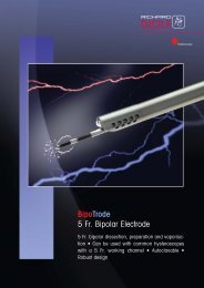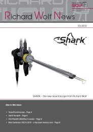
brochure - Richard Wolf
brochure - Richard Wolf
brochure - Richard Wolf

You also want an ePaper? Increase the reach of your titles
YUMPU automatically turns print PDFs into web optimized ePapers that Google loves.
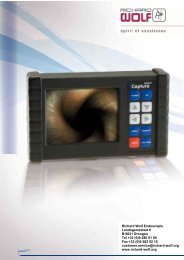
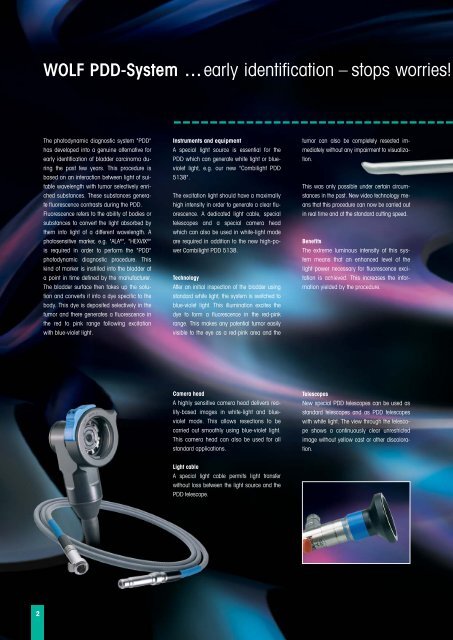
WOLF PDD-System …early identification – stops worries!<br />
The photodynamic diagnostic system "PDD"<br />
has developed into a genuine alternative for<br />
early identification of bladder carcinoma during<br />
the past few years. This procedure is<br />
based on an interaction between light of suitable<br />
wavelength with tumor selectively enriched<br />
substances. These substances generate<br />
fluorescence contrasts during the PDD.<br />
Fluorescence refers to the ability of bodies or<br />
substances to convert the light absorbed by<br />
them into light of a different wavelength. A<br />
photosensitive marker, e.g. "ALA ® ", "HEXVIX ® "<br />
is required in order to perform the "PDD"<br />
photodynamic diagnostic procedure. This<br />
kind of marker is instilled into the bladder at<br />
a point in time defined by the manufacturer.<br />
The bladder surface then takes up the solution<br />
and converts it into a dye specific to the<br />
body. This dye is deposited selectively in the<br />
tumor and there generates a fluorescence in<br />
the red to pink range following excitation<br />
with blue-violet light.<br />
Instruments and equipment<br />
A special light source is essential for the<br />
PDD which can generate white light or blueviolet<br />
light, e.g. our new "Combilight PDD<br />
5138".<br />
The excitation light should have a maximally<br />
high intensity in order to generate a clear fluorescence.<br />
A dedicated light cable, special<br />
telescopes and a special camera head<br />
which can also be used in white-light mode<br />
are required in addition to the new high-power<br />
Combilight PDD 5138.<br />
Technology<br />
After an initial inspection of the bladder using<br />
standard white light, the system is switched to<br />
blue-violet light. This illumination excites the<br />
dye to form a fluorescence in the red-pink<br />
range. This makes any potential tumor easily<br />
visible to the eye as a red-pink area and the<br />
tumor can also be completely resected immediately<br />
without any impairment to visualization.<br />
This was only possible under certain circumstances<br />
in the past. New video technology means<br />
that this procedure can now be carried out<br />
in real time and at the standard cutting speed.<br />
Benefits<br />
The extreme luminous intensity of this system<br />
means that an enhanced level of the<br />
light power necessary for fluorescence excitation<br />
is achieved. This increases the information<br />
yielded by the procedure.<br />
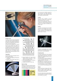
Camera head<br />
A highly sensitive camera head delivers reality-based<br />
images in white-light and blueviolet<br />
mode. This allows resections to be<br />
carried out smoothly using blue-violet light.<br />
This camera head can also be used for all<br />
standard applications.<br />
Telescopes<br />
New special PDD telescopes can be used as<br />
standard telescopes and as PDD telescopes<br />
with white light. The view through the telescope<br />
shows a continuously clear unrestricted<br />
image without yellow cast or other discoloration.<br />
Light cable<br />
A special light cable permits light transfer<br />
without loss between the light source and the<br />
PDD telescope.<br />
2