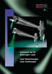
brochure - Richard Wolf
brochure - Richard Wolf
brochure - Richard Wolf

You also want an ePaper? Increase the reach of your titles
YUMPU automatically turns print PDFs into web optimized ePapers that Google loves.
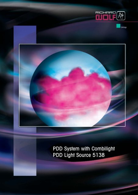
PDD System with Combilight<br />
PDD Light Source 5138<br />
Urology
WOLF PDD-System …early identification – stops worries!<br />
The photodynamic diagnostic system "PDD"<br />
has developed into a genuine alternative for<br />
early identification of bladder carcinoma during<br />
the past few years. This procedure is<br />
based on an interaction between light of suitable<br />
wavelength with tumor selectively enriched<br />
substances. These substances generate<br />
fluorescence contrasts during the PDD.<br />
Fluorescence refers to the ability of bodies or<br />
substances to convert the light absorbed by<br />
them into light of a different wavelength. A<br />
photosensitive marker, e.g. "ALA ® ", "HEXVIX ® "<br />
is required in order to perform the "PDD"<br />
photodynamic diagnostic procedure. This<br />
kind of marker is instilled into the bladder at<br />
a point in time defined by the manufacturer.<br />
The bladder surface then takes up the solution<br />
and converts it into a dye specific to the<br />
body. This dye is deposited selectively in the<br />
tumor and there generates a fluorescence in<br />
the red to pink range following excitation<br />
with blue-violet light.<br />
Instruments and equipment<br />
A special light source is essential for the<br />
PDD which can generate white light or blueviolet<br />
light, e.g. our new "Combilight PDD<br />
5138".<br />
The excitation light should have a maximally<br />
high intensity in order to generate a clear fluorescence.<br />
A dedicated light cable, special<br />
telescopes and a special camera head<br />
which can also be used in white-light mode<br />
are required in addition to the new high-power<br />
Combilight PDD 5138.<br />
Technology<br />
After an initial inspection of the bladder using<br />
standard white light, the system is switched to<br />
blue-violet light. This illumination excites the<br />
dye to form a fluorescence in the red-pink<br />
range. This makes any potential tumor easily<br />
visible to the eye as a red-pink area and the<br />
tumor can also be completely resected immediately<br />
without any impairment to visualization.<br />
This was only possible under certain circumstances<br />
in the past. New video technology means<br />
that this procedure can now be carried out<br />
in real time and at the standard cutting speed.<br />
Benefits<br />
The extreme luminous intensity of this system<br />
means that an enhanced level of the<br />
light power necessary for fluorescence excitation<br />
is achieved. This increases the information<br />
yielded by the procedure.<br />
Camera head<br />
A highly sensitive camera head delivers reality-based<br />
images in white-light and blueviolet<br />
mode. This allows resections to be<br />
carried out smoothly using blue-violet light.<br />
This camera head can also be used for all<br />
standard applications.<br />
Telescopes<br />
New special PDD telescopes can be used as<br />
standard telescopes and as PDD telescopes<br />
with white light. The view through the telescope<br />
shows a continuously clear unrestricted<br />
image without yellow cast or other discoloration.<br />
Light cable<br />
A special light cable permits light transfer<br />
without loss between the light source and the<br />
PDD telescope.<br />
2
Urology<br />
New<br />
Flexible PDD video cystoscope<br />
Specially designed for extremely gentle and atraumatic follow-up<br />
(checking for recurrences) after resection of bladder tumours.<br />
Due to its flexibility, the instrument allows the user an excellent<br />
overview of the entire bladder structure. The special shape of the<br />
distal tip and small sheath diameter ensure an absolutely atraumatic<br />
intervention.<br />
The flexible PDD video cystoscope can be connected easily to<br />
the previous <strong>Richard</strong> <strong>Wolf</strong> PDD system and can also be used<br />
with standard cystoscopes with white light.<br />
Combilight PDD light source<br />
New xenon high-power light source with 300 watt for use in<br />
the PDD. Delivers enhanced image visualization and tissue<br />
differentiation combined with improved user-friendliness.<br />
This light source can also be used as a white-light source<br />
for standard interventions.<br />
4
PDD System with Combilight<br />
PDD Light Source 5138<br />
spirit of excellence<br />
Combilight PDD 5138 Set<br />
High-power light source for photodynamic diagnostic<br />
system "PDD", early identification of<br />
bladder carcinomas, switchable between white<br />
and blue-violet light incl. anti-bleaching filter<br />
comprising:<br />
Light source Combilight PDD 5138 (5138.101),<br />
lamp module with 300 watt (2431.111),<br />
system cable (103.03), power cable 3 m<br />
(2440.03) , CAN-BUS connecting cable<br />
0.5 m (103.701),<br />
pedal switch (2030.105)..............5138.1011<br />
Panoview telescope "PDD"<br />
Ø 4 mm, free of distortion<br />
0°, with universal eyepiece ..............8650.514<br />
Panoview telescope "PDD"<br />
Ø 4 mm, free of distortion<br />
12°, with universal eyepiece ............8654.531<br />
Panoview telescope "PDD"<br />
Ø 4 mm, free of distortion,<br />
30°, with universal eyepiece ............8654.522<br />
Panoview telescope "PDD"<br />
Ø 4 mm, free of distortion<br />
70°, with universal eyepiece ............8650.515<br />
Printed on paper based on cellulose which has been bleached without the use of chlorine.<br />
Flexible PDD video cystoscope<br />
oblique distal tip 9.8 Fr., sheath 15.9 Fr.,<br />
working and irrigation channel 6 Fr., deflection<br />
210° up , 150° down (in total 360°),<br />
WL 400 mm, with integrated suction valve<br />
and fixed light cable<br />
including:<br />
Leak tester with bayonet connector (163.903),<br />
steri-gas valve (163.904), cleaning brush<br />
(7264.691) and case, control lever action<br />
towards distal; deflection down,<br />
PAL version ................................730900142<br />
Urological camera head<br />
for photodynamic diagnostic system "PDD"<br />
with 1CCD ENDOCAM 5520, PAL color<br />
system, integrated wide-angle lens,<br />
rotatable endoscope standard locking<br />
mechanism, cable length 3 m<br />
focal length f = 22 mm ............5520.833<br />
Fluid light cable<br />
Recommended accessories:<br />
Flat-screen monitor 19"<br />
for pin-sharp endo images............5370.019<br />
Base leg ..............................5370.0190<br />
Remote control ........................5520.401<br />
As above however with control lever action<br />
towards distal; deflection up,<br />
PAL version ................................730900642<br />
Ø 3 mm, 2.3 m long ................4070.253<br />
Endocam controller 5520<br />
can also be used with standard<br />
Types in NTSC version on request<br />
camera heads ..........................5520.201<br />
Usable with all standard cystoscopes and standard resectoscopes.<br />
RICHARD WOLF GmbH · 75434 Knittlingen · PF 1164 · Telephone +49 70 43 35-0 · Telefax +49 70 43 35-300 · GERMANY · info@richard-wolf.com · www.richard-wolf.com<br />
Specifications subject to change without notice.<br />
D 677.IX.08.GB.2 www.stuetzlepartner.de<br />
AUSTRIA · BELGIUM / NETHERLANDS · FRANCE · GERMANY · INDIA · U.A.E. · UK · USA