

Brain and Sense Organ Anatomy and Histology of the ... - Lannoo Lab
Brain and Sense Organ Anatomy and Histology of the ... - Lannoo Lab
Brain and Sense Organ Anatomy and Histology of the ... - Lannoo Lab

You also want an ePaper? Increase the reach of your titles
YUMPU automatically turns print PDFs into web optimized ePapers that Google loves.
88 J.T. EASTMAN AND M.J. LANNOO<br />
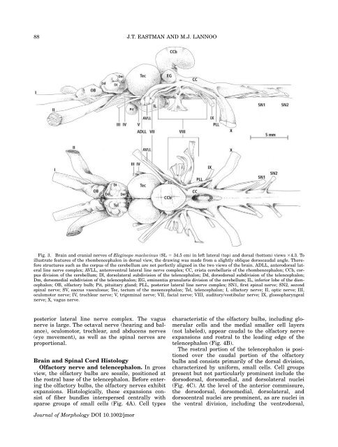
Fig. 3. <strong>Brain</strong> <strong>and</strong> cranial nerves <strong>of</strong> Eleginops maclovinus (SL 5 34.5 cm) in left lateral (top) <strong>and</strong> dorsal (bottom) views 34.3. To<br />
illustrate features <strong>of</strong> <strong>the</strong> rhombencephalon in dorsal view, <strong>the</strong> drawing was made from a slightly oblique dorsocaudal angle. Therefore<br />
structures such as <strong>the</strong> corpus <strong>of</strong> <strong>the</strong> cerebellum are not perfectly aligned in <strong>the</strong> two views <strong>of</strong> <strong>the</strong> brain. ADLL, anterodorsal lateral<br />
line nerve complex; AVLL, anteroventral lateral line nerve complex; CC, crista cerebellaris <strong>of</strong> <strong>the</strong> rhombencephalon; CCb, corpus<br />
division <strong>of</strong> <strong>the</strong> cerebellum; Dl, dorsolateral subdivision <strong>of</strong> <strong>the</strong> telencephalon; Dd, dorsodorsal subdivision <strong>of</strong> <strong>the</strong> telencephalon;<br />
Dm, dorsomedial subdivision <strong>of</strong> <strong>the</strong> telencephalon; EG, eminentia granularis division <strong>of</strong> <strong>the</strong> cerebellum; IL, inferior lobe <strong>of</strong> <strong>the</strong> diencephalon;<br />
OB, olfactory bulb; Pit, pituitary gl<strong>and</strong>; PLL, posterior lateral line nerve complex; SN1, first spinal nerve; SN2, second<br />
spinal nerve; SV, saccus vasculosus; Tec, tectum <strong>of</strong> <strong>the</strong> mesencephalon; Tel, telencephalon; I, olfactory nerve; II, optic nerve; III,<br />
oculomotor nerve; IV, trochlear nerve; V, trigeminal nerve; VII, facial nerve; VIII, auditory/vestibular nerve; IX, glossopharyngeal<br />
nerve; X, vagus nerve.<br />
posterior lateral line nerve complex. The vagus<br />
nerve is large. The octaval nerve (hearing <strong>and</strong> balance),<br />
oculomotor, trochlear, <strong>and</strong> abducens nerves<br />
(eye movement), as well as <strong>the</strong> spinal nerves are<br />
proportional.<br />
<strong>Brain</strong> <strong>and</strong> Spinal Cord <strong>Histology</strong><br />
Olfactory nerve <strong>and</strong> telencephalon. In gross<br />
view, <strong>the</strong> olfactory bulbs are sessile, positioned at<br />
<strong>the</strong> rostral base <strong>of</strong> <strong>the</strong> telencephalon. Before entering<br />
<strong>the</strong> olfactory bulbs, <strong>the</strong> olfactory nerves exhibit<br />
expansions. Histologically, <strong>the</strong>se expansions consist<br />
<strong>of</strong> fiber bundles interspersed centrally with<br />
sparse groups <strong>of</strong> small cells (Fig. 4A). Cell types<br />
characteristic <strong>of</strong> <strong>the</strong> olfactory bulbs, including glomerular<br />
cells <strong>and</strong> <strong>the</strong> medial smaller cell layers<br />
(not labeled), appear caudal to <strong>the</strong> olfactory nerve<br />
expansions <strong>and</strong> rostral to <strong>the</strong> leading edge <strong>of</strong> <strong>the</strong><br />
telencephalon (Fig. 4B).<br />
The rostral portion <strong>of</strong> <strong>the</strong> telencephalon is positioned<br />
over <strong>the</strong> caudal portion <strong>of</strong> <strong>the</strong> olfactory<br />
bulbs <strong>and</strong> consists primarily <strong>of</strong> <strong>the</strong> dorsal division,<br />
characterized by uniform, small cells. Cell groups<br />
present but not particularly prominent include <strong>the</strong><br />
dorsodorsal, dorsomedial, <strong>and</strong> dorsolateral nuclei<br />
(Fig. 4C). At <strong>the</strong> level <strong>of</strong> <strong>the</strong> anterior commissure,<br />
<strong>the</strong> dorsodorsal, dorsomedial, dorsolateral, <strong>and</strong><br />
dorsocentral nuclei are prominent, as are nuclei in<br />
<strong>the</strong> ventral division, including <strong>the</strong> ventrodorsal,<br />
Journal <strong>of</strong> Morphology DOI 10.1002/jmor


