

Brain and Sense Organ Anatomy and Histology of the ... - Lannoo Lab
Brain and Sense Organ Anatomy and Histology of the ... - Lannoo Lab
Brain and Sense Organ Anatomy and Histology of the ... - Lannoo Lab

You also want an ePaper? Increase the reach of your titles
YUMPU automatically turns print PDFs into web optimized ePapers that Google loves.
BRAIN AND SENSE ORGANS OF ELEGINOPS MACLOVINUS 91<br />
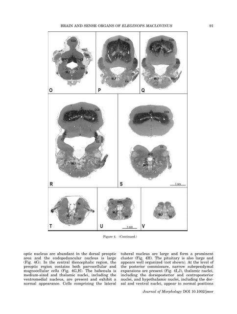
Figure 4.<br />
(Continued.)<br />
optic nucleus are abundant in <strong>the</strong> dorsal preoptic<br />
area <strong>and</strong> <strong>the</strong> endopeduncular nucleus is large<br />
(Fig. 4G). In <strong>the</strong> central diencephalic region, <strong>the</strong><br />
preoptic region contains both parvocellular <strong>and</strong><br />
magnocellular cells (Fig. 4G,H). The habenula is<br />
medium-sized <strong>and</strong> thalamic nuclei, including <strong>the</strong><br />
ventromedial nucleus, are present <strong>and</strong> exhibit a<br />
normal appearance. Cells comprising <strong>the</strong> lateral<br />
tuberal nucleus are large <strong>and</strong> form a prominent<br />
cluster (Fig. 4H). The pituitary is also large <strong>and</strong><br />
appears well organized (not shown). At <strong>the</strong> level <strong>of</strong><br />
<strong>the</strong> posterior commissure, narrow subependymal<br />
expansions are present (Fig. 4I,J), thalamic nuclei,<br />
including <strong>the</strong> dorsoposterior <strong>and</strong> centroposterior<br />
nuclei, <strong>and</strong> hypothalamic nuclei, including <strong>the</strong> dorsal<br />
<strong>and</strong> ventral nuclei, appear in normal positions<br />
Journal <strong>of</strong> Morphology DOI 10.1002/jmor


