eucaryotic cell division: mitosis and meiosis - Los Angeles Mission ...
eucaryotic cell division: mitosis and meiosis - Los Angeles Mission ...
eucaryotic cell division: mitosis and meiosis - Los Angeles Mission ...

Create successful ePaper yourself
Turn your PDF publications into a flip-book with our unique Google optimized e-Paper software.
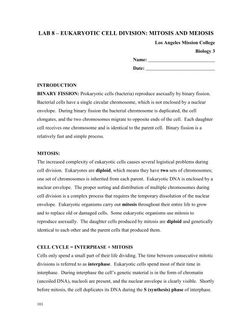
LAB 8 – EUKARYOTIC CELL DIVISION: MITOSIS AND MEIOSIS<br />
<strong>Los</strong> <strong>Angeles</strong> <strong>Mission</strong> College<br />
Biology 3<br />
Name: ___________________________<br />
Date: ____________________________<br />
INTRODUCTION<br />
BINARY FISSION: Prokaryotic <strong>cell</strong>s (bacteria) reproduce asexually by binary fission.<br />
Bacterial <strong>cell</strong>s have a single circular chromosome, which is not enclosed by a nuclear<br />
envelope. During binary fission the bacterial chromosome is duplicated, the <strong>cell</strong><br />
elongates, <strong>and</strong> the two chromosomes migrate to opposite ends of the <strong>cell</strong>. Each daughter<br />
<strong>cell</strong> receives one chromosome <strong>and</strong> is identical to the parent <strong>cell</strong>. Binary fission is a<br />
relatively fast <strong>and</strong> simple process.<br />
MITOSIS:<br />
The increased complexity of eukaryotic <strong>cell</strong>s causes several logistical problems during<br />
<strong>cell</strong> <strong>division</strong>. Eukaryotes are diploid, which means they have two sets of chromosomes;<br />
one set of chromosomes is inherited from each parent. Eukaryotic DNA is enclosed by a<br />
nuclear envelope. The proper sorting <strong>and</strong> distribution of multiple chromosomes during<br />
<strong>cell</strong> <strong>division</strong> is a complex process that requires the temporary dissolution of the nuclear<br />
envelope. Eukaryotic organisms carry out <strong>mitosis</strong> throughout their entire life to grow<br />
<strong>and</strong> to replace old or damaged <strong>cell</strong>s. Some eukaryotic organisms use <strong>mitosis</strong> to<br />
reproduce asexually. The daughter <strong>cell</strong>s produced by <strong>mitosis</strong> are diploid <strong>and</strong> genetically<br />
identical to each other <strong>and</strong> the parent <strong>cell</strong>s that produced them.<br />
CELL CYCLE = INTERPHASE + MITOSIS<br />
Cells only spend a small part of their life dividing. The time between consecutive mitotic<br />
<strong>division</strong>s is referred to as interphase. Eukaryotic <strong>cell</strong>s spend most of their time in<br />
interphase. During interphase the <strong>cell</strong>’s genetic material is in the form of chromatin<br />
(uncoiled DNA), nucleoli are present, <strong>and</strong> the nuclear envelope is clearly visible. Shortly<br />
before <strong>mitosis</strong>, the <strong>cell</strong> duplicates its DNA during the S (synthesis) phase of interphase.<br />
101
Mitosis can be divided into four distinct phases:<br />
I. Prophase: Nuclear envelope <strong>and</strong> nucleoli disappear. Chromatin condenses into<br />
chromosomes, which are made up of two identical sister chromatids joined by a<br />
centromere. In animal <strong>cell</strong>s, centrioles start migrating to opposite ends of the <strong>cell</strong><br />
(centrioles are not present in plant <strong>cell</strong>s). The mitotic spindle forms <strong>and</strong> begins to move<br />
chromosomes towards the center of the <strong>cell</strong>.<br />
II. Metaphase: Brief stage in which chromosomes line up in the equatorial plane of the<br />
<strong>cell</strong>. In animal <strong>cell</strong>s, one pair of centrioles are visible at both ends of the <strong>cell</strong>. The<br />
mitotic spindle is fully formed.<br />
III. Anaphase: Sister chromatids begin to separate, becoming individual chromosomes,<br />
which begin to migrate to opposite ends of the <strong>cell</strong>.<br />
IV. Telophase: A full set of chromosomes reaches each pole of the <strong>cell</strong>. The mitotic<br />
spindle begins to disappear. The nucleus <strong>and</strong> nucleoli begin to reappear. Chromosomes<br />
begin to unravel into chromatin.<br />
Cytokinesis or cytoplasmic <strong>division</strong> usually occurs at the end of telophase.<br />
In plant <strong>cell</strong>s cytokinesis is accomplished by the formation of a <strong>cell</strong> plate.<br />
Animal <strong>cell</strong>s separate by forming a cleavage furrow.<br />
MITOSIS EXERCISE:<br />
1. Examine prepared microscope slides of both animal <strong>cell</strong>s (whitefish blastula) <strong>and</strong><br />
plant <strong>cell</strong>s (onion/allium root tip). Even though the <strong>cell</strong>s in these tissues are rapidly<br />
dividing, most of the <strong>cell</strong>s you will observe will be in interphase (between <strong>cell</strong><br />
<strong>division</strong>s). Using your microscope, scan the slides to find a <strong>cell</strong> in interphase <strong>and</strong><br />
each one of the four stages of <strong>mitosis</strong>. Draw a schematic representation of your<br />
observations for both plant <strong>and</strong> animal <strong>cell</strong>s at each stage in the space provided<br />
below. Indicate <strong>and</strong> clearly label the important features or events of each stage.<br />
2. Using the Micro-Slide Viewers, examine the prepared microslides of:<br />
A. Plant Mitosis: Onion (Allium) Root Tip. The diploid number of chromosomes of<br />
allium is 16. The total magnification of the images is 1000 X.<br />
B. Animal Mitosis: Ascaris egg sac. The diploid number of chromosomes of ascaris is 4.<br />
The total magnification of the images is 750 X.<br />
102
Animal Cell (Whitefish Blastula)<br />
Magnification: _________<br />
Plant Cell (Onion Root Tip)<br />
Magnification: __________<br />
Interphase<br />
Prophase<br />
Metaphase<br />
Anaphase<br />
Telophase<br />
103
MEIOSIS:<br />
During sexual reproduction in eukaryotes, a haploid sperm <strong>cell</strong> fuses with a haploid<br />
egg <strong>cell</strong> to produce a diploid zygote or fertilized egg. In most species, it is very important<br />
that the offspring produced by fertilization have the same number of chromosomes as the<br />
parents. Even a single extra or missing chromosome can be lethal or extremely<br />
deleterious to an individual (e.g.: Down’s syndrome in humans). Meiosis is a special type<br />
of <strong>cell</strong> <strong>division</strong> that produces haploid gametes (sperm <strong>cell</strong>s or ova). Meiosis only occurs<br />
in an individual’s gonads, during their reproductive years.<br />
Meiosis involves two <strong>cell</strong> <strong>division</strong>s <strong>and</strong> ultimately produces four haploid gametes.<br />
The haploid gametes produced by <strong>meiosis</strong> are different from each other as well as from<br />
the parent <strong>cell</strong>s due to the crossing over of genetic material between homologous<br />
chromosomes <strong>and</strong> the r<strong>and</strong>om distribution of homologous chromosomes.<br />
Meiosis is different in males <strong>and</strong> females:<br />
Spermatogenesis: In males four functional sperm <strong>cell</strong>s are produced by <strong>meiosis</strong>.<br />
Oogenesis: Due to unequal distribution of cytoplasm in during <strong>meiosis</strong>, one large<br />
functional egg (ova) <strong>and</strong> three small polar bodies are produced.<br />
STAGES OF MEIOSIS<br />
MEIOSIS I: Reductive Division (Diploid Haploid)<br />
Prophase I: Similar to prophase of <strong>mitosis</strong> with one important difference:<br />
Crossing Over: Pairs of homologous chromosomes synapse together to form tetrads<br />
<strong>and</strong> exchange genetic information (DNA). Crossing over creates new, recombinant<br />
chromosomes.<br />
Homologous chromosomes code for the same genetic information <strong>and</strong> are of the same<br />
size, but are different because one comes from an individual’s mother, while the other<br />
comes from the individual’s father.<br />
Metaphase I: Brief stage in which tetrads line up in the equatorial plane of the <strong>cell</strong><br />
Anaphase I: Homologous chromosomes separate <strong>and</strong> migrate to opposite ends of <strong>cell</strong>.<br />
Telophase I: A full set of chromosomes reaches each pole of the <strong>cell</strong>.<br />
The <strong>cell</strong>s produced are haploid, only contain half of the original number of<br />
chromosomes.<br />
104
Interphase may be very brief or absent between <strong>meiosis</strong> I <strong>and</strong> <strong>meiosis</strong> II.<br />
MEIOSIS II: Very similar to <strong>mitosis</strong>.<br />
Prophase II: DNA condenses into chromosomes. No crossing over occurs.<br />
Metaphase II: Individual chromosomes line up in the equatorial plane of the <strong>cell</strong>.<br />
Anaphase II: Chromatids separate <strong>and</strong> begin to migrate to opposite poles of the <strong>cell</strong>.<br />
Telophase II <strong>and</strong> Cytokinesis: A full set of chromosomes reaches each pole of the <strong>cell</strong> .<br />
Four different gametes are produced.<br />
MEIOSIS EXERCISE<br />
1. Stages of Meiosis: Using the chromosome bead models construct a single pair of<br />
homologous chromosomes, each with two sister chromatids. Use red for maternal<br />
chromosome <strong>and</strong> yellow for paternal chromosome). Use the magnets for centromeres<br />
<strong>and</strong> attach 10 beads to each end of the small tubes.<br />
To illustrate crossing over, exchange several of the beads between your chromosomes.<br />
Represent each stage of <strong>meiosis</strong> <strong>and</strong> clearly depict <strong>and</strong> label the behavior of<br />
chromosomes below. Use different colors (ink <strong>and</strong> pencil or blue <strong>and</strong> red) to indicate<br />
maternal <strong>and</strong> paternal chromosomes.<br />
MEIOSIS I<br />
Important Events<br />
Prophase I<br />
Tetrad formation<br />
<strong>and</strong> crossing over<br />
Metaphase I<br />
Alignment of tetrads<br />
105
Anaphase I<br />
Important Events<br />
Separation of<br />
homologous<br />
chromosomes<br />
Telophase I <strong>and</strong><br />
Cytokinesis<br />
Two haploid <strong>cell</strong>s<br />
produced<br />
MEIOSIS II<br />
Prophase II<br />
Metaphase II<br />
Alignment of<br />
individual<br />
chromosomes<br />
Anaphase II<br />
Telophase II <strong>and</strong><br />
Cytokinesis<br />
Chromatids separate<br />
Four gametes<br />
produced<br />
106
2. Independent assortment of chromosomes: Chromosomes are shuffled r<strong>and</strong>omly <strong>and</strong><br />
distributed into daughter <strong>cell</strong>s independently from one another during <strong>meiosis</strong> I.<br />
Independent assortment creates a staggering number of possible gamete combinations as<br />
the number of chromosomes in a <strong>cell</strong> increases.<br />
The number of possible gametes generated by independent assortment alone is 2 n , where<br />
n is the haploid number of chromosomes in a <strong>cell</strong>. A human <strong>cell</strong> can produce over 8<br />
million different gametes by independent assortment 2 23 = 8.3 million.<br />
Additionally, crossing over further increases the number of possible gametes generated<br />
by an individual.<br />
INDEPENDENT ASSORTMENT EXERCISE<br />
Independent Assortment in Meiosis:<br />
Using the chromosome bead models, find all of the possible gametes formed by a <strong>cell</strong><br />
with one, two, <strong>and</strong> three pairs of homologous chromosomes.<br />
Use red for maternal chromosomes <strong>and</strong> yellow for paternal chromosomes.<br />
Note: We will disregard the production of recombinant chromosomes by crossing<br />
over for this portion of the exercise.<br />
A. A <strong>cell</strong> with one pair of homologous chromosomes (Diploid number 2)<br />
Chromosomes # 1 have 10 beads on each end of centromere.<br />
Use the following abbreviations to represent the chromosomes:<br />
M1 = Maternal chromosome # 1<br />
P1 = Paternal chromosome # 1<br />
Possible Gametes Produced by Cell:<br />
107
B. A <strong>cell</strong> with two pairs of homologous chromosomes (Diploid number 4)<br />
Chromosomes # 1 have 10 beads on each end of centromere (M1 <strong>and</strong> P1).<br />
Chromosomes # 2 have 5 beads on each end of centromere (M2 <strong>and</strong> P2).<br />
Possible Gametes Produced by Cell:<br />
C. A <strong>cell</strong> with three pairs of homologous chromosomes (Diploid number 6)<br />
Chromosomes # 1 have 10 beads on each end of centromere (M1 <strong>and</strong> P1).<br />
Chromosomes # 2 have 5 beads on each end of centromere (M2 <strong>and</strong> P2).<br />
Chromosomes # 3 have 3 beads on each end of centromere (M3 <strong>and</strong> P3).<br />
Possible Gametes Produced by Cell:<br />
Question: How many gametes would be produced by independent assortment alone, in a<br />
<strong>cell</strong> with 7 pairs of homologous chromosomes? __________________<br />
108