
Create successful ePaper yourself
Turn your PDF publications into a flip-book with our unique Google optimized e-Paper software.
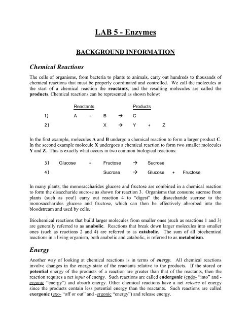
<strong>LAB</strong> 5 - <strong>Enzymes</strong><br />
Chemical Reactions<br />
BACKGROUND INFORMATION<br />
The cells of organisms, from bacteria to plants to animals, carry out hundreds to thousands of<br />
chemical reactions that must be properly coordinated and controlled. We call the molecules at<br />
the start of a chemical reaction the reactants, and the resulting molecules are called the<br />
products. Chemical reactions can be represented as shown below:<br />
Reactants<br />
Products<br />
1) A + B C<br />
2) X Y + Z<br />
In the first example, molecules A and B undergo a chemical reaction to form a larger product C.<br />
In the second example molecule X undergoes a chemical reaction to form two smaller molecules<br />
Y and Z. This is exactly what occurs in two common biological reactions:<br />
3) Glucose + Fructose Sucrose<br />
4) Sucrose Glucose + Fructose<br />
In many plants, the monosaccharides glucose and fructose are combined in a chemical reaction<br />
to form the disaccharide sucrose as shown for reaction 3. Organisms that consume sucrose from<br />
plants (such as you!) carry out reaction 4 to “digest” the disaccharide sucrose to the<br />
monosaccharides glucose and fructose, which can then be effectively absorbed into the<br />
bloodstream and used by cells.<br />
Biochemical reactions that build larger molecules from smaller ones (such as reactions 1 and 3)<br />
are generally referred to as anabolic. Reactions that break down larger molecules into smaller<br />
ones (such as reactions 2 and 4) are referred to as catabolic. The sum of all biochemical<br />
reactions in a living organism, both anabolic and catabolic, is referred to as metabolism.<br />
Energy<br />
Another way of looking at chemical reactions is in terms of energy. All chemical reactions<br />
involve changes in the energy state of the reactants relative to the products. If the stored or<br />
potential energy of the products of a reaction are greater than that of the reactants, then the<br />
reaction requires a net input of energy. Such reactions are called endergonic (endo- “into” and -<br />
ergonic “energy”) and absorb energy. Other chemical reactions have a net release of energy<br />
since the products contain less potential energy than the reactants. Such reactions are called<br />
exergonic (exo- “off or out” and -ergonic “energy”) and release energy.
ENDERGONIC: amino acid-1 + amino acid-2… + ENERGY polypeptide<br />
EXERGONIC: gasoline + O 2 CO 2 + H 2 O + ENERGY<br />
As indicated above, the synthesis of a polypeptide from free amino acids is a complex series of<br />
chemical reactions that requires energy and thus is endergonic. There is more stored energy in a<br />
polypeptide than in the free amino acids from which it is made. The burning of gasoline, on the<br />
other hand, is a chemical reaction that releases energy and thus is exergonic. There is less stored<br />
energy in the CO 2 and water products than in the reactants gasoline and oxygen. As a general<br />
rule, anabolic reactions such as polypeptide synthesis are endergonic and catabolic reactions<br />
are exergonic, with exergonic reactions providing the needed energy for endergonic ones.<br />
Activation Energy and Catalysts<br />
Another important aspect of chemical reactions and energy is the concept of activation energy<br />
(E a ). Regardless of whether a chemical reaction is endergonic or exergonic, every chemical<br />
reaction requires a certain amount of energy to get the reaction started. It is clear that the<br />
burning of gasoline is an exergonic reaction, however gasoline doesn’t just burn spontaneously,<br />
it requires some sort of energy input to “spark” the reaction. This is why car engines have spark<br />
plugs, to supply the necessary activation energy to burn gasoline in a very controlled manner.<br />
When you strike a match on a rough surface and then light a candle you are doing the same<br />
thing, providing the necessary activation energy to get each of these exergonic reactions started.<br />
Chemical reactions in biological systems generally occur at a negligible rate by themselves.<br />
Without changing the amount of reactants, the only ways to increase the rate of a chemical<br />
reaction are to 1) increase the temperature (which is what you do when you light a match, burn a<br />
candle, or burn gasoline), or 2) introduce a catalyst. Catalysts are substances that interact<br />
directly with chemical reactants and position them so that they react more easily. No increase in<br />
temperature is necessary. Catalysts actually lower the activation energy requirement for a<br />
reaction, allowing it to occur much more readily as illustrated in this graph:<br />
2
<strong>Enzymes</strong><br />
Since it would be impossible for living cells to control and coordinate their many biochemical<br />
reactions by adjusting the temperature, cells rely on biological catalysts. The biological<br />
catalysts in cells are proteins called enzymes, and just about every biochemical reaction in a cell<br />
has its own specifically shaped enzyme catalyst. By controlling the production and activity of<br />
enzymes (which are encoded by genes), cells control and coordinate their biochemical activity,<br />
i.e., their metabolism.<br />
As with any catalyst, an enzyme works by binding and positioning the reactant(s) for a specific<br />
reaction in a way that lowers the activation energy. The biochemical reactant(s) that a given<br />
enzyme binds to is referred to as its substrate, and the part of the enzyme that binds the substrate<br />
is called the active site. The diagram below illustrates this for the enzyme sucrase and its<br />
substrate sucrose (the suffix –ase denotes an enzyme, whereas –ose denotes a carbohydrate):<br />
As you can see, the enzyme sucrase, a protein, binds directly to its substrate sucrose and<br />
positions it so the covalent bond between the monosaccharides glucose and fructose is strained in<br />
a way that lowers the activation energy enough to break the bond. This yields the products<br />
glucose and fructose, which are then released. The enzyme is free to repeat this process,<br />
catalyzing the reaction over and over again until it is no longer active.<br />
Like any protein, the action of an enzyme is dependent upon its unique three-dimensional shape.<br />
Anything that causes an enzyme to adopt a non-functional shape is said to denature the enzyme.<br />
Factors that can denature an enzyme and cause it to become non-functional include changes in<br />
temperature, pH and salt concentration. For example, most human enzymes have evolved to<br />
function best at normal cellular conditions: 37 o C, pH 7.4 and 0.9% NaCl. If the temperature,<br />
pH or salt concentration deviates significantly from the “normal” state, enzymes and other<br />
proteins will begin to denature and lose their function. This is largely why high fevers and<br />
deviations in pH (acidosis, alkalosis), for example, can be so dangerous.<br />
In today’s lab you will examine the functions of three digestive enzymes and test the effect of<br />
denaturing conditions on one of these enzymes…<br />
3
DIGESTIVE ENZYME FUNCTION<br />
In the first three exercises you will observe how three different digestive enzymes catalyze<br />
biochemical reactions that break down their substrates into smaller molecules. Below is a list of<br />
each enzyme with its substrate and resulting product(s):<br />
Enzyme Substrate Product<br />
lipase triglycerides fatty acids, glycerol<br />
trypsin proteins smaller polypeptides<br />
amylase starch glucose<br />
Each of these digestive enzymes is produced in the pancreas as part of a cocktail of digestive<br />
enzymes in what we call “pancreatic juice”. Pancreatic juice is released into the first section of<br />
the small intestine, the duodenum, when it receives partially digested food called chyme from<br />
the stomach. The enzymes contained in pancreatic juice will complete the chemical digestion of<br />
a meal so that the monomeric nutrients it contains (e.g., amino acids, monosaccharide sugars,<br />
fatty acids) can be absorbed.<br />
Digestion of Triglycerides by the Enzyme Lipase<br />
The first enzyme you will examine is pancreatic lipase. Lipase is produced by the pancreas to<br />
catalyze the break down of lipids such as triglycerides into free fatty acids and glycerol:<br />
Triglycerides are the main form of lipid found in animal fats and vegetable oils, however they<br />
must be digested to fatty acids and glycerol via pancreatic lipase to be absorbed. Since lipids are<br />
not soluble in water they will form large droplets to minimize contact with the surrounding<br />
aqueous environment. This limits their interaction with lipase thus making their digestion very<br />
slow and inefficient. To avoid this problem, your liver produces bile, a greenish fluid with<br />
properties similar to soap that helps to emulsify lipids, i.e., break them up into smaller droplets.<br />
Bile is stored in the gall bladder until your partially digested meal reaches the duodenum. This<br />
triggers the release of bile into the duodenum to help emulsify the lipids.<br />
In the laboratory, digestion of triglycerides to fatty acids and glycerol can be detected by a<br />
decrease in pH. As their name implies, fatty acids are acidic (they release H + into solution) due<br />
to their carboxyl groups. The release of fatty acids from neutral triglycerides will thus result in<br />
an increase in the H + concentration (i.e., lowering of the pH value). In the following exercise,<br />
you will detect such changes in pH by using a pH indicator that changes color in response to pH.<br />
4
*Exercise 1 – Digestion of cream by lipase<br />
In this exercise you will test the ability of pancreatic lipase to digest triglycerides in cream, both<br />
with and without bile. The cream contains litmus, a pH indicator that turns red under acidic<br />
conditions, blue under basic conditions, and is purple at neutral pH. The “litmus cream” you will<br />
use is a neutral lavender color and will gradually turn reddish-pink if free fatty acids are released<br />
due to the digestion of triglycerides. Your reactions should be performed as follows:<br />
1. label four test tubes 1, 2, 3 & 4 and add 2 ml of litmus cream to each tube<br />
2. add a pinch (~0.02 g or 20 mg) of bile powder to tubes 2 & 4 only<br />
3. add 0.5 ml of water to tubes 1 & 2 and 0.5 ml of 1% lipase solution to tubes 3 & 4<br />
4. mix each tube well and incubate them in a 37 o C water bath for 1 hour<br />
5. record the results on your worksheet and answer the associated questions<br />
NOTE: For exercises 1 & 2, begin the next exercise while the current one is incubating.<br />
Digestion of Proteins by the Enzyme Trypsin<br />
The next enzyme you will examine is trypsin, one of many enzymes your body produces to<br />
digest or break down proteins. Trypsin will catalyze the breakage of peptide bonds in proteins at<br />
lysine and arginine amino acid residues. This results in larger polypeptides being broken down<br />
into smaller polypeptides (commonly referred to as “peptides”).<br />
The protein source you will subject to trypsin digestion is gelatin. Gelatin consists primarily of<br />
the protein collagen extracted from animal bones and other connective tissues. At room<br />
temperature and below, gelatin is a semisolid gel due to interactions between the collagen fibers<br />
that form a fishnet-like structure. Trypsin will partially digest the collagen fibers, disrupting<br />
their interaction and causing the gelatin to liquefy and remain liquid, even at cool temperatures.<br />
*Exercise 2 – Digestion of gelatin by trypsin<br />
To demonstrate the ability of trypsin to catalyze the partial digestion of gelatin, you will carry<br />
out two reactions as described below. If digestion of the gelatin has occurred, you will see that<br />
the gelatin remains liquid even on ice:<br />
1. obtain two tubes of molten gelatin from the 37 o C water bath and label them 1 & 2<br />
2. add 0.5 ml of water to tube 1 and 0.5 ml of 1% trypsin solution to tube 2<br />
3. mix well and place both tubes in the 37 o C water bath for 30 minutes<br />
4. place both tubes on ice for 15 minutes (or long enough for tube 1 to solidify)<br />
5. invert each tube to see if the gelatin is liquid or solid and record on your worksheet<br />
5
Digestion of Starch by the Enzyme Amylase<br />
Starch is a large polymer of the monosaccharide glucose. In order for your body to obtain<br />
glucose from the starch you eat, it must be digested by the enzyme amylase:<br />
Amylase is present in human saliva as well as pancreatic juice. As you learned in the previous<br />
lab, starch can be detected by the addition of a small amount of iodine solution. If starch is<br />
present the sample will turn dark blue or black when iodine solution is added, if there is no starch<br />
then the sample should be a clear light brown color. The complete digestion of starch to free<br />
glucose should result in no dark blue or black color when iodine solution is added. In the next<br />
exercise you will use iodine solution to determine if starch is digested by amylase.<br />
*Exercise 3 – Digestion of starch by amylase<br />
In this exercise you will set up three reaction tubes. Two reactions will serve as controls, one<br />
lacking the enzyme and the other lacking starch. The third reaction will contain both the enzyme<br />
amylase and its substrate, starch. Your three reactions should be performed as follows:<br />
1. label three test tubes 1, 2 & 3<br />
2. add 0.5 ml of water to tube 1 and 2.5 ml of water to tube 2<br />
3. add 2.5 ml of starch solution to tubes 1 & 3<br />
4. add 0.5 ml of 1% amylase solution to tubes 2 & 3<br />
5. mix well and incubate each tube at room temperature for 10 minutes<br />
6. add 2 drops of iodine to each tube, mix and record the results on your worksheet<br />
NOTE: Save your control tubes from Exercise 3 for use in Exercise 4.<br />
Effect of pH on Enzyme Function<br />
As you learned earlier, for an enzyme to function properly it must be in its native conformation<br />
or shape. If anything causes the enzyme to become misshapen or denatured, it will no longer<br />
function as a catalyst since it can no longer bind properly to its substrate. Changes in<br />
temperature, pH and salt concentration all can denature an enzyme and inhibit its activity. To<br />
illustrate this, we will focus on the effects of changes in pH.<br />
6
Pancreatic enzymes such as lipase, trypsin and amylase normally carry out their catalytic<br />
activities at a relatively neutral pH 7 to pH 8. To test the effect of pH on the activity of such<br />
enzymes, you will test the activity of the enzyme amylase at a variety of pH values…<br />
*Exercise 4 – Effect of pH on amylase activity<br />
In this exercise you will carry out five amylase reactions much like you did in Exercise 3,<br />
however each reaction will occur at a different pH. The control tubes from Exercise 3 can be<br />
used as controls for this experiment. Perform this experiment as described below:<br />
1. label five test tubes 2, 4, 7, 10 & 12 for the pH values you will be testing<br />
2. add 2.5 ml of starch solution to each tube<br />
3. add 1 ml of the corresponding pH buffer to each tube (e.g., add pH 7 buffer to tube 7)<br />
4. add 0.5 ml of 1% amylase solution to each tube<br />
5. mix well and incubate each tube at room temperature for 10 minutes<br />
6. add 2 drops of iodine to each tube and mix<br />
7. record the color of each reaction on your worksheet, and score* each reaction for<br />
amylase activity as indicated below:<br />
0 – no amylase activity (completely dark blue/black)<br />
1 – a little amylase activity (dark but not completely dark blue/black)<br />
2 – significant amylase activity (slightly darkened)<br />
3 – high amylase activity (clear light brown)<br />
8. graph your results (pH vs amylase activity)<br />
* Use the control tubes from Exercise 3 to help you score each reaction. The tube with no<br />
enzyme represents a score of “0”, and the tube with no starch represents a score of “3”.<br />
7
<strong>LAB</strong>ORATORY 5 WORKSHEET<br />
Ex. 1 – Digestion of cream by lipase<br />
Your control tubes are the tubes without lipase. Why is it<br />
important to include the second control (tube #2)?<br />
Which of your reactions was most acidic?<br />
What pH indicator allowed you to observe changes in pH?<br />
Name ________________________<br />
Section_______________________<br />
tube # color<br />
1<br />
2<br />
3<br />
4<br />
What product caused the pH to drop in the acidic reactions?<br />
Did bile alone (without lipase) result in triglyceride digestion?<br />
Why or why not?<br />
Did bile aid the digestion of triglycerides in the cream? If so, what is its role in this reaction?<br />
Ex. 2 – Digestion of gelatin by trypsin<br />
State your hypothesis for this experiment and identify the control.<br />
Which of your reactions, if any, remained liquid after incubating on ice? ___________________<br />
Explain your results below:<br />
Human beings can make only 12 of the 20 amino acids needed to build proteins, the other 8 are<br />
referred to as essential amino acids since they can only be obtained from food. Gelatin is<br />
mostly collagen extracted from animal connective tissues and contains little to none of the<br />
essential amino acids methionine and tryptophan. Is gelatin by itself a nutritious protein source?<br />
Why or why not?
Ex. 3 – Digestion of starch by amylase<br />
Tubes 1 and 2 served as controls with or without starch.<br />
Explain your result for tube 3 with starch and amylase:<br />
tube # color starch?<br />
1<br />
2<br />
3<br />
When starch is digested, what product is produced?<br />
“Blood sugar” is actually blood glucose. Why does eating starch raise your blood sugar level?<br />
Would a person with diabetes be better off eating a scoop of mashed potatoes or ice cream?<br />
Why?<br />
Ex. 4 – Effect of pH on amylase activity<br />
State your hypothesis for this experiment:<br />
pH<br />
2<br />
4<br />
7<br />
10<br />
12<br />
color<br />
amylase<br />
activity<br />
At what pH value does amylase work best? ________ worst? ________<br />
Did these results support or refute your hypothesis? _____________<br />
A person with a blood pH below 7.35 will be diagnosed with acidosis. What effect might an<br />
abnormal pH such as this have on proteins in the blood?