

Witmer, L. M., and R. C. Ridgely. 2008. The paranasal air sinuses of ...
Witmer, L. M., and R. C. Ridgely. 2008. The paranasal air sinuses of ...
Witmer, L. M., and R. C. Ridgely. 2008. The paranasal air sinuses of ...

You also want an ePaper? Increase the reach of your titles
YUMPU automatically turns print PDFs into web optimized ePapers that Google loves.
VOL. 291 NO. 11 NOVEMBER 2008<br />
THE ANATOMICAL RECORD NOVEMBER 2008 VOL. 291-NO. 11-PAGES 1343- 1572<br />
AN OFFICIAL PUBLICATION OF THE AMERICAN ASSOCIATION OF ANATOMISTS KURT H. ALBERTINE • EDITOR-IN-CHIEF<br />
A R<br />
<strong>The</strong>AnatomicalRecord<br />
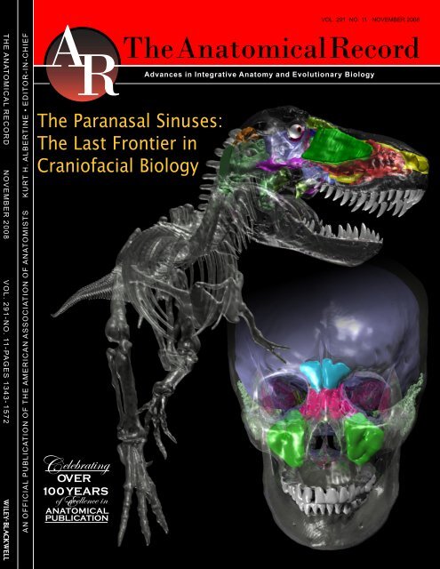
<strong>The</strong> Paranasal Sinuses:<br />
<strong>The</strong> Last Frontier in<br />
Crani<strong>of</strong>acial Biology<br />
elebrating C OVER<br />
100 YEARS<br />
<strong>of</strong>Excellence in<br />
ANATOMICAL<br />
PUBLICATION<br />
Advances in Integrative Anatomy <strong>and</strong> Evolutionary Biology
THE ANATOMICAL RECORD 291:1362–1388 (2008)<br />
<strong>The</strong> Paranasal Air Sinuses <strong>of</strong> Predatory<br />
<strong>and</strong> Armored Dinosaurs (Archosauria:<br />
<strong>The</strong>ropoda <strong>and</strong> Ankylosauria) <strong>and</strong> <strong>The</strong>ir<br />
Contribution to Cephalic Structure<br />
LAWRENCE M. WITMER* AND RYAN C. RIDGELY<br />
Department <strong>of</strong> Biomedical Sciences, College <strong>of</strong> Osteopathic Medicine,<br />
Ohio University, Athens, Ohio<br />
ABSTRACT<br />
<strong>The</strong> <strong>paranasal</strong> <strong>air</strong> <strong>sinuses</strong> <strong>and</strong> nasal cavities were studied along with<br />
other cephalic spaces (brain cavity, paratympanic <strong>sinuses</strong>) in certain dinosaurs<br />
via CT scanning <strong>and</strong> 3D visualization to document the anatomy<br />
<strong>and</strong> examine the contribution <strong>of</strong> the <strong>sinuses</strong> to the morphological organization<br />
<strong>of</strong> the head as a whole. Two representatives each <strong>of</strong> two dinosaur<br />
clades are compared: the theropod saurischians Majungasaurus <strong>and</strong><br />
Tyrannosaurus <strong>and</strong> the ankylosaurian ornithischians Panoplosaurus <strong>and</strong><br />
Euoplocephalus. <strong>The</strong>ir extant archosaurian outgroups, birds <strong>and</strong> crocodilians<br />
(exemplified by ostrich <strong>and</strong> alligator), display a diversity <strong>of</strong> <strong>paranasal</strong><br />
<strong>sinuses</strong>, yet they share only a single homologous antorbital sinus, which<br />
in birds has an important subsidiary diverticulum, the suborbital sinus.<br />
Both <strong>of</strong> the theropods had a large antorbital sinus that pneumatized<br />
Anatomical abbreviations used (taxonomic representation<br />
indicated in parentheses): <strong>air</strong>way 5 main nasal <strong>air</strong>way (respiratory<br />
region <strong>of</strong> the nasal cavity; all); antorb 5 antorbital sinus<br />
(archosaurs); a<strong>of</strong>en 5 internal antorbital fenestra in the skull;<br />
the external antorbital fenestra is the rim around the antorbital<br />
fossa (Majungasaurus, Tyrannosaurus); caudal loop 5 caudal<br />
loop <strong>of</strong> the nasal <strong>air</strong>way (Panoplosaurus, Euoplocephalus); ch 5<br />
choana (all); dalv 5 dorsal alveolar canal, transmitting<br />
branches <strong>of</strong> the maxillary nerves <strong>and</strong> large vessels (Euoplocephalus);<br />
con 5 conchal spaces in the <strong>air</strong>way <strong>of</strong> the ostrich,<br />
where the mucosal nasal conchae reside; ect 5 ectopterygoid<br />
sinus (source <strong>of</strong> diverticulum uncertain, probably not from<br />
antorbital sinus; Tyrannosaurus); endocast 5 cranial endocast<br />
<strong>of</strong> brain cavity (all); eth 5 ethmoidal sinus (human); fr 5 frontal<br />
sinus (human; in ostrich, frontal portion <strong>of</strong> fronto-ethmoidal<br />
sinus; Majungasaurus); ialv 5 interalveolar <strong>sinuses</strong> (a maxillary<br />
sinus, from antorbital sinus via other maxillary <strong>sinuses</strong>;<br />
Tyrannosaurus); jug 5 jugal sinus (from antorbital sinus;<br />
Tyrannosaurus); lac 5 lacrimal sinus proper (from antorbital<br />
sinus in nonavian theropods including most birds but from suborbital<br />
sinus in ostrich); lacm 5 medial lacrimal sinus (from<br />
antorbital sinus in nonavian theropods); mant 5 maxillary<br />
antral sinus (a maxillary sinus, from antorbital sinus; Tyrannosaurus);<br />
max 5 maxillary sinus (human, alligator, theropods—<br />
nonhomologous); mes 5 mesethmoidal portion <strong>of</strong> fronto-ethmoidal<br />
sinus (ostrich); mfen 5 maxillary fenestra <strong>of</strong> skull (Tyrannosaurus);<br />
mnas 5 medial nasal canal, transmitting the medial<br />
nasal branches <strong>of</strong> the ophthalmic nerve <strong>and</strong> enlarged medial<br />
nasal branches <strong>of</strong> the ethmoidal vessels (Euoplocephalus); nar<br />
5 nostril (fossil skulls); nas 5 nasal sinus (from antorbital<br />
sinus; Majungasaurus); npdu 5 nasopharyngeal duct (alligator);<br />
nvas 5 neurovascular canals in the premaxilla derived<br />
principally from the medial nasal canal (Euoplocephalus); olf 5<br />
olfactory region <strong>of</strong> the nasal cavity (all); orbit 5 orbit or eye<br />
socket (fossil skulls); pf 5 prefrontal sinus (alligator); pal 5 palatine<br />
sinus (alligator, Tyrannosaurus, Panoplosaurus, Euoplocephalus—not<br />
homologous); pmax 5 promaxillary sinus (a maxillary<br />
sinus, from antorbital sinus; Tyrannosaurus); pter 5 pterygoid<br />
sinus (from nasopharyngeal duct in alligator, from<br />
suborbital sinus in ostrich); pterpal 5 pterygopalatine sinus <strong>of</strong><br />
nasopharyngeal duct (alligator); pv 5 postvestibular sinus (alligator);<br />
rostral loop 5 rostral loop <strong>of</strong> the nasal <strong>air</strong>way (Panoplosaurus,<br />
Euoplocephalus); sph 5 sphenoidal sinus (human); squ 5 squamosal<br />
sinus (perhaps from antorbital sinus via suborbital sinus;<br />
Tyrannosaurus); sub 5 suborbital sinus (from antorbital sinus in<br />
theropods, including ostrich); tymp 5 main middle ear cavity <strong>and</strong><br />
paratympanic <strong>sinuses</strong> (all); * 5 position <strong>of</strong> the putative ‘‘<strong>paranasal</strong><br />
aperture,’’ which is not demonstrably separate either externally or<br />
internallyfromthetruenarialaperture(Euoplocephalus).<br />
Grant sponsor: National Science Foundation; Grant numbers:<br />
NSF BSR-9112070, NSF IBN-9601174, NSF IBN-0343744, NSF<br />
IOB-0517257; Grant sponsor: Ohio University College <strong>of</strong> Osteopathic<br />
Medicine.<br />
*Correspondence to: Lawrence M. <strong>Witmer</strong>, Department <strong>of</strong><br />
Biomedical Sciences, College <strong>of</strong> Osteopathic Medicine, Ohio University,<br />
Athens, OH 45701. Fax: 740-593-2400.<br />
E-mail: witmerL@ohio.edu<br />
Received 22 April 2008; Accepted 23 April 2008<br />
DOI 10.1002/ar.20794<br />
Published online in Wiley InterScience (www.interscience.wiley.<br />
com).<br />
Ó 2008 WILEY-LISS, INC.
DINOSAUR PARANASAL AIR SINUSES<br />
many <strong>of</strong> the facial <strong>and</strong> palatal bones as well as a birdlike suborbital<br />
sinus. Given that the suborbital sinus interleaves with jaw muscles, the<br />
<strong>paranasal</strong> <strong>sinuses</strong> <strong>of</strong> at least some theropods (including birds) were<br />
actively ventilated rather than being dead-<strong>air</strong> spaces. Although many<br />
ankylosaurians have been thought to have had extensive <strong>paranasal</strong><br />
<strong>sinuses</strong>, most <strong>of</strong> the snout is instead (<strong>and</strong> surprisingly) <strong>of</strong>ten occupied by<br />
a highly convoluted <strong>air</strong>way. Digital segmentation, coupled with 3D visualization<br />
<strong>and</strong> analysis, allows the positions <strong>of</strong> the <strong>sinuses</strong> to be viewed in<br />
place within both the skull <strong>and</strong> the head <strong>and</strong> then measured volumetrically.<br />
<strong>The</strong>se quantitative data allow the first reliable estimates <strong>of</strong> dinosaur head<br />
mass <strong>and</strong> an assessment <strong>of</strong> the potential savings in mass afforded by the<br />
<strong>sinuses</strong>. Anat Rec, 291:1362–1388, <strong>2008.</strong> Ó 2008 Wiley-Liss, Inc.<br />
1363<br />
Key words: dinosaur; theropod; ankylosaur; bird; crocodilian;<br />
human; <strong>paranasal</strong> sinus; computed tomography;<br />
gross anatomy<br />
Paranasal <strong>air</strong> <strong>sinuses</strong> are seemingly ubiquitous features<br />
<strong>of</strong> mammals, <strong>and</strong> studies <strong>of</strong> mammalian <strong>paranasal</strong><br />
<strong>sinuses</strong>—particularly those <strong>of</strong> humans (Fig. 1) <strong>and</strong> other<br />
primates—are diverse in both taxonomic <strong>and</strong> biological<br />
scope, impacting debates pertaining to systematics, biomechanics,<br />
physiology, development, medicine, <strong>and</strong> paleontology,<br />
among others (e.g., see articles in this issue<br />
<strong>and</strong> in Koppe et al., 1999). Mammals are not alone, however,<br />
in having <strong>air</strong>-filled epithelial diverticula <strong>of</strong> the<br />
nasal cavity that pneumatize the facial skeleton. Archosaurs<br />
are another highly pneumatic clade. Archosauria<br />
is the sauropsid clade comprised <strong>of</strong> birds <strong>and</strong> crocodilians<br />
today, <strong>and</strong> including such extinct Mesozoic forms as<br />
nonavian dinosaurs, pterosaurs, <strong>and</strong> a variety <strong>of</strong> basal<br />
taxa. <strong>The</strong> <strong>paranasal</strong> <strong>sinuses</strong> <strong>of</strong> mammals <strong>and</strong> archosaurs<br />
are not homologous (<strong>Witmer</strong>, 1995b), <strong>and</strong>, although various<br />
extracapsular epithelial diverticula have been<br />
described for different tetrapod groups (e.g., amphibians,<br />
squamate reptiles), only mammals <strong>and</strong> archosaurs<br />
display pneumatic invasion <strong>of</strong> the bones <strong>of</strong> the face <strong>and</strong><br />
cranium (<strong>Witmer</strong>, 1999). In archosaurs, pneumatic diverticula<br />
may arise from all parts <strong>of</strong> the nasal cavity,<br />
including the nasal vestibule (e.g., some hadrosaurid<br />
<strong>and</strong> ankylosaurid dinosaurs) <strong>and</strong> nasopharyngeal duct<br />
(e.g., crocodilians), but, as in mammals, the nasal cavity<br />
proper (cavum nasi proprium) is the source <strong>of</strong> the major<br />
<strong>paranasal</strong> <strong>air</strong> sinus, called the antorbital sinus because<br />
it is lodged within the bony antorbital cavity (<strong>Witmer</strong>,<br />
1990, 1995b, 1997a,b, 1999; Hill et al., 2003).<br />
Archosaurian <strong>and</strong> mammalian <strong>paranasal</strong> <strong>sinuses</strong> are<br />
quite different from each other in that, whereas mammalian<br />
<strong>sinuses</strong> (Fig. 1) tend to be almost fully enclosed<br />
within bone (connected by typically narrow ostia), archosaur<br />
<strong>sinuses</strong> tend to be much more open <strong>and</strong> less constrained.<br />
<strong>The</strong> archosaurian antorbital sinus is usually<br />
partially enclosed within the lacrimal bone caudally <strong>and</strong><br />
maxilla rostrally <strong>and</strong> variably floored by the palatine<br />
bone <strong>and</strong> ro<strong>of</strong>ed by the nasal bone. <strong>The</strong> antorbital sinus<br />
itself has subsidiary diverticula that invade <strong>and</strong> pneumatize<br />
many <strong>of</strong> the surrounding bones, although such<br />
accessory cavities are best developed in theropod dinosaurs,<br />
including birds (see <strong>Witmer</strong>, 1997a,b). In most<br />
taxa the antorbital sinus is exposed laterally, being covered<br />
only by skin. Moreover, in birds the antorbital<br />
sinus has a subsidiary diverticulum (the suborbital or<br />
‘‘infraorbital’’ sinus) that extends caudally from the<br />
antorbital cavity into the orbit where it is juxtaposed<br />
between the eyeball, jaw muscles, <strong>and</strong> other structures<br />
(Bang <strong>and</strong> Wenzel, 1985; <strong>Witmer</strong>, 1990, 1995b; Evans,<br />
1996). New evidence suggests that such a suborbital<br />
sinus is found in at least the theropodan ancestors <strong>of</strong><br />
birds, if not even more broadly among archosaurs<br />
(<strong>Witmer</strong>, 1997a; Sampson <strong>and</strong> <strong>Witmer</strong>, 2007). Thus, the<br />
<strong>paranasal</strong> <strong>sinuses</strong> <strong>of</strong> most archosaurs are not the familiar<br />
blind sacs housed within bony chambers <strong>of</strong> mammals<br />
but rather are more expansive <strong>and</strong> relate directly to<br />
(i.e., contact) a diversity <strong>of</strong> other anatomical systems.<br />
This article seeks to explore not just the morphology<br />
<strong>of</strong> select dinosaur <strong>paranasal</strong> <strong>air</strong> <strong>sinuses</strong> but also the<br />
relationship to other anatomical systems, such as<br />
the <strong>air</strong>way, the olfactory chamber <strong>of</strong> the nasal cavity, the<br />
paratympanic <strong>air</strong> <strong>sinuses</strong>, the orbital contents, <strong>and</strong><br />
the brain <strong>and</strong> endocranial cavity, among others. Examining<br />
the contribution <strong>of</strong> the <strong>paranasal</strong> <strong>sinuses</strong> to the<br />
architecture <strong>of</strong> the head as a whole is now possible,<br />
thanks to the development <strong>of</strong> computed tomography<br />
(CT scanning) coupled with 3D computer visualization.<br />
<strong>The</strong>se new approaches allow many different anatomical<br />
structures <strong>of</strong> extinct <strong>and</strong> extant animals alike to be<br />
viewed in place within the skull or head, as well as to be<br />
analyzed quantitatively. <strong>The</strong> power <strong>of</strong> 3D visualization<br />
will be used to illustrate the anatomy rather than<br />
lengthy morphological description. After looking briefly<br />
at the <strong>sinuses</strong> <strong>of</strong> the modern relatives <strong>of</strong> dinosaurs, the<br />
focus will turn to two predatory dinosaurs with which<br />
the authors have extensive experience <strong>and</strong> on which<br />
they have published previously: the Cretaceous abelisaurid<br />
theropod from Madagascar, Majungasaurus crenatissimus<br />
(<strong>Witmer</strong> et al., 2004; Sampson <strong>and</strong> <strong>Witmer</strong>,<br />
2007), <strong>and</strong> the Cretaceous coelurosaur Tyrannosaurus<br />
rex from North America (<strong>Witmer</strong>, 1997a; <strong>Witmer</strong> <strong>and</strong><br />
<strong>Ridgely</strong>, 2005; <strong>Witmer</strong> et al., 2008). <strong>The</strong>se two theropods<br />
allow a look at a dinosaur system in which <strong>paranasal</strong><br />
pneumaticity is relatively well understood, <strong>and</strong> which<br />
can be integrated into a more comprehensive picture <strong>of</strong><br />
cephalic anatomy. Two North American Cretaceous armored<br />
ankylosaurian dinosaurs also will be investigated:<br />
the nodosaurid Panoplosaurus mirus <strong>and</strong> the ankylosaurid<br />
Euoplocephalus tutus. <strong>The</strong>se provide the opportunity<br />
to investigate a classically problematic nasal <strong>and</strong>
1364 WITMER AND RIDGELY<br />
Fig. 1. Paranasal <strong>sinuses</strong> <strong>and</strong> other cephalic components <strong>of</strong> a<br />
human (Homo sapiens, OUVC 10503) based on CT scanning followed<br />
by segmentation <strong>and</strong> 3D visualization. Bone is rendered semitransparent.<br />
A: Left anterodorsolateral view. B: Anterior view. C: Left lateral<br />
view. D–G: isolated <strong>paranasal</strong> <strong>sinuses</strong>. D: Left anterodorsolateral view<br />
corresponding to A. E: Anterior view corresponding to B. F: Dorsal<br />
view. G: Left lateral view corresponding to C. <strong>The</strong> paratympanic<br />
<strong>sinuses</strong> <strong>and</strong> endosseous labyrinth are also visualized. Scale bars 5 2cm.
<strong>paranasal</strong> sinus system, <strong>and</strong>, it is hoped, shed some new<br />
light, although as will be seen, these armored dinosaurs<br />
have truly bizarre systems. Finally, the new analytical<br />
capabilities provided by CT scanning will be used to calculate<br />
volumes <strong>and</strong> masses for cephalic structures in the<br />
two theropods, providing not only the first reliable estimates<br />
<strong>of</strong> head mass but also an assessment <strong>of</strong> the<br />
impact <strong>of</strong> the <strong>paranasal</strong> <strong>sinuses</strong> on head mass.<br />
MATERIALS AND METHODS<br />
Materials<br />
<strong>The</strong> dinosaur sample largely focuses on four main<br />
taxa. (1) Archosauria, Dinosauria, <strong>The</strong>ropoda, Abelisauridae,<br />
Majungasaurus crenatissimus; Field Museum <strong>of</strong><br />
Natural History (FMNH, Chicago) PR2100; collected<br />
from the Upper Cretaceous (Maastrichtian, 66–71 Ma)<br />
Maevarano Formation <strong>of</strong> northwestern Madagascar<br />
(Krause et al., 2007). (2) Archosauria, Dinosauria, <strong>The</strong>ropoda,<br />
Coelurosauria, Tyrannosauridae, Tyrannosaurus<br />
rex; FMNH PR2081 (as well as a restored, one-thirdscale<br />
sculpture <strong>of</strong> FMNH PR2081 crafted by Brian<br />
Cooley), American Museum <strong>of</strong> Natural History (AMNH,<br />
New York City) FR 5117, Black Hills Institute (BHI, Hill<br />
City, SD) 3033, <strong>and</strong> an unnumbered Carnegie Museum<br />
<strong>of</strong> Natural History (Pittsburgh) skull; collected from the<br />
Upper Cretaceous (Maastrichtian, 66–71 Ma) Hell Creek<br />
<strong>and</strong> Lance Formations <strong>of</strong> Montana, Wyoming, <strong>and</strong> South<br />
Dakota. (3) Archosauria, Dinosauria, Ornithischia,<br />
Ankylosauria, Nodosauridae, Panoplosaurus mirus;<br />
Royal Ontario Museum (ROM, Toronto) 1215 [note:<br />
referral to P. mirus follows Coombs (1978), Carpenter<br />
(1990), <strong>and</strong> Vickaryous et al. (2004), although Russell<br />
(1940) <strong>and</strong> Ryan <strong>and</strong> Evans (2005) referred it to Edmontonia<br />
rugosidens]; collected from the Upper Cretaceous<br />
(Campanian, 83–71 Ma) Dinosaur Park Formation <strong>of</strong><br />
Alberta. (4) Archosauria, Dinosauria, Ornithischia,<br />
Ankylosauria, Ankylosauridae, Euoplocephalus tutus;<br />
AMNH FR 5405; collected from the Upper Cretaceous<br />
(Campanian, 83–71 Ma) Dinosaur Park Formation <strong>of</strong><br />
Alberta. Two additional ankylosaur specimens became<br />
available late enough in the study that it was not possible<br />
to perform the same level <strong>of</strong> analysis <strong>and</strong> visualization,<br />
although important details were assessed. One<br />
specimen is another skull <strong>of</strong> E. tutus (AMNH FR 5403),<br />
<strong>and</strong> the other is a skull <strong>of</strong> Edmontonia rugosidens<br />
(AMNH FR 5381), a nodosaurid closely related to Panoplosaurus.<br />
As with the other ankylosaurs in the sample,<br />
these specimens derive from the Dinosaur Park Formation<br />
<strong>of</strong> Alberta.<br />
Inferences about the unpreserved traits <strong>of</strong> extinct dinosaur<br />
taxa are grounded in the extant phylogenetic<br />
bracket approach (<strong>Witmer</strong>, 1995a) whereby extant outgroups<br />
(in this case, birds <strong>and</strong> crocodilians) provide critical<br />
data on s<strong>of</strong>t tissues <strong>and</strong> their osteological correlates.<br />
Although numerous extant birds <strong>and</strong> crocodilians were<br />
examined, data are presented here on a characteristic<br />
representative <strong>of</strong> each. (1) Archosauria, Suchia, Crocodylia,<br />
Alligatoridae, Alligator mississippiensis (American<br />
alligator); Ohio University Vertebrate Collections<br />
(OUVC) 9761; fresh carcass <strong>of</strong> adult (total skull length:<br />
371 mm) obtained from the Rockefeller Wildlife Refuge,<br />
Gr<strong>and</strong> Chenier, Louisiana. (2) Archosauria, Dinosauria,<br />
<strong>The</strong>ropoda, Aves, Ratitae, Struthio camelus (ostrich);<br />
OUVC 10491; fresh head <strong>and</strong> neck <strong>of</strong> adult (total skull<br />
DINOSAUR PARANASAL AIR SINUSES<br />
length: 182 mm) purchased from a commercial source.<br />
For further comparison <strong>and</strong> illustration, a human skull<br />
(Homo sapiens, OUVC 10503) was also analyzed. Figure 2<br />
presents the phylogenetic relationships <strong>of</strong> the taxa mentioned<br />
in this article.<br />
Reference was also made to an existing series <strong>of</strong> avian<br />
specimens in which the <strong>paranasal</strong> <strong>sinuses</strong> were injected<br />
with latex followed by removal <strong>of</strong> s<strong>of</strong>t tissue (for methods,<br />
see <strong>Witmer</strong>, 1995b). <strong>The</strong>se skull-sinus preparations<br />
included the following specimens: juvenile (6 weeks old)<br />
ostrich, Struthio camelus (OUVC 10504); six adult<br />
domestic chicken, Gallus gallus (OUVC 10259–10264);<br />
one hatchling (OUVC 10254) <strong>and</strong> two adult (OUVC<br />
10257–10258) domestic goose, Anser anser; four adult<br />
domestic ducks, Anas platyrhynchos (OUVC 10248–<br />
10251); <strong>and</strong> one adult ring-billed gull, Larus delawarensis<br />
(OUVC 10308).<br />
CT Scanning <strong>and</strong> 3D Visualization<br />
1365<br />
Other than the latex-injected specimens, all <strong>of</strong> the<br />
above, both extant <strong>and</strong> extinct, were subjected to CT<br />
scanning at O’Bleness Memorial Hospital, Athens, Ohio,<br />
using a General Electric (GE) LightSpeed Ultra Multislice<br />
CT scanner equipped with the Extended Hounsfield<br />
option (which greatly improves resolvability <strong>of</strong> detail<br />
from dense objects such as fossils by extending the<br />
dynamic range <strong>of</strong> images as much 16-fold) <strong>and</strong> a ‘‘bowtie’’<br />
filter (which decreases beam-hardening artifacts).<br />
All specimens were scanned helically at a slice thickness<br />
<strong>of</strong> 625 mm, 120–140 kV, <strong>and</strong> 200–300 mA. <strong>The</strong> raw scan<br />
data were reconstructed using a bone algorithm. Data<br />
were output from the scanners in DICOM format, <strong>and</strong><br />
then imported into Amira 3.1.1 or 4.1.2 (Mercury-TGS,<br />
Chelmsford, MA) for viewing, analysis, <strong>and</strong> visualization.<br />
<strong>The</strong> only exceptions to the above protocol were<br />
FMNH PR2081 (scanned elsewhere; see Brochu, 2003),<br />
the Carnegie Museum Tyrannosaurus (scanned at<br />
NASA’s Marshall Space Flight Center in Alabama), <strong>and</strong><br />
BHI 3033. All CT data, regardless <strong>of</strong> source, were analyzed<br />
on 32- <strong>and</strong> 64-bit PC workstations with 4 GB <strong>of</strong><br />
RAM <strong>and</strong> nVidia Quadro FX 3000 or 4500 video cards<br />
<strong>and</strong> running Micros<strong>of</strong>t Windows XP Pr<strong>of</strong>essional, Windows<br />
XP Pr<strong>of</strong>essional x64, or Linux 2.6.18 (Debian 4.0<br />
distribution). Structures <strong>of</strong> interest (e.g., <strong>paranasal</strong><br />
<strong>sinuses</strong>, cranial endocast, otic labyrinth, paratympanic<br />
<strong>sinuses</strong>, etc.) were highlighted <strong>and</strong> digitally extracted<br />
using Amira’s segmentation tools for quantification <strong>and</strong><br />
visualization.<br />
<strong>The</strong> theropod studies each require additional explanation.<br />
As described in Sampson <strong>and</strong> <strong>Witmer</strong> (2007), the<br />
skull <strong>of</strong> Majungasaurus used here (FMNH PR2100) was<br />
discovered as largely disarticulated bony elements.<br />
Many <strong>of</strong> the individual fossil elements were CT scanned,<br />
as was a cast <strong>of</strong> the full skull, which had been<br />
assembled from the individual cast elements. In Amira,<br />
the CT datasets from the fossil elements were then registered<br />
(aligned) to the dataset <strong>of</strong> the skull cast, which<br />
thus allowed the <strong>sinuses</strong> segmented from the fossil elements<br />
to be ‘‘plugged into’’ their proper places in the full<br />
skull. As noted above, not all <strong>air</strong> <strong>sinuses</strong> in archosaurs<br />
are fully enclosed in bone, <strong>and</strong> thus the skull <strong>and</strong> structures<br />
segmented in Amira were imported into the 3D<br />
modeling s<strong>of</strong>tware Maya 8.5 (Autodesk, San Rafael, CA) to<br />
model the antorbital sinus <strong>and</strong> its suborbital diverticulum,
1366 WITMER AND RIDGELY<br />
Fig. 2. Diagram <strong>of</strong> phylogenetic relationships <strong>of</strong> the taxa mentioned in the text. <strong>The</strong> focal taxa are indicated<br />
in boldface type. Topology derives from Hill et al. (2003), Holtz et al. (2004), Vickaryous et al.<br />
(2004), Bininda-Emonds et al. (2007), <strong>and</strong> Livezey <strong>and</strong> Zusi (2007).<br />
as well as the middle ear sac (based on a series <strong>of</strong> anatomical<br />
criteria that will be presented elsewhere). A similar<br />
approach was used for Tyrannosaurus, although,<br />
whereas the Majungasaurus system represents a single<br />
specimen, the Tyrannosaurus system represents a composite<br />
based on structures segmented or modeled from<br />
multiple specimens <strong>and</strong> then all digitally inserted into<br />
the restored sculpture.<br />
Supplemental visualizations as well as the native CT<br />
data for some <strong>of</strong> the specimens are available on the<br />
authors’ website: www.ohio.edu/witmerlab.<br />
Mass Estimation <strong>of</strong> <strong>The</strong>ropod Dinosaur Heads<br />
Novel data on mass <strong>of</strong> the fleshed-out heads in the<br />
two theropods in the sample are presented here. <strong>The</strong>se<br />
were calculated by generating volumes for various cephalic<br />
components from the CT data <strong>and</strong> then converting<br />
these volumes to masses. More specifically, the skull<br />
models were digitally ‘‘wrapped’’ with a skin, taking into<br />
account jaw muscle bulges (from Holliday, 2006) but<br />
ignoring the cervicocephalic musculature; total head volume<br />
was calculated from this skin surface. <strong>The</strong> head<br />
was modeled with the jaws completely adducted such<br />
that the oral cavity is a potential (not a real) space,<br />
which is appropriate based on the authors’ findings from<br />
CT data <strong>of</strong> a broad diversity <strong>of</strong> extant amniotes. Skull<br />
volume was generated directly from the CT scans <strong>of</strong> the<br />
Majungasaurus <strong>and</strong> Tyrannosaurus skull casts (see<br />
above); for Majungasaurus, the vomer <strong>and</strong> right pterygoid<br />
were digitally reconstructed, <strong>and</strong> this volume was<br />
added to the total. Based on digital segmentation, the<br />
volumes <strong>of</strong> all <strong>of</strong> the <strong>paranasal</strong> <strong>and</strong> paratympanic<br />
<strong>sinuses</strong>, as well as <strong>of</strong> the cranial endocast (brain cavity)<br />
were calculated, <strong>and</strong> these were then subtracted from<br />
the skull cast volumes to get a very realistic volume for<br />
the bone comprising the skull. In truth, these bone volumes<br />
are very slight overestimates, because it was not<br />
possible to consider the minute intertrabecular spaces<br />
<strong>and</strong> tiny neurovascular canals within the bone; this<br />
source <strong>of</strong> error is regarded as negligible <strong>and</strong> probably<br />
within measurement error <strong>and</strong> natural individual
DINOSAUR PARANASAL AIR SINUSES<br />
TABLE 1. Volumes, tissue densities, <strong>and</strong> masses for head structural components <strong>and</strong> the head itself <strong>of</strong><br />
Majungasaurus crenatissimus under three different states <strong>of</strong> pneumaticity<br />
Head with all pneumatic<br />
<strong>sinuses</strong><br />
Head without <strong>paranasal</strong><br />
<strong>sinuses</strong> a<br />
Head without <strong>paranasal</strong><br />
or paratympanic <strong>sinuses</strong> a<br />
Volume (cm 3 ) Density (g/cm 3 ) Mass (g) Density (g/cm 3 ) Mass (g) Density (g/cm 3 ) Mass (g)<br />
Bone b 5909.5 1.35 7977.8 1.35 7977.8 1.35 7977.8<br />
Cranial endocast 126.2 1.036 130.7 1.036 130.7 1.036 130.7<br />
Nasal cavity<br />
Airway 643.7 0.0 0.0 0.0 0.0 0.0 0.0<br />
Olfactory region 1230.3 0.0 0.0 0.0 0.0 0.0 0.0<br />
Paranasal <strong>sinuses</strong><br />
Antorbital sinus 777.8 0.0 0.0 1.35 1050.0 1.35 1050<br />
Maxillary sinus 5.3 0.0 0.0 1.35 7.2 1.35 7.2<br />
Lacrimal sinus proper 37.2 0.0 0.0 1.35 50.2 1.35 50.2<br />
Medial lacrimal sinus 9.0 0.0 0.0 1.35 12.2 1.35 12.2<br />
Nasal sinus 429.2 0.0 0.0 1.35 579.4 1.35 579.4<br />
Frontal sinus 86.1 0.0 0.0 1.35 116.2 1.35 116.2<br />
Suborbital sinus 403.3 0.0 0.0 1.05 423.5 1.05 423.5<br />
Middle ear cavity 716.4 0.0 0.0 0.0 0.0 0.0 0.0<br />
Paratympanic <strong>sinuses</strong> 29.5 0.0 0.0 0.0 0.0 1.35 39.8<br />
S<strong>of</strong>t tissue 22850.8 1.05 23993.3 1.05 23993.3 1.05 23993.3<br />
Total head a 33254.3 – 32101.8 – 34340.5 – 34380.3<br />
Skull mass a – – 7977.8 – 9793.0 – 9832.8<br />
a For the calculations <strong>of</strong> head mass <strong>and</strong> skull mass in the absence <strong>of</strong> pneumatic <strong>sinuses</strong>, the various sinus cavities are<br />
assigned the density <strong>of</strong> bone.<br />
b Includes restored vomer <strong>and</strong> right pterygoid.<br />
1367<br />
variation, but it does represent a target for future refinements<br />
in technique. <strong>The</strong> form <strong>of</strong> the suborbital sinus,<br />
which is not enclosed in bone (see below), was modeled<br />
in Amira <strong>and</strong> Maya based on anatomical l<strong>and</strong>marks in<br />
the fossils <strong>and</strong> the structure <strong>of</strong> the sinus in birds. Subtracting<br />
the volumes <strong>of</strong> the bony skull, <strong>air</strong> <strong>sinuses</strong>, <strong>and</strong><br />
endocast from total head volume gives the volume <strong>of</strong> the<br />
remaining s<strong>of</strong>t tissue.<br />
To convert volumes to masses, volume (cm 3 ) was multiplied<br />
by density (g/cm 3 ). Ignoring the thin sinus epithelium,<br />
<strong>air</strong> sinus density was taken as zero, as was the<br />
resulting mass. Density <strong>of</strong> the cranial endocast was<br />
assigned the density <strong>of</strong> brain tissue, using the commonly<br />
used 1.036 g/cm 3 value (e.g., <strong>Witmer</strong> et al., 2003). <strong>The</strong><br />
s<strong>of</strong>t-tissue volume in life included a heterogeneous mix<br />
<strong>of</strong> muscle, fat, nerves, vessels, <strong>and</strong> so forth, but muscle<br />
certainly predominated. Common literature values for<br />
muscle density (e.g., Urbanchek et al., 2001) are 1.06 g/<br />
cm 3 , <strong>and</strong> thus, for the ‘‘s<strong>of</strong>t-tissue’’ density value used<br />
here, that muscle value was arbitrarily reduced to 1.05<br />
g/cm 3 to account for fat <strong>and</strong> other tissue types. <strong>The</strong> bone<br />
density values in the literature are somewhat unsatisfactory<br />
in that they tend to be derived from small cubes<br />
<strong>of</strong> mammalian bone <strong>of</strong> particular types (e.g., compact,<br />
trabecular, otic), whereas skulls include virtually all<br />
bone types as well as teeth. Consequently, whole-skull<br />
density values were generated for a range <strong>of</strong> avian, crocodilian,<br />
<strong>and</strong> mammalian skulls by dividing the mass <strong>of</strong><br />
the skull (as weighed on a digital balance) by the volume<br />
<strong>of</strong> the skull (as determined by CT scanning to be consistent<br />
with the fossil sample). <strong>The</strong> resulting bone density<br />
values ranged from 0.5 g/cm 3 (barn owl, Tyto alba) to 1.7<br />
g/cm 3 (Adelie penguin, Pygoscelis adeliae), with a total<br />
sample mean <strong>of</strong> 1.2 g/cm 3 . However, the avian sample<br />
was excluded because birds lack teeth, <strong>and</strong> consequently<br />
the mean <strong>of</strong> the remaining sample (1.35 g/cm 3 ) was<br />
used. This value is still somewhat lower than typical<br />
values for bone density in the literature (e.g., Currey,<br />
1984), suggesting that whole skulls have relatively lower<br />
densities than bone explants from the appendicular skeleton.<br />
For example, Yang et al. (2002, p 313) reported<br />
‘‘normal human bone density" as 1.85 g/cm 3 , yet the<br />
whole-skull density calculated here for humans was<br />
1.1 g/cm 3 .<br />
In addition to simply estimating head mass, the contribution<br />
<strong>of</strong> pneumaticity to total head mass (thus<br />
assessing any weight savings) was estimated by doing<br />
calculations in which the <strong>paranasal</strong> <strong>sinuses</strong> were considered<br />
to be bone by assigning the sinus volumes the density<br />
<strong>of</strong> bone (except for the suborbital sinus which was<br />
assigned the density <strong>of</strong> s<strong>of</strong>t tissue). Similar calculations<br />
considering the head to be completely without any pneumatic<br />
<strong>sinuses</strong> (yet retaining the main nasal <strong>and</strong> middle<br />
ear cavities) were made by assigning bone density to the<br />
paratympanic as well as the <strong>paranasal</strong> <strong>sinuses</strong>. For comparison,<br />
similar calculations were also made for the<br />
human skull in the sample. <strong>The</strong> volumes <strong>and</strong> masses <strong>of</strong><br />
all relevant structures for Majungasaurus, Tyrannosaurus,<br />
<strong>and</strong> Homo sapiens are presented in Tables 1–3,<br />
respectively.<br />
RESULTS AND OBSERVATIONS<br />
<strong>The</strong> Modern Archosaurian Condition: Alligator<br />
<strong>and</strong> Ostrich<br />
<strong>The</strong> extant relatives <strong>of</strong> dinosaurs are particularly relevant,<br />
not only because they can be directly examined<br />
(e.g., via dissection, medical imaging) for the detailed<br />
relationships between the s<strong>of</strong>t tissues <strong>and</strong> the skeleton,<br />
but also because, being close phylogenetic relatives,<br />
their attributes have a greater likelihood <strong>of</strong> being homologous<br />
to those <strong>of</strong> dinosaurs (<strong>Witmer</strong>, 1995a). For example,<br />
the bony antorbital cavities <strong>of</strong> extant birds <strong>and</strong>
1368 WITMER AND RIDGELY<br />
TABLE 2. Volumes, tissue densities, <strong>and</strong> masses for head structural components <strong>and</strong> the head itself <strong>of</strong><br />
Tyrannosaurus rex under three different states <strong>of</strong> pneumaticity<br />
Head with all pneumatic<br />
<strong>sinuses</strong><br />
Head without <strong>paranasal</strong><br />
<strong>sinuses</strong> a<br />
Head without <strong>paranasal</strong><br />
or paratympanic <strong>sinuses</strong> a<br />
Volume (cm 3 ) Density (g/cm 3 ) Mass (g) Density (g/cm 3 ) Mass (g) Density (g/cm 3 ) Mass (g)<br />
Bone 122709.8 1.35 165658.2 1.35 165658.2 1.35 165658.2<br />
Cranial endocast b 1174.9 1.036 1217.2 1.036 1217.2 1.036 1217.2<br />
Nasal cavity<br />
Airway c 10740.0 0.0 0.0 0.0 0.0 0.0 0.0<br />
Olfactory region c 19057.8 0.0 0.0 0.0 0.0 0.0 0.0<br />
Paranasal <strong>sinuses</strong><br />
Antorbital sinus c 6766.1 0.0 0.0 1.35 9134.2 1.35 9134.2<br />
Maxillary <strong>sinuses</strong> d 7772.5 0.0 0.0 1.35 10492.9 1.35 10492.9<br />
Lacrimal sinus proper e 1177.1 0.0 0.0 1.35 1589.1 1.35 1589.1<br />
Medial lacrimal sinus e 136.2 0.0 0.0 1.35 183.9 1.35 183.9<br />
Jugal sinus f 1031.3 0.0 0.0 1.35 1392.2 1.35 1392.2<br />
Palatine sinus c 1082.5 0.0 0.0 1.35 1461.4 1.35 1461.4<br />
Squamosal sinus f 1377.4 0.0 0.0 1.35 1859.5 1.35 1859.5<br />
Suborbital sinus c 9710.6 0.0 0.0 1.05 10196.1 1.05 10196.1<br />
Middle ear cavity c 13791.3 0.0 0.0 0.0 0.0 0.0 0.0<br />
Paratympanic <strong>sinuses</strong><br />
Braincase <strong>sinuses</strong> b 2254.3 0.0 0.0 0.0 0.0 1.35 3043.3<br />
Quadrate sinus e 482.1 0.0 0.0 0.0 0.0 1.35 650.8<br />
Articular sinus f 743.2 0.0 0.0 0.0 0.0 1.35 1003.3<br />
Ectopterygoid sinus e 1641.3 0.0 0.0 0.0 0.0 1.35 2215.8<br />
S<strong>of</strong>t tissue 332050.1 1.05 348652.6 1.05 348652.6 1.05 348652.6<br />
Total head a 533698.5 – 515528.0 – 551837.3 – 558750.5<br />
Skull mass a – – 165658.2 – 191771.4 – 198684.6<br />
a For the calculations <strong>of</strong> head mass <strong>and</strong> skull mass in the absence <strong>of</strong> pneumatic <strong>sinuses</strong>, the various sinus cavities are<br />
assigned the density <strong>of</strong> bone.<br />
b Segmented from AMNH 5117.<br />
c Restored one-third scale sculpture <strong>of</strong> FMNH PR2081.<br />
d Segmented from BHI 3033; includes promaxillary recess, maxillary antrum, <strong>and</strong> interalveolar recesses.<br />
e Segmented from Carnegie museum skull.<br />
f Segmented from FMNH PR2081.<br />
TABLE 3. Volumes, tissue densities, <strong>and</strong> masses for head structural components <strong>and</strong> the head itself<br />
<strong>of</strong> Homo sapiens under three different states <strong>of</strong> pneumaticity<br />
Head with all<br />
pneumatic <strong>sinuses</strong><br />
Head without<br />
<strong>paranasal</strong> <strong>sinuses</strong> a<br />
Head without <strong>paranasal</strong><br />
or paratympanic <strong>sinuses</strong> a<br />
Volume (cm 3 ) Density (g/cm 3 ) Mass (g) Density (g/cm 3 ) Mass (g) Density (g/cm 3 ) Mass (g)<br />
Bone 577.3 1.1 b 635.0 1.1 b 635.0 1.1 b 635.0<br />
Cranial endocast 1178.7 1.036 1221.1 1.036 1221.1 1.036 1221.1<br />
Nasal cavity 45.1 0.0 0.0 0.0 0.0 0.0 0.0<br />
Paranasal <strong>sinuses</strong><br />
Maxillary sinus 29.5 0.0 0.0 1.1 b 32.4 1.1 b 32.4<br />
Frontal sinus 3.2 0.0 0.0 1.1 b 3.5 1.1 b 3.5<br />
Sphenoid sinus 8.7 0.0 0.0 1.1 b 9.6 1.1 b 9.6<br />
Ethmoidal <strong>sinuses</strong> 7.1 0.0 0.0 1.1 b 7.8 1.1 b 7.8<br />
Middle ear cavity 0.4 0.0 0.0 0.0 0.0 0.0 0.0<br />
Paratympanic <strong>sinuses</strong> 11.5 0.0 0.0 0.0 0.0 1.1 b 12.6<br />
S<strong>of</strong>t tissue 739.3 1.05 776.3 1.05 776.3 1.05 776.3<br />
Total head a 2600.8 – 2632.4 – 2685.7 – 2698.3<br />
Skull mass a – – 635.0 – 688.3 – 700.9<br />
a For the calculations <strong>of</strong> head mass <strong>and</strong> skull mass in the absence <strong>of</strong> pneumatic <strong>sinuses</strong>, the various sinus cavities are<br />
assigned the density <strong>of</strong> bone.<br />
b Bone density for this specimen <strong>of</strong> H. sapiens (OUVC 10503) was determined empirically from this specimen itself, <strong>and</strong> so<br />
this value is used rather than the estimate generated from a larger, more diverse sample used for the dinosaurs.<br />
crocodilians—despite dramatic differences stemming<br />
from over 230 million years <strong>of</strong> divergent evolution—<br />
house a homologous <strong>paranasal</strong> <strong>air</strong> sinus (the antorbital<br />
sinus), suggesting that the antorbital cavity <strong>of</strong> dinosaurs<br />
likewise housed the same homologous antorbital <strong>air</strong><br />
sinus (<strong>Witmer</strong>, 1997a). Nevertheless, long divergent evolution<br />
<strong>of</strong> the clades leading to modern birds <strong>and</strong> crocodilians<br />
has produced significant differences. <strong>The</strong> structure
DINOSAUR PARANASAL AIR SINUSES<br />
<strong>of</strong> the <strong>paranasal</strong> <strong>sinuses</strong> in extant archosaurs has been<br />
described in detail previously (see <strong>Witmer</strong>, 1990, 1995b,<br />
1999; <strong>and</strong> references therein), <strong>and</strong> will only be summarized<br />
here, although the opportunity is taken to provide<br />
new visualizations (Figs. 3 <strong>and</strong> 4) that also demonstrate<br />
the relationship <strong>of</strong> the <strong>paranasal</strong> <strong>sinuses</strong> to other anatomical<br />
systems (e.g., brain cavity, tympanic cavity <strong>and</strong><br />
its <strong>sinuses</strong>).<br />
Alligator mississippiensis (e.g., OUVC 9761) is a good<br />
representative <strong>of</strong> the extant crocodilian condition (Fig.<br />
3), although different species have somewhat different<br />
<strong>sinuses</strong> (Wegner, 1958; <strong>Witmer</strong>, 1995b). Perhaps the<br />
most remarkable attribute <strong>of</strong> extant crocodilian <strong>paranasal</strong><br />
<strong>sinuses</strong> is that the antorbital sinus (the ‘‘caviconchal<br />
sinus’’ <strong>of</strong> the old literature) is enclosed laterally<br />
within bone; that is, the antorbital fenestra, the most<br />
quintessentially archosaurian character, is apomorphically<br />
lost, both ontogenetically (<strong>Witmer</strong>, 1995b) <strong>and</strong> phylogenetically<br />
(<strong>Witmer</strong>, 1997a). <strong>The</strong> antorbital sinus, however,<br />
remains, <strong>and</strong> in some ways it is much like the<br />
mammalian maxillary sinus in being largely enclosed<br />
within the maxillary bone. <strong>The</strong> antorbital sinus in large<br />
alligators (such as OUVC 9761) has a medial diverticulum<br />
inflating the palatal process <strong>of</strong> the maxilla (Fig. 3).<br />
Crocodilians have a range <strong>of</strong> other <strong>paranasal</strong> <strong>sinuses</strong><br />
arising from the nasal cavity proper, such as, in alligators,<br />
the postvestibular sinus <strong>and</strong> the prefrontal sinus<br />
(Fig. 3). <strong>The</strong> nasal <strong>air</strong>way is very long in crocodilians,<br />
owing largely to their extensive secondary palate. <strong>The</strong><br />
<strong>air</strong>way enters the long nasopharyngeal duct, formed by<br />
the vomers, palatines, <strong>and</strong> pterygoids, on its way to the<br />
pharynx where it opens at the secondary choana. Along<br />
the way, the nasopharyngeal duct gives rise to several<br />
<strong>paranasal</strong> <strong>sinuses</strong>, such as in large alligators, the vomerine<br />
bullar sinus, the pterygopalatine bullar sinus, <strong>and</strong><br />
the pterygoid sinus (for illustration <strong>of</strong> other such <strong>sinuses</strong>,<br />
see Wegner, 1958; <strong>Witmer</strong>, 1995b, 1999). Paranasal <strong>sinuses</strong><br />
arising from the nasopharyngeal duct appear to be restricted<br />
to the lineage leading to crocodilians <strong>and</strong> are<br />
absent in mammals (<strong>Witmer</strong>, 1999; <strong>and</strong> references therein).<br />
Birds are particularly relevant to the issue <strong>of</strong> dinosaur<br />
<strong>paranasal</strong> <strong>sinuses</strong>, because birds are themselves evolutionarily<br />
nested within the clade <strong>of</strong> theropod dinosaurs—that<br />
is, birds are dinosaurs. As basal, large-bodied<br />
modern birds, ratites such as ostriches (Struthio<br />
camelus, OUVC 10491; Fig. 4) are potentially good<br />
models for nonavian theropods (<strong>and</strong>, as it turns out,<br />
ostriches are f<strong>air</strong>ly typical for birds with regard to <strong>paranasal</strong><br />
<strong>sinuses</strong>). Birds share only a single <strong>paranasal</strong> sinus<br />
with crocodilians, the antorbital sinus, which is, in fact,<br />
the only <strong>paranasal</strong> <strong>air</strong> sinus that can be homologized<br />
across Archosauria (<strong>Witmer</strong>, 1997a). In comparison with<br />
most archosaur groups, the avian antorbital cavity (<strong>and</strong><br />
hence the sinus within) is relatively small in volume,<br />
largely as a result <strong>of</strong> expansion <strong>of</strong> the nasal vestibule <strong>and</strong><br />
eyeball, which together compress the <strong>paranasal</strong> space<br />
(<strong>Witmer</strong>, 1995b). Nevertheless, through its many diverticula<br />
(<strong>Witmer</strong>, 1990), the antorbital sinus pneumatizes<br />
much <strong>of</strong> the surrounding skeleton. For example, the antorbital<br />
sinus has a ventromedial diverticulum that pneumatizes<br />
the maxillary palatal process; although a similar<br />
maxillary sac was reported above in alligators, the two are<br />
not homologous.<br />
<strong>The</strong> most voluminous diverticulum <strong>of</strong> the antorbital<br />
sinus is the suborbital sinus, which in ostriches is connected<br />
more directly to the maxillary sac than to the<br />
main antorbital sinus (Fig. 4). This relationship pertains<br />
also to the juvenile ostrich (OUVC 10504) in the latexinjected<br />
sample, but all <strong>of</strong> the other birds in the sample<br />
in which the <strong>sinuses</strong> were injected with latex show the<br />
situation where the suborbital sinus emerges directly<br />
from the antorbital sinus. <strong>The</strong> suborbital sinus in all the<br />
birds studied here has a number <strong>of</strong> subsidiary diverticula,<br />
the most consistent ones being a lacrimal sac<br />
(pneumatizing the lacrimal bone in Struthio; Fig. 4), a<br />
preocular sac in front <strong>of</strong> the eyeball, <strong>and</strong> an intermuscular<br />
sac that interleaves between different bellies <strong>of</strong> the<br />
jaw adductor musculature (e.g., components <strong>of</strong> the pterygoideus,<br />
protractor, <strong>and</strong> adductor m<strong>and</strong>ibulae externus<br />
muscles; Holliday <strong>and</strong> <strong>Witmer</strong>, 2007). Moreover, in Struthio<br />
(<strong>and</strong> most other ratites) there is a prominent sac<br />
that lies atop the pterygoid bone (which is thus pneumatized<br />
by it) <strong>and</strong> then passes dorsally over the basipterygoid<br />
processes to project into the middle ear region,<br />
although it does not communicate with the tympanic<br />
cavity (Fig. 4).<br />
Struthio <strong>and</strong> probably other birds have another <strong>paranasal</strong><br />
<strong>air</strong> sinus in addition to the antorbital sinus. <strong>The</strong><br />
fronto-ethmoidal sinus, reported here for the first time,<br />
derives as a diverticulum from the nasal cavity proper<br />
near its caudodorsal apex, within the olfactory region <strong>of</strong><br />
the nasal cavity (Fig. 4). <strong>The</strong> sinus ostium is topographically<br />
similar in position to the spheno-ethmoidal recess<br />
<strong>of</strong> human anatomy, but the two are certainly not homologous.<br />
From this region, both the frontal bone <strong>and</strong> the<br />
mesethmoid bone (an ossification <strong>of</strong> the cartilaginous<br />
septum) are pneumatized by the fronto-ethmoidal sinus.<br />
<strong>Witmer</strong> (1990, 1995b) previously suggested that these<br />
bones were pneumatized by a diverticulum <strong>of</strong> the antorbital<br />
sinus, but CT scanning now shows that this is<br />
not the case for Struthio <strong>and</strong> perhaps not for other birds<br />
either.<br />
Extinct Nonavian <strong>The</strong>ropods: Majungasaurus<br />
<strong>and</strong> Tyrannosaurus<br />
1369<br />
Given that birds are theropod dinosaurs, it should not<br />
be surprising that the <strong>paranasal</strong> <strong>sinuses</strong> <strong>of</strong> extinct<br />
theropods, such as Majungasaurus <strong>and</strong> Tyrannosaurus,<br />
resemble those <strong>of</strong> the ostrich more than those <strong>of</strong> the alligator.<br />
<strong>The</strong> <strong>paranasal</strong> <strong>air</strong> <strong>sinuses</strong> <strong>of</strong> theropods in general<br />
were surveyed previously (<strong>Witmer</strong>, 1997a,b), <strong>and</strong> the<br />
reader is referred to those analyses for an account <strong>of</strong> the<br />
diversity <strong>of</strong> theropod pneumatic accessory cavities. Likewise,<br />
Sampson <strong>and</strong> <strong>Witmer</strong> (2007) provided detailed<br />
descriptions <strong>of</strong> the individual pneumatic spaces <strong>of</strong><br />
Majungasaurus, which will not be repeated here.<br />
Instead, those previous studies will be used as a springboard,<br />
<strong>and</strong> integrate these findings with new analyses<br />
based on new visualizations <strong>of</strong> all the pneumatic structures<br />
together <strong>and</strong> in place. In general, the antorbital<br />
<strong>paranasal</strong> systems <strong>of</strong> probably all theropods resemble<br />
that outlined above for the ostrich. That is, there is a<br />
well developed (in some cases, enormous) antorbital cavity<br />
bounded by the maxilla, lacrimal, <strong>and</strong> palatine, <strong>and</strong><br />
also <strong>of</strong>ten the jugal (zygomatic <strong>of</strong> mammalian anatomy)<br />
<strong>and</strong>/or nasal bones. As in extant theropods (i.e., birds),<br />
the bony antorbital cavity is open laterally such that, in<br />
life, the external antorbital fenestra was covered only<br />
by skin. Although once controversial, there is now
1370 WITMER AND RIDGELY<br />
Fig. 3. Paranasal <strong>sinuses</strong> <strong>and</strong> other cephalic components <strong>of</strong> an American alligator (Alligator mississippiensis,<br />
OUVC 9761) based on CT scanning followed by segmentation <strong>and</strong> 3D visualization. Bone is rendered<br />
semitransparent. A: Left lateral view. B: Left rostrodorsolateral view. C: Dorsal view. D: Ventral<br />
view. Scale bars 5 2 cm.
DINOSAUR PARANASAL AIR SINUSES<br />
1371<br />
Fig. 4. Paranasal <strong>sinuses</strong> <strong>and</strong> other cephalic components <strong>of</strong> an ostrich<br />
(Struthio camelus, OUVC 10491) based on CT scanning followed<br />
by segmentation <strong>and</strong> 3D visualization. Bone is rendered semitransparent.<br />
A: Rostral view. B: Dorsal view. C: Left lateral view. D: Ventral<br />
view. E: Isolated <strong>paranasal</strong> <strong>sinuses</strong> in left rostrodorsolateral view. F:<br />
Left rostrodorsolateral view. Scale bars 5 2 cm.
1372 WITMER AND RIDGELY<br />
Fig. 5. Paranasal <strong>sinuses</strong> <strong>and</strong> other cephalic components <strong>of</strong><br />
Majungasaurus crenatissimus (FMNH PR2100) based on CT scanning<br />
followed by segmentation <strong>and</strong> 3D visualization. Bone is rendered<br />
semitransparent (except in C), as is the nasal cavity (<strong>air</strong>way <strong>and</strong> olfactory<br />
region). A: Left lateral view. B: Left rostrodorsolateral view. C:<br />
Skull in left lateral view. D: Ventral view. E: Rostral view. F: Dorsal<br />
view. Scale bars 5 5 cm.
DINOSAUR PARANASAL AIR SINUSES<br />
1373<br />
Figure 5.<br />
(continued)<br />
abundant evidence that the antorbital cavity <strong>of</strong> extinct<br />
archosaurs was causally linked to the presence <strong>of</strong> the<br />
antorbital <strong>paranasal</strong> <strong>air</strong> sinus, just as in extant archosaurs,<br />
<strong>and</strong> some <strong>of</strong> the strongest evidence comes from<br />
theropods where there are numerous examples <strong>of</strong> accessory<br />
cavities that open directly into the antorbital cavity<br />
(<strong>Witmer</strong>, 1997a). <strong>The</strong>se accessory cavities have the same<br />
smooth-walled, strutted appearance <strong>of</strong> pneumatic cav-
1374 WITMER AND RIDGELY<br />
ities as seen in extant archosaurs <strong>and</strong> mammals, <strong>and</strong><br />
the well-preserved fossils <strong>of</strong> Majungasaurus serve well<br />
as an exemplar.<br />
In Majungasaurus (Fig. 5), the antorbital sinus occupied<br />
the main antorbital cavity, bounded by the maxilla,<br />
jugal, lacrimal, nasal, <strong>and</strong> palatine bones. Majungasaurus<br />
<strong>and</strong> other abelisaurids are unusual among theropods<br />
in that the facial bones are highly sculptured due to<br />
mineralization <strong>of</strong> the overlying periosteum <strong>and</strong> dermis<br />
(Sampson <strong>and</strong> <strong>Witmer</strong>, 2007). This mineralization <strong>of</strong> the<br />
integument had the effect <strong>of</strong> somewhat diminishing the<br />
size <strong>of</strong> the external antorbital fenestra because <strong>of</strong> overgrowth<br />
at the bony margins. This overgrowth also eliminated<br />
the smooth fossa on the lateral surfaces <strong>of</strong> many<br />
<strong>of</strong> the surrounding bones caused by the sinus epithelium<br />
<strong>and</strong> retained in most other theropods. In Majungasaurus,<br />
the pneumatic fossa is retained on only the rostral<br />
portion <strong>of</strong> the maxilla <strong>and</strong> small parts <strong>of</strong> the nasal <strong>and</strong><br />
lacrimal. As reconstructed here for the first time, the<br />
epithelial antorbital sinus was a more or less lenticular<br />
structure, presumably flattened laterally where it would<br />
have been covered by skin <strong>and</strong> peaked medially as it<br />
conformed to the <strong>air</strong>way (the peak represents the vomeropterygoid<br />
or choanal process <strong>of</strong> the palatine bone).<br />
<strong>The</strong> antorbital sinus <strong>of</strong> Majungasaurus had five demonstrable<br />
subsidiary diverticula (Fig. 5). (1) A very<br />
small maxillary sac extended from the rostral vertex <strong>of</strong><br />
the antorbital sinus into the ascending ramus <strong>of</strong> the<br />
maxilla. Most theropods had much larger pneumatic<br />
<strong>sinuses</strong> in the maxilla, <strong>and</strong> the generally small space in<br />
abelisaurids is probably a primitive attribute. (2) What<br />
represents a dramatic derived character for Majungasaurus,<br />
even among abelisaurids, is the extensive <strong>paranasal</strong><br />
sinus in the nasal bones. <strong>The</strong> nasals are fused in<br />
Majungasaurus, <strong>and</strong> the element is markedly inflated<br />
by the sinus, which entered the bone laterally at its<br />
mid-length via a large pneumatic foramen. <strong>The</strong> nasal<br />
sinus was incompletely partitioned by struts <strong>and</strong> septa,<br />
resulting in its lobular form. (3) At its caudodorsal vertex,<br />
the antorbital sinus sent a diverticulum into the lacrimal<br />
bone. In most theropods, the lacrimal pneumatic<br />
aperture is visible laterally, but overgrowth <strong>of</strong> bone by<br />
mineralization <strong>of</strong> the integument obscured the aperture<br />
in Majungasaurus, diverting it to open rostrally. <strong>The</strong><br />
lacrimal sinus proper exp<strong>and</strong>ed within the body <strong>of</strong> the<br />
bone, <strong>and</strong>, again, incomplete bony partitions produced<br />
3–4 rounded pneumatic chambers. (4) <strong>The</strong> lacrimal bone<br />
received another, separate diverticulum from the antorbital<br />
sinus. This medial lacrimal sinus is relatively small<br />
in Majungasaurus, as it is in most theropods. (5) <strong>The</strong><br />
final diverticulum <strong>of</strong> the antorbital sinus to be considered<br />
here is the suborbital sinus, extending caudally<br />
into the orbit. <strong>The</strong> evidence for the suborbital sinus is<br />
the weakest simply because the diverticulum is not fully<br />
enclosed within bone, <strong>and</strong> the details <strong>of</strong> its shape indicated<br />
in Fig. 5 are partly conjectural (modeled on the<br />
avian sac) <strong>and</strong> partly based on the space available after<br />
jaw adductor musculature is reconstructed (Holliday,<br />
2006). However, there is evidence for a preocular sac <strong>of</strong><br />
the suborbital sinus in Majungasaurus in that there is a<br />
canal connecting the lacrimal sinus proper with the orbit<br />
(well dorsal to <strong>and</strong> separate from the nasolacrimal<br />
canal). Such a canal has been identified in other theropods<br />
(e.g., Allosaurus fragilis; <strong>Witmer</strong>, 1997a; Sampson <strong>and</strong><br />
<strong>Witmer</strong>, 2007). Moreover, other theropods (e.g., dromaeosaurids;<br />
see <strong>Witmer</strong>, 1997a) show further evidence for a<br />
suborbital diverticulum, such as pneumatic apertures on<br />
the dorsal surfaces <strong>of</strong> certain palatal bones, much as noted<br />
above for the <strong>sinuses</strong> within the pterygoids <strong>of</strong> ostriches.<br />
<strong>The</strong>re is no positive evidence in Majungasaurus or<br />
currently any other theropod for the other <strong>paranasal</strong><br />
sinus reported above for birds, the fronto-ethmoidal<br />
sinus. Nevertheless, Majungasaurus indeed appears to<br />
have had <strong>air</strong> <strong>sinuses</strong> within the frontal bones, although<br />
they are problematic for a variety <strong>of</strong> reasons (Sampson<br />
<strong>and</strong> <strong>Witmer</strong>, 2007). Not only are they variable among<br />
specimens (they happen to be largest in FMNH PR2100;<br />
Fig. 5), but the source <strong>of</strong> the pneumatic diverticulum is<br />
not entirely clear. <strong>The</strong>re are no pneumatic apertures in<br />
the frontals that would be consistent with a fronto-ethmoidal<br />
sinus, <strong>and</strong> in fact the best c<strong>and</strong>idates for pneumatic<br />
ostia are apertures associated with the articular<br />
surface where the frontal sutures to the lacrimal. This<br />
scenario would require that the <strong>paranasal</strong> sinus in the<br />
lacrimal would have crossed the suture to pneumatize<br />
the frontal. Cases <strong>of</strong> cross-sutural pneumatization<br />
abound in mammals, crocodilians, <strong>and</strong> birds (the ‘‘extramural’’<br />
pneumatization <strong>of</strong> <strong>Witmer</strong>, 1990). <strong>The</strong>re is<br />
some evidence for this hypothesis in Majungasaurus<br />
(Sampson <strong>and</strong> <strong>Witmer</strong>, 2007), but requires further testing<br />
with additional fossil material. Significantly, frontal<br />
<strong>sinuses</strong> were identified in another theropod (Ceratosaurus,<br />
a close relative <strong>of</strong> abelisaurids; <strong>Witmer</strong> et al., 2004;<br />
S<strong>and</strong>ers <strong>and</strong> Smith, 2005; Sampson <strong>and</strong> <strong>Witmer</strong>, 2007),<br />
<strong>and</strong>, armed with a CT scanner <strong>and</strong> the proper search<br />
image, more cases may be discovered, although frontal<br />
<strong>sinuses</strong> can be shown definitively to be absent in a number<br />
<strong>of</strong> theropods that the authors have sampled.<br />
In Tyrannosaurus (Fig. 6), the antorbital sinus <strong>and</strong><br />
its subsidiary diverticula are generally organized in a<br />
similar fashion to those <strong>of</strong> Majungasaurus <strong>and</strong> other<br />
theropods. For example, the antorbital sinus again was<br />
a relatively extensive but mediolaterally thin sac that<br />
extended to the margins <strong>of</strong> the external antorbital fenestra,<br />
bounded largely by the maxilla, lacrimal, <strong>and</strong> jugal<br />
bones. Unlike Majungasaurus, the nasal bone does not<br />
participate in the antorbital cavity in tyrannosaurids,<br />
<strong>and</strong> so is not pneumatic. This variability in the presence<br />
<strong>of</strong> <strong>paranasal</strong> <strong>sinuses</strong> within the nasal bone characterizes<br />
theropods as a whole, <strong>and</strong> even close relatives may have<br />
different states (e.g., among velociraptorine dromaeosaurid<br />
maniraptorans, Deinonychus antirrhopus has a<br />
nasal sinus whereas Velociraptor mongoliensis lacks it).<br />
<strong>The</strong> antorbital sinus <strong>of</strong> Tyrannosaurus is roughly triangular<br />
in lateral view, <strong>and</strong> a diverticulum extends into<br />
bone at each vertex. <strong>The</strong> promaxillary sinus, located at<br />
the rostral vertex, will be discussed along with the other<br />
maxillary <strong>sinuses</strong> in the next paragraph. <strong>The</strong> jugal sinus<br />
was located at the caudoventral vertex, <strong>and</strong> excavated a<br />
large aperture in the jugal bone before pneumatizing the<br />
body <strong>and</strong> rami <strong>of</strong> the bone. <strong>The</strong> lacrimal diverticulum<br />
proper evaginated at the caudodorsal vertex <strong>of</strong> the<br />
antorbital sinus, just as it did in Majungasaurus.<br />
Indeed, the lacrimal sinus proper was among the most<br />
consistent <strong>paranasal</strong> <strong>sinuses</strong> in theropods, <strong>and</strong> Tyrannosaurus<br />
exhibits an extensive series <strong>of</strong> interconnected<br />
chambers within the body <strong>of</strong> the lacrimal, as well as a<br />
large medial lacrimal sinus.<br />
<strong>The</strong> presence <strong>of</strong> antorbital sinus diverticula into the<br />
maxilla is also almost universal in theropods, but
whereas Majungasaurus had only a very small sinus,<br />
Tyrannosaurus had extensive maxillary <strong>sinuses</strong> (Fig. 6).<br />
Moreover, Tyrannosaurus displays the derived condition<br />
<strong>of</strong> having had two separate diverticula into the maxilla<br />
(<strong>Witmer</strong>, 1997a,b). As mentioned, the promaxillary sinus<br />
evaginated from the rostral vertex <strong>of</strong> the antorbital<br />
sinus, passing through an aperture in the maxilla to<br />
excavate a series <strong>of</strong> bony chambers known collectively as<br />
the promaxillary recess. In Tyrannosaurus, the promaxillary<br />
recess is huge, strutted, <strong>and</strong> septate, <strong>and</strong> pneumatizes<br />
much <strong>of</strong> the ascending ramus. Just caudal to the<br />
promaxillary sinus, another antorbital sinus diverticulum<br />
evaginated medially into the maxilla. This diverticulum<br />
produced a large aperture (the maxillary fenestra)<br />
<strong>and</strong> excavated a bony cavity known as the maxillary<br />
antrum. Although the promaxillary recess <strong>and</strong> maxillary<br />
antrum <strong>of</strong> most disarticulated tyrannosaurid maxillae<br />
appear to be open medially, intact specimens (e.g.,<br />
FMNH PR2081; Brochu, 2003) reveal that these <strong>sinuses</strong><br />
were covered with a thin lamina <strong>of</strong> bone medially, such<br />
that the contralateral bony chambers virtually touched<br />
each other (separated only by the cartilaginous septum)<br />
<strong>and</strong> diverted the nasal <strong>air</strong>way dorsally over the sinus<br />
chambers. <strong>The</strong> promaxillary <strong>and</strong> maxillary antral<br />
<strong>sinuses</strong> also had a series <strong>of</strong> diverticula directed ventrally<br />
into the body <strong>of</strong> the maxilla between the teeth (the interalveolar<br />
recesses; <strong>Witmer</strong>, 1997a). <strong>The</strong> maxillary antral<br />
sinus had yet another diverticulum, passing caudally<br />
through an aperture in the back wall <strong>of</strong> the antrum (the<br />
postantral fenestra) to reach the palatine bone, which it<br />
invaded through one or more apertures. <strong>The</strong> resulting<br />
palatine <strong>sinuses</strong> <strong>of</strong> most Tyrannosaurus specimens<br />
inflated the bone to the point that it <strong>of</strong>ten seems puffy<br />
<strong>and</strong> misshapen. Thus, <strong>air</strong> reached the palatine bone <strong>of</strong><br />
tyrannosaurids via a circuitous route: from the nasal<br />
cavity to the antorbital sinus to the maxillary antral<br />
sinus <strong>and</strong> finally to the palatine sinus.<br />
<strong>The</strong> final diverticulum <strong>of</strong> the antorbital sinus is the<br />
suborbital sinus (Fig. 6), the precise form <strong>of</strong> which, as in<br />
Majungasaurus, is somewhat speculative, because it<br />
largely passed between s<strong>of</strong>t tissues. Again as in Majungasaurus,<br />
there is good evidence for a preocular sac <strong>of</strong><br />
the suborbital sinus in Tyrannosaurus in that there is a<br />
canal connecting the orbit with a pneumatic sinus in the<br />
lacrimal (the medial lacrimal sinus, in this case); Molnar<br />
(1991) had interpreted this canal as the nasolacrimal<br />
canal, but the latter takes a different course (through<br />
the lacrimal’s rostral ramus) in all theropods, including<br />
tyrannosaurids. <strong>Witmer</strong> (1997a,b) noted the presence <strong>of</strong><br />
two problematic pneumatic cavities in theropods, both <strong>of</strong><br />
which Tyrannosaurus had. <strong>The</strong> first is in the squamosal<br />
bone (also found in ornithomimosaurs; Fig. 6). <strong>The</strong> cavity<br />
is clearly pneumatic in that it is partially partitioned<br />
by struts <strong>and</strong> septa. <strong>The</strong> problem is whether the pneumatic<br />
diverticulum derives from the suborbital diverticulum<br />
<strong>of</strong> the antorbital sinus or from the nearby paratympanic<br />
<strong>sinuses</strong>. As <strong>Witmer</strong> (1997a,b) discussed, there is<br />
insufficient evidence to make a clear choice, but a caudodorsal<br />
intermuscular diverticulum <strong>of</strong> the suborbital<br />
sinus is perhaps more likely. <strong>The</strong> second cavity is in the<br />
ectopterygoid bone (Fig. 6). Again, this cavity is clearly<br />
pneumatic (see also <strong>Witmer</strong> <strong>and</strong> <strong>Ridgely</strong>, in press), but<br />
the source <strong>of</strong> the pneumatizing diverticulum is even<br />
more uncertain.<br />
DINOSAUR PARANASAL AIR SINUSES<br />
1375<br />
In summary, the <strong>paranasal</strong> <strong>air</strong> <strong>sinuses</strong> <strong>of</strong> nonavian<br />
theropod dinosaurs, as typified by Majungasaurus <strong>and</strong><br />
Tyrannosaurus, are very extensive, pneumatizing many<br />
or most <strong>of</strong> the facial <strong>and</strong> palatal bones, <strong>and</strong>, in some<br />
cases (e.g., the nasal <strong>of</strong> Majungasaurus, the palatine <strong>of</strong><br />
Tyrannosaurus), positively inflating the bones. Moreover,<br />
the systems are remarkably complex. Despite there<br />
being just a single demonstrable <strong>paranasal</strong> sinus arising<br />
from the nasal cavity proper (the antorbital sinus), there<br />
are numerous subsidiary diverticula <strong>of</strong> that one sinus,<br />
which may themselves have subsidiary diverticula. In<br />
Tyrannosaurus, the end result is as many as 10 named<br />
<strong>paranasal</strong> <strong>sinuses</strong>.<br />
Armored Dinosaurs: Panoplosaurus <strong>and</strong><br />
Euoplocephalus<br />
<strong>The</strong> snouts <strong>of</strong> both nodosaurid (e.g., Panoplosaurus)<br />
<strong>and</strong> ankylosaurid (e.g., Euoplocephalus) ankylosaurians<br />
are highly transformed compared with the theropods<br />
discussed earlier. <strong>The</strong> challenge <strong>of</strong> ankylosaurs is that,<br />
being armored dinosaurs, their skulls are covered with<br />
thickened ro<strong>of</strong>ing bones <strong>and</strong> ornamented dermal ossifications<br />
(osteoderms) that are fused to the skull <strong>and</strong> close<br />
the external antorbital fenestra. As a result, their skulls<br />
have <strong>of</strong>ten seemed as impregnable to scientific study as<br />
they were to predatory attack, requiring broken, incomplete,<br />
or sawn specimens to provide information on internal<br />
structure. Nevertheless, paleontologists have<br />
always regarded ankylosaurs as having had sometimes<br />
extensive <strong>paranasal</strong> <strong>air</strong> <strong>sinuses</strong>. For example, in the initial<br />
announcement naming the group, Brown (1908, p<br />
188–190) observed ‘‘many large continuous chambers in<br />
the upper part <strong>of</strong> the skull [<strong>of</strong> Ankylosaurus]... that are<br />
bilaterally symmetrical <strong>and</strong> may have been <strong>air</strong> chambers,<br />
comparable to the <strong>sinuses</strong> in Proboscidean [i.e., elephant]<br />
skulls.’’ Since that time, numerous researchers<br />
have identified sometimes complex <strong>sinuses</strong> in various<br />
ankylosaurs (e.g., Maryańska, 1977; Coombs, 1978;<br />
Tumanova, 1987; Coombs <strong>and</strong> Maryańska, 1990; <strong>Witmer</strong>,<br />
1997a,b). CT scanning has opened up new opportunities<br />
(Hill et al., 2003; Vickaryous <strong>and</strong> Russell, 2003; Vickaryous<br />
et al., 2004; Kilbourne <strong>and</strong> Carpenter, 2005; Vickaryous,<br />
2006), but the present study is the first to go<br />
beyond looking at CT slices to use digital segmentation<br />
tools <strong>and</strong> 3D visualization. <strong>The</strong>se approaches shed new<br />
light on the course <strong>of</strong> the nasal <strong>air</strong>way <strong>and</strong> the disposition<br />
<strong>of</strong> the <strong>paranasal</strong> <strong>sinuses</strong>.<br />
Nodosaurids such as Panoplosaurus (Fig. 7) are generally<br />
regarded as more generalized or ‘‘primitive’’ than<br />
ankylosaurids (Coombs <strong>and</strong> Maryańska, 1990; Hill et al.,<br />
2003; Vickaryous et al., 2004), in part because nodosaurids<br />
were thought to lack the complicated nasal cavities<br />
<strong>and</strong> <strong>paranasal</strong> <strong>sinuses</strong> <strong>of</strong> ankylosaurids (Coombs, 1978;<br />
Coombs <strong>and</strong> Maryańska, 1990). More recent studies<br />
seemed to confirm that indeed the <strong>air</strong>way was a simple<br />
straight tube running from nostril to choana, although<br />
maybe there was a small <strong>paranasal</strong> <strong>air</strong> sinus laterally<br />
within the maxilla (<strong>Witmer</strong>, 1997a; Vickaryous et al.,<br />
2004; Vickaryous, 2006; see comments below on the<br />
authors’ preliminary findings on Edmontonia). However,<br />
the CT-based studies <strong>of</strong> Panoplosaurus (ROM 1215) presented<br />
here suggest that the nasal <strong>air</strong>way <strong>of</strong> this nodosaurid<br />
was much more complicated than previously<br />
thought. Completely enclosed in bone, the <strong>air</strong>way <strong>of</strong>
1376 WITMER AND RIDGELY<br />
Fig. 6. Paranasal <strong>sinuses</strong> <strong>and</strong> other cephalic components <strong>of</strong> Tyrannosaurus<br />
rex (skull based on FMNH PR2081; s<strong>of</strong>t-tissue components<br />
from several specimens, see text) based on CT scanning followed by<br />
segmentation <strong>and</strong> 3D visualization. Bone is rendered semitransparent<br />
(except in D), as is the nasal cavity (<strong>air</strong>way <strong>and</strong> olfactory region). A:<br />
Left lateral view. B: Rostral view. C: Left rostrodorsolateral view. D:<br />
Skull in left lateral view. E: Dorsal view. F: Ventral view. G: Right side<br />
<strong>of</strong> sagittally sectioned head in medial view. Scale bars 5 20 cm.
DINOSAUR PARANASAL AIR SINUSES<br />
1377<br />
Figure 6.<br />
(continued)<br />
ROM 1215 takes a series <strong>of</strong> twists <strong>and</strong> turns that ultimately<br />
comprise two separate 3608 loops, each in a different<br />
plane. <strong>The</strong> course <strong>of</strong> the <strong>air</strong>way will be described<br />
in relation to the ‘‘alert’’ or habitual posture <strong>of</strong> the head,<br />
which is strongly down-turned (Fig. 7), as reconstructed<br />
from the orientation <strong>of</strong> the lateral semicircular canal <strong>of</strong><br />
the endosseous labyrinth (for justification, see <strong>Witmer</strong><br />
et al., 2003, 2008; Sereno et al., 2007). Starting rostrally
1378 WITMER AND RIDGELY<br />
Fig. 7. Paranasal <strong>sinuses</strong> <strong>and</strong> other cephalic components <strong>of</strong> Panoplosaurus<br />
mirus (ROM 1215) based on CT scanning followed by segmentation<br />
<strong>and</strong> 3D visualization. Bone is rendered semitransparent<br />
(except in A). A: Skull in left lateral view. B: Rostral view. C: Left lateral<br />
view. D: Dorsal view. E: Right side <strong>of</strong> sagittally sectioned head in<br />
medial view with s<strong>of</strong>t-tissue components isolated. F: Ventral view. G:<br />
Left rostrodorsolateral view. H: Isolated <strong>and</strong> semitransparent nasal<br />
cavity in left rostrodorsolateral view, revealing the course <strong>of</strong> the nasal<br />
<strong>air</strong>way (arrow). I: Same in left lateral view. Scale bars 5 5cm.
DINOSAUR PARANASAL AIR SINUSES<br />
1379<br />
Figure 7.<br />
(continued)<br />
at the nostril, the <strong>air</strong>way ascends directly caudodorsally<br />
adjacent to the median septum. It then begins the rostral<br />
loop, turning laterally <strong>and</strong> then rostroventromedially,<br />
completing the loop directly below the ascending<br />
tract. <strong>The</strong> <strong>air</strong>way then ascends again, passing caudodorsolaterally,<br />
after which it makes the second loop, arcing<br />
caudoventromedially to the choana. This unexpected<br />
pattern <strong>of</strong> complex looping is remarkably symmetrical,
1380 WITMER AND RIDGELY<br />
<strong>and</strong> the osteological evidence is very clear, in that there<br />
are a series <strong>of</strong> bony lamina segregating the various<br />
loops.<br />
<strong>The</strong> next question becomes, what is the status <strong>of</strong> any<br />
<strong>paranasal</strong> <strong>sinuses</strong> in the ROM 1215 skull <strong>of</strong> Panoplosaurus?<br />
Virtually all <strong>of</strong> the nasal cavity space rostral to the<br />
choana can be attributed to the main nasal <strong>air</strong>way. Perhaps<br />
the part <strong>of</strong> the nasal cavity medial to the caudal<br />
loop <strong>and</strong> rostral to the choana could be regarded as a<br />
sinus based on the fact that it is somewhat out <strong>of</strong> the<br />
course <strong>of</strong> the main <strong>air</strong>way. Indeed, there could have<br />
been cartilaginous subdivision <strong>of</strong> that chamber (unpreserved<br />
in the fossil), although there is no real evidence<br />
for it, <strong>and</strong> the chamber is fully confluent with the main<br />
nasal cavity. <strong>The</strong> best case for <strong>paranasal</strong> <strong>sinuses</strong> in Panoplosaurus<br />
(at least ROM 1215) comes from the region<br />
behind the choana. This space is here regarded as the olfactory<br />
region <strong>of</strong> the nasal cavity (as opposed to the respiratory<br />
region rostral to it) based on the presence<br />
within this chamber <strong>of</strong> a complex <strong>and</strong> symmetrical series<br />
<strong>of</strong> delicate, <strong>of</strong>ten scroll-like laminae, resembling the olfactory<br />
turbinates <strong>of</strong> many amniotes. Maryańska (1977)<br />
<strong>and</strong> Tumanova (1987) previously identified similar ‘‘ethmoturbinals’’<br />
in some Asian ankylosaurids. Moreover,<br />
this chamber in ROM 1215 directly contacts the region<br />
where the olfactory lobes <strong>of</strong> the brain would have been<br />
located (Fig. 7). <strong>The</strong> relevance for <strong>paranasal</strong> <strong>sinuses</strong> is<br />
that this olfactory chamber communicates with chambers<br />
within the palatine bone, which itself has a large<br />
aperture opening into the choanal region. This palatine<br />
aperture has previously been regarded as leading to<br />
sinus chambers in ankylosaurids (Maryańska, 1977;<br />
Tumanova, 1987; Hill et al., 2003; Vickaryous <strong>and</strong> Russell,<br />
2003; Vickaryous et al., 2004), but Panoplosaurus<br />
(ROM 1215) represents its first record for nodosaurids.<br />
<strong>The</strong>se findings for Panoplosaurus (ROM 1215) st<strong>and</strong><br />
in stark contrast to those <strong>of</strong> <strong>Witmer</strong> (1997a) <strong>and</strong> Vickaryous<br />
(2006; see also Vickaryous et al., 2004), who identified<br />
a simple straight <strong>air</strong>way <strong>and</strong> a small <strong>paranasal</strong><br />
sinus in the presumably very closely related nodosaurid<br />
Edmontonia. <strong>Witmer</strong>’s (1997a) interpretation can be<br />
largely discounted, because it was based on a single<br />
transverse section through a broken specimen (AMNH<br />
FR 3076), <strong>and</strong> in fact, the arrangement <strong>of</strong> the nasal cavity<br />
in the section agrees very well with the caudal loop<br />
<strong>of</strong> the <strong>air</strong>way observed here for ROM 1215, suggesting<br />
that AMNH FR 3076 may have had a similarly looped<br />
<strong>air</strong>way. Vickaryous’ (2006) interpretations <strong>of</strong> a straight<br />
<strong>air</strong>way <strong>and</strong> <strong>paranasal</strong> sinus were based on CT slices<br />
through a well-preserved Edmontonia skull (AMNH FR<br />
5381), the same skull that was scanned late in the<br />
present study <strong>and</strong> for which preliminary findings are<br />
available. <strong>The</strong>se findings largely affirm Vickaryous’<br />
observations, <strong>and</strong> the differences cannot be attributed to<br />
ontogeny (ROM 1215 <strong>and</strong> AMNH FR 5381 are similarly<br />
sized), pathology, or preservation. Rather, we suggest<br />
that these are real (<strong>and</strong> potentially pr<strong>of</strong>ound) differences<br />
between ROM 1215 <strong>and</strong> AMNH FR 5381. However,<br />
although Vickaryous (2006, p 1011) stated that AMNH<br />
FR 5381 shows ‘‘no further signs <strong>of</strong> subdivision, internal<br />
bracing, or conchae within either the nasal cavities or<br />
<strong>paranasal</strong> sinus cavities,’’ the new findings reveal thin<br />
bony (or mineralized) laminae within the main nasal<br />
cavity, as well as suggestive heterogeneities in the<br />
enclosed rock matrix, that may indicate that some complexity<br />
<strong>of</strong> the <strong>air</strong>way may have been present but not<br />
fully mineralized. Also, the new scan data show that<br />
AMNH FR 5381 indeed has olfactory conchae similar to<br />
those reported here for Panoplosaurus. Significantly,<br />
Vickaryous (2006) suggested that the <strong>paranasal</strong> sinus <strong>of</strong><br />
Edmontonia connected not with the nasal cavity proper<br />
but rather with the nasal vestibule via an aperture separate<br />
from but adjacent to the nostril. He identified (<strong>and</strong><br />
we can confirm) this ‘‘<strong>paranasal</strong> aperture’’ in Edmontonia<br />
skulls other than AMNH FR 5381. However, AMNH<br />
FR 5381 itself lacks the aperture <strong>and</strong>, according to the<br />
new scan data, the <strong>paranasal</strong> sinus joins the main nasal<br />
cavity well behind the vestibule. <strong>The</strong> ROM 1215 skull <strong>of</strong><br />
Panoplosaurus also clearly lacks such a <strong>paranasal</strong> aperture.<br />
Unfortunately, to the knowledge <strong>of</strong> the authors,<br />
skulls with a demonstrable <strong>paranasal</strong> aperture have not<br />
been CT scanned.<br />
Similar apertures within the nasal vestibule are well<br />
known in some ankylosaurids (Maryańska, 1977), but,<br />
as Hill et al. (2003) documented in their CT-based study<br />
<strong>of</strong> Pinacosaurus, these narial apertures do not open into<br />
a ‘‘<strong>paranasal</strong> sinus’’ adjacent to the nasal cavity proper<br />
(i.e., the condition Vickaryous postulated for the nodosaurid<br />
Edmontonia). Rather, these narial apertures open<br />
into a large <strong>air</strong> sinus restricted to <strong>and</strong> inflating the premaxillary<br />
bone <strong>and</strong>, in particular, its palatal process.<br />
Thus, these premaxillary <strong>sinuses</strong> indeed constitute <strong>paranasal</strong><br />
<strong>air</strong> <strong>sinuses</strong> but are <strong>of</strong> a variety that is very rare in<br />
amniotes, namely, a diverticulum from the nasal vestibule<br />
rather than from the more common source <strong>of</strong> the<br />
nasal cavity proper.<br />
<strong>The</strong> ankylosaurid Euoplocephalus (Fig. 8) presents a<br />
situation very similar to that for Panoplosaurus in that<br />
full segmentation <strong>and</strong> 3D visualization <strong>of</strong> the CT data<br />
produced results that require a revision <strong>of</strong> previous<br />
notions <strong>of</strong> nasal anatomy. As noted above, many<br />
researchers have interpreted ankylosaurids as having<br />
had numerous <strong>paranasal</strong> <strong>air</strong> <strong>sinuses</strong>, as well as a more<br />
complex <strong>air</strong>way that made a sagittal S-loop through the<br />
snout. Most <strong>of</strong> these observations were made based on<br />
broken specimens, as well as a transversely sawn specimen<br />
<strong>of</strong> Euoplocephalus (AMNH FR 5403) that formed<br />
the basis <strong>of</strong> Coombs (1978) very influential work.<br />
<strong>Witmer</strong> (1997a) studied the same specimen <strong>and</strong> affirmed<br />
Coombs’ observations <strong>and</strong> interpretations. Vickaryous<br />
<strong>and</strong> Russell (2003; see also Vickaryous et al., 2004) presented<br />
important new CT data (publishing five slices) <strong>of</strong><br />
a different specimen <strong>of</strong> Euoplocephalus, again supporting<br />
the S-loop <strong>air</strong>way <strong>and</strong> <strong>paranasal</strong> <strong>air</strong> <strong>sinuses</strong>. <strong>The</strong><br />
new CT data for AMNH FR 5405 presented here, as<br />
well the CT data for the sawn specimen (AMNH FR<br />
5403) generated late in the present study, generally<br />
agree with other specimens <strong>and</strong> Vickaryous’ published<br />
slices, suggesting that these specimens <strong>of</strong> Euoplocephalus<br />
are all anatomically consistent.<br />
<strong>The</strong> results for AMNH FR 5405 presented here, however,<br />
support neither the S-loop <strong>air</strong>way nor the maxillary<br />
sinus <strong>of</strong> Coombs (1978), <strong>Witmer</strong> (1997a), or Vickaryous<br />
<strong>and</strong> Russell (2003). <strong>The</strong> new findings suggest that<br />
the <strong>air</strong>way <strong>of</strong> Euoplocephalus took an almost absurdly<br />
complex looping pathway (Fig. 8), by comparison making<br />
the double 3608 loops <strong>of</strong> Panoplosaurus look relatively<br />
simple. However, there are perhaps some fundamental<br />
similarities in that Euoplocephalus also can be interpreted<br />
as having rostral <strong>and</strong> caudal loops <strong>of</strong> the <strong>air</strong>way.
Again, the course <strong>of</strong> the <strong>air</strong>way will be described with<br />
the skull oriented in the alert posture (i.e., with the lateral<br />
semicircular canal horizontal), which produces a<br />
somewhat more down-turned posture than typically portrayed.<br />
Previous workers had suggested that the S-loop<br />
<strong>air</strong>way took a dorsomedial course, hugging the median<br />
septum <strong>and</strong> skull ro<strong>of</strong> before turning rostrally <strong>and</strong> ventrally<br />
to curve around palatal shelves on its way to the<br />
choana. According to the new findings, that dorsomedial<br />
course is initially true, but the <strong>air</strong>way then encounters a<br />
lamina <strong>of</strong> bone that diverts it ventrolaterally. It then<br />
makes a quick rostral turn, passing dorsomedially after<br />
which it then passes rostrally again before taking a long<br />
caudoventrolateral course though the maxilla before<br />
making another rostrodorsal loop. [It is worth noting<br />
here that this maxillary course <strong>of</strong> the <strong>air</strong>way represents<br />
the ‘‘maxillary sinus’’ <strong>of</strong> previous authors. In fact,<br />
<strong>Witmer</strong>’s (1997a, p 31) photograph <strong>of</strong> AMNH FR 5403<br />
<strong>and</strong> Vickaryous <strong>and</strong> Russell’s (2003) CT slices both display<br />
the ‘‘maxillary sinus’’ as being horizontally pinched<br />
if not fully subdivided, no doubt reflecting the rostrodorsal<br />
loop at the caudal end <strong>of</strong> the maxillary course <strong>of</strong> the<br />
<strong>air</strong>way.] <strong>The</strong> looping just described in some ways is comparable<br />
to the rostral loop <strong>of</strong> the <strong>air</strong>way described above<br />
for Panoplosaurus, albeit much more complex. Picking<br />
up the course <strong>of</strong> the <strong>air</strong>way, it enters a caudal loop that<br />
is more directly comparable to that <strong>of</strong> Panoplosaurus.<br />
<strong>The</strong> <strong>air</strong>way comes out <strong>of</strong> the rostrodorsal loop within the<br />
maxilla <strong>and</strong> passes caudodorsomedially. As it approaches<br />
the midline, the <strong>air</strong>way turns directly caudally adjacent<br />
to the median septum <strong>and</strong> below the skull ro<strong>of</strong> (the caudal<br />
dorsomedial portion <strong>of</strong> the <strong>air</strong>way in the old S-loop<br />
model), passing through part <strong>of</strong> the olfactory region<br />
(again as defined by the presence <strong>of</strong> a chamber containing<br />
scroll-like turbinates adjacent to the olfactory lobes<br />
<strong>of</strong> the brain) before turning rostroventrally on its way to<br />
the choana. Preliminary segmentation <strong>of</strong> the <strong>air</strong>way <strong>of</strong><br />
AMNH FR 5403 shows that it is virtually identical to<br />
that just described for AMNH FR 5405.<br />
Thus, there are potentially no typical <strong>paranasal</strong><br />
<strong>sinuses</strong> within the snout <strong>of</strong> Euoplocephalus (apart from<br />
the <strong>sinuses</strong> within the palatine that lead to the olfactory<br />
chamber, as in Panoplosaurus <strong>and</strong> many other ankylosaurians;<br />
see above). Instead <strong>of</strong> <strong>sinuses</strong>, the snout<br />
houses a highly convoluted <strong>air</strong>way. However, an apparent<br />
incongruity arises with the identification in Euoplocephalus<br />
<strong>of</strong> a premaxillary ‘‘<strong>paranasal</strong> aperture’’ for the<br />
‘‘maxillary sinus’’ (Coombs <strong>and</strong> Maryańska, 1990; Vickaryous<br />
<strong>and</strong> Russell, 2003; Vickaryous et al., 2004). This<br />
aperture would be similar to the ‘‘<strong>paranasal</strong> aperture’’<br />
identified for some specimens <strong>of</strong> Edmontonia by Vickaryous<br />
(2006) or the narial apertures that lead into the<br />
premaxillary <strong>sinuses</strong> in Pinacosaurus (Hill et al., 2003).<br />
<strong>The</strong>re are, however, no premaxillary <strong>sinuses</strong> in Euoplocephalus.<br />
According to the findings presented here, the<br />
putative aperture opens into the <strong>air</strong>way <strong>and</strong> indeed into<br />
that portion that passes through the maxilla. However,<br />
on closer inspection <strong>of</strong> AMNH FR 5405, the ‘‘aperture’’ is<br />
not truly separated from the bony nostril on either side<br />
but rather is confluent with it. Thus, it is possible that<br />
the supposed <strong>paranasal</strong> aperture is just a part <strong>of</strong> the<br />
true nasal opening. Indeed, it is not entirely clear that<br />
other Euoplocephalus have a second aperture within the<br />
narial region. Nevertheless, the incongruity remains,<br />
pending resolution with other fossil specimens.<br />
DINOSAUR PARANASAL AIR SINUSES<br />
Finally, a medial channel in the snout was discovered<br />
that passes caudodorsally from the rostralmost part <strong>of</strong><br />
the <strong>air</strong>way directly to the caudal dorsomedial part <strong>of</strong> the<br />
<strong>air</strong>way, <strong>and</strong> it might seem that this channel could<br />
‘‘short-circuit’’ our convoluted <strong>air</strong>way. However, when<br />
followed rostrally, this median channel leads to a series<br />
<strong>of</strong> neurovascular canals in the premaxilla, suggesting<br />
instead that this channel conducts the medial nasal<br />
branches <strong>of</strong> the ophthalmic nerve <strong>and</strong> ethmoidal vessels.<br />
Indeed, these structures take a similar course in extant<br />
birds <strong>and</strong> crocodilians (<strong>Witmer</strong>, 1995b). <strong>The</strong> medial nasal<br />
neurovascular channel is quite large in diameter, suggesting<br />
that the vascular component was extensive. Preliminary<br />
analysis <strong>of</strong> AMNH 5403 confirms that this<br />
medial channel is best interpreted as a neurovascular<br />
canal. This interpretation agrees with the finding <strong>of</strong> a<br />
very large dorsal alveolar canal in AMNH FR 5405,<br />
which also must have conducted large vessels along with<br />
the maxillary nerve. Taken together, it appears that the<br />
nasal cavity <strong>and</strong> <strong>air</strong>way had a very rich blood supply.<br />
In summary, CT scanning followed by segmentation<br />
<strong>and</strong> 3D visualization <strong>of</strong> the nasal systems <strong>of</strong> some ankylosaurians<br />
dramatically changes the assessment <strong>of</strong> <strong>paranasal</strong><br />
<strong>sinuses</strong> in this clade. Panoplosaurus <strong>and</strong> Euoplocephalus<br />
apparently lacked <strong>paranasal</strong> sinus diverticula<br />
from the respiratory portion <strong>of</strong> the nasal cavity proper,<br />
although they had pneumatic apertures in the palatine<br />
bones leading to chambers within the olfactory region.<br />
On the other h<strong>and</strong>, <strong>and</strong> no less significantly, both taxa<br />
were found to have had complex nasal <strong>air</strong>ways, in each<br />
case taking a highly convoluted course through the<br />
nasal cavity. Although rostral <strong>and</strong> caudal loops <strong>of</strong> the<br />
<strong>air</strong>way can be described for both taxa, homology <strong>of</strong> these<br />
loops cannot be assessed until more taxa are sampled.<br />
<strong>The</strong> caudal loops are quite similar, but the rostral loops<br />
have some important differences. Comparing the lengths<br />
<strong>of</strong> the <strong>air</strong>ways between the Coombs (1978) models <strong>and</strong><br />
the new ones proposed here yields some striking differences.<br />
For Panoplosaurus, the <strong>air</strong>way reconstructed<br />
using a Coombs model is 206 mm, whereas the <strong>air</strong>way<br />
in the new model measures 479 mm—a 232% increase.<br />
For Euoplocephalus, the Coombs <strong>air</strong>way is 250 mm,<br />
whereas the new <strong>air</strong>way is 790 mm—a 316% increase. It<br />
is worth emphasizing the caveat that the interpretations<br />
here are hypotheses based on the authors interpretation<br />
<strong>of</strong> the CT data which, when dealing with fossils, are not<br />
always as clear as would be desired. Nevertheless, the<br />
symmetry is striking, suggesting that these interpretations<br />
are largely correct. Further CT scanning <strong>of</strong> other<br />
specimens, coupled with segmentation <strong>and</strong> 3D visualization,<br />
is necessary to test our novel hypotheses <strong>and</strong> to<br />
assess how widely they pertain. <strong>The</strong> seemingly contrary<br />
finding <strong>of</strong> a simpler <strong>air</strong>way in Edmontonia—yet still<br />
presenting some evidence for internal subdivision<br />
(incomplete bony laminae)—raises the question <strong>of</strong> whether<br />
variation in mineralization <strong>of</strong> nasal structures could make<br />
a complicated, convoluted <strong>air</strong>way appear to be simple.<br />
Head Mass Calculations for Majungasaurus<br />
<strong>and</strong> Tyrannosaurus<br />
1381<br />
Using the methods described above, mass <strong>of</strong> the head<br />
was calculated for the two theropod dinosaurs in our<br />
sample, as well as the human (Tables 1–3). Based on the<br />
reconstruction <strong>of</strong> pneumatic <strong>sinuses</strong> discussed here
1382 WITMER AND RIDGELY<br />
Fig. 8. Paranasal <strong>sinuses</strong> <strong>and</strong> other cephalic components <strong>of</strong> Euoplocephalus<br />
tutus (AMNH FR 5405) based on CT scanning followed by<br />
segmentation <strong>and</strong> 3D visualization. Bone is rendered semitransparent<br />
(except in A). A: Skull in left lateral view. B: Rostral view. C: Left lateral<br />
view. D: Dorsal view. E: Right side <strong>of</strong> sagittally sectioned head in<br />
medial view with s<strong>of</strong>t-tissue components isolated. F: Ventral view. G:<br />
Left rostrodorsolateral view. H: Isolated <strong>and</strong> semitransparent nasal<br />
cavity in left rostrodorsolateral view, revealing the course <strong>of</strong> the nasal<br />
<strong>air</strong>way (arrow). I: Same in left lateral view. Scale bars 5 5cm.
DINOSAUR PARANASAL AIR SINUSES<br />
1383<br />
Figure 8.<br />
(continued)<br />
(Figs. 5 <strong>and</strong> 6), the heads <strong>of</strong> Majungasaurus <strong>and</strong> Tyrannosaurus<br />
weighed 32.1 <strong>and</strong> 515.5 kg, respectively. <strong>The</strong><br />
bony skulls (meaning real bone, not fossilized bone)<br />
would have weighed 8.0 <strong>and</strong> 16.6 kg, respectively. <strong>The</strong><br />
tables also list the volumes <strong>of</strong> the various <strong>paranasal</strong> <strong>and</strong><br />
paratympanic <strong>sinuses</strong>. Table 4 presents the results <strong>of</strong><br />
the calculations comparing the proportion <strong>of</strong> the head<br />
<strong>and</strong> skull occupied by <strong>sinuses</strong>. Tables 1–4 also present
1384 WITMER AND RIDGELY<br />
TABLE 4. Proportional comparisons <strong>of</strong> sinus contributions to head <strong>and</strong> skull volume <strong>and</strong> mass in<br />
Majungasaurus, Tyrannosaurus, <strong>and</strong> Homo<br />
Majungasaurus Tyrannosaurus Homo<br />
Paranasal sinus volume a /skull volume b 0.184 0.131 0.076<br />
Paranasal sinus volume/head volume 0.052 0.054 0.019<br />
Paratympanic sinus volume/skull volume b 0.004 0.035 0.018<br />
Paratympanic sinus volume/head volume 0.001 0.010 0.004<br />
Cranial endocast volume/head volume 0.004 0.002 0.453<br />
Skull mass (g) [per cent] savings due to <strong>paranasal</strong> <strong>sinuses</strong> c 1818.2 [0.185] 26113.2 [0.136] 53.3 [0.077]<br />
Skull mass (g) [per cent] savings due to all pneumaticity d 1855.0 [0.189] 33026.4 [0.166] 65.9 [0.094]<br />
Head mass (g) [per cent] savings due to <strong>paranasal</strong> <strong>sinuses</strong> e 2238.7 [0.065] 36309.3 [0.066] 53.3 [0.020]<br />
Head mass (g) [per cent] savings due to all pneumaticity f 2278.5 [0.066] 43222.5 [0.077] 65.9 [0.024]<br />
a Paranasal sinus volume excludes the suborbital sinus.<br />
Skull volume includes the volume <strong>of</strong> intraosseous <strong>sinuses</strong>.<br />
c Calculated as mass <strong>of</strong> skull with <strong>paranasal</strong> <strong>sinuses</strong> a converted to bone relative to skull mass with <strong>sinuses</strong> <strong>air</strong>-filled.<br />
d Calculated as mass <strong>of</strong> skull with all <strong>sinuses</strong> converted to bone relative to skull mass with <strong>sinuses</strong> <strong>air</strong>-filled.<br />
e Calculated as mass <strong>of</strong> head with <strong>paranasal</strong> <strong>sinuses</strong> a converted to bone relative to head mass with <strong>sinuses</strong> <strong>air</strong>-filled.<br />
f Calculated as mass <strong>of</strong> head with all <strong>sinuses</strong> converted to bone relative to head mass with <strong>sinuses</strong> <strong>air</strong>-filled.<br />
information on the differences in masses for both heads<br />
<strong>and</strong> skulls based on (1) whether the <strong>paranasal</strong> <strong>sinuses</strong><br />
are filled with <strong>air</strong> or bone <strong>and</strong> (2) whether the paratympanic<br />
as well as the <strong>paranasal</strong> <strong>sinuses</strong> are filled with bone.<br />
<strong>The</strong>se data provide an estimate <strong>of</strong> the savings in mass due<br />
to the presence <strong>of</strong> <strong>air</strong> <strong>sinuses</strong> for these different species.<br />
<strong>The</strong>se savings are more explicitly summarized in Table 4<br />
<strong>and</strong> explored more fully in the discussion.<br />
DISCUSSION<br />
Active Ventilation <strong>of</strong> Dinosaur<br />
Paranasal Air Sinuses<br />
<strong>The</strong> inferred presence <strong>of</strong> the suborbital diverticulum<br />
in theropod dinosaurs, such as Majungasaurus <strong>and</strong><br />
Tyrannosaurus, signals a fundamentally different functional<br />
organization <strong>of</strong> the <strong>paranasal</strong> <strong>sinuses</strong> than that<br />
observed in mammals. Mammalian <strong>paranasal</strong> <strong>sinuses</strong><br />
are dead-<strong>air</strong> spaces that, with the exception <strong>of</strong> a typically<br />
small ostium connecting the epithelial diverticulum<br />
to the nasal cavity proper, are cul-de-sacs more or less<br />
fully enclosed within bone. As a result, <strong>air</strong> moves<br />
between the nasal cavity <strong>and</strong> sinus very slowly <strong>and</strong><br />
largely by diffusion. In birds, however, such as the ostrich<br />
showcased here (Fig. 4), the suborbital sinus<br />
extends caudally out <strong>of</strong> the antorbital cavity <strong>and</strong> passes<br />
into the orbit where it interleaves with a diversity <strong>of</strong> tissues,<br />
most importantly the jaw muscles. Because the<br />
suborbital sinus directly contacts these muscles (<strong>of</strong>ten<br />
insinuating itself between the muscle bellies), the sinus<br />
epithelium is affected by the adduction/abduction cycles<br />
<strong>of</strong> the jaws. Positive <strong>and</strong> negative pressures, respectively,<br />
are experienced within the suborbital sinus <strong>air</strong><br />
space, which then transfers those pressures throughout<br />
the system <strong>and</strong> back to the antorbital sinus ostium<br />
where <strong>air</strong> would flow back <strong>and</strong> forth. Thus, unlike mammals,<br />
the <strong>paranasal</strong> <strong>sinuses</strong> <strong>of</strong> birds <strong>and</strong> apparently at<br />
least some extinct dinosaurs were actively ventilated,<br />
with jaw movements acting like a bellows pump to move<br />
<strong>air</strong> through the system (<strong>Witmer</strong>, 1997a,b, 1999). To date,<br />
there have been no in vivo experimental studies <strong>of</strong> this<br />
system, although it is readily demonstrated on cadaveric<br />
birds, <strong>and</strong>, anecdotally, the authors have observed the<br />
skin overlying the antorbital sinus pulsating in phase<br />
with m<strong>and</strong>ibular movements in live domestic turkey.<br />
More to the point, there does not appear to be any<br />
mechanism that would prevent active ventilation; that<br />
is, it seems to be an inherent physical design characteristic<br />
<strong>of</strong> the system, regardless <strong>of</strong> whether it confers any<br />
advantage.<br />
Active ventilation may have been widespread in<br />
extinct archosaurs in that many clades have skulls with<br />
a general construction that is similar to that <strong>of</strong> theropods.<br />
Again, the suborbital sinus does not routinely<br />
pneumatize bone in extant birds or extinct theropods,<br />
<strong>and</strong> so its presence is difficult to infer outside <strong>of</strong> the few<br />
cases where positive evidence exists (e.g., Majungasaurus,<br />
Tyrannosaurus, dromaeosaurids; see also <strong>Witmer</strong>,<br />
1997a) <strong>and</strong> those taxa phylogenetically nested between<br />
them. It is known that some archosaurs lack this system.<br />
<strong>The</strong> <strong>paranasal</strong> <strong>sinuses</strong> <strong>of</strong> extant crocodilians, for<br />
example, are generally mammal-like in being cul-desacs.<br />
Likewise, the ankylosaurians discussed here could<br />
not have had a suborbital sinus because their nasal cavities<br />
<strong>and</strong> antorbital regions are largely walled <strong>of</strong>f from<br />
the orbit, <strong>and</strong> this is true for many other ornithischian<br />
dinosaur clades (<strong>Witmer</strong>, 1997a). And even in those taxa<br />
with active sinus ventilation, the intraosseous components<br />
(e.g., lacrimal <strong>sinuses</strong> <strong>of</strong> theropods, fronto-ethmoid<br />
<strong>sinuses</strong> <strong>of</strong> birds) would still be cul-de-sacs, the exception<br />
being the nasal sinus <strong>of</strong> Majungasaurus which potentially<br />
was ventilated via the communication it afforded<br />
between the contralateral antorbital <strong>sinuses</strong>. It is tempting<br />
to read biological meaning into these differences<br />
between clades in possession <strong>of</strong> the suborbital sinus <strong>and</strong><br />
active ventilation, but they could relate more to constraints<br />
<strong>of</strong> skull architecture than sinus function. Moreover,<br />
the physiological impact <strong>of</strong> active ventilation <strong>of</strong><br />
<strong>sinuses</strong> is unknown. However, active ventilation opens<br />
up the possibility <strong>of</strong> evaporative cooling, <strong>and</strong> research in<br />
the authors’ laboratory reveals a dense vascular network<br />
associated with the antorbital <strong>and</strong> suborbital <strong>sinuses</strong> in<br />
modern birds. Thus, active ventilation could play some<br />
role in thermal biology.
DINOSAUR PARANASAL AIR SINUSES<br />
1385<br />
Paranasal Sinuses in the Context <strong>of</strong> Dinosaur<br />
Head Anatomy<br />
Paranasal <strong>sinuses</strong> do not exist in isolation, but rather<br />
are integral components <strong>of</strong> head anatomy. For example,<br />
the <strong>paranasal</strong> <strong>sinuses</strong> are not just the ‘‘spaces between<br />
the braces’’ (DuBrul, 1998, p 49), but actually inflate<br />
bones in a number <strong>of</strong> archosaur taxa such that they are<br />
larger than they might be otherwise. Examples include<br />
the nasal <strong>of</strong> Majungasaurus <strong>and</strong> the palatine <strong>and</strong> maxillary<br />
<strong>sinuses</strong> <strong>of</strong> Tyrannosaurus. In these cases, pneumatization<br />
actually increases the mass <strong>of</strong> the inflated<br />
element, because the amount <strong>of</strong> bone tissue must be<br />
increased to enclose the diverticulum. <strong>The</strong>re may be<br />
functional or architectural constraints (or selection pressures)<br />
dictating a decoupling <strong>of</strong> internal <strong>and</strong> external<br />
bone form (<strong>Witmer</strong>, 1997a), <strong>and</strong> pneumatization may<br />
have provided the mechanism to displace bone surfaces.<br />
To continue these examples, pneumatic inflation <strong>of</strong> the<br />
nasal <strong>of</strong> Majungasaurus may have been the mechanism<br />
to increase the size <strong>of</strong> a visual display organ. For Tyrannosaurus,<br />
pneumatic inflation <strong>of</strong> the maxillary <strong>sinuses</strong><br />
may have been the mechanism to modify the structure<br />
<strong>of</strong> the respiratory portion <strong>of</strong> the nasal cavity by constricting<br />
the <strong>air</strong>way such that it passed through the dorsal<br />
portion <strong>of</strong> the snout (perhaps in association with <strong>air</strong>streaming<br />
dynamics). Likewise, pneumatization provides<br />
a mechanism to alter the form <strong>and</strong> hence the biomechanical<br />
properties <strong>of</strong> bones. For example, pneumatization<br />
<strong>of</strong> the lacrimal bone <strong>of</strong> Tyrannosaurus changes the<br />
cross-sections <strong>of</strong> its rami from being relatively flat (the<br />
primitive condition) to being circular <strong>and</strong> hollow.<br />
Although Molnar (2000) argued that the <strong>sinuses</strong> might<br />
weaken the skull <strong>of</strong> Tyrannosaurus, these shape changes<br />
would increase the second moment <strong>of</strong> area <strong>and</strong> polar<br />
moment <strong>of</strong> inertia <strong>and</strong> hence enhance bending <strong>and</strong> torsional<br />
rigidity, respectively, which, given the potential<br />
bite forces generated by Tyrannosaurus (Erickson et al.,<br />
1996; Meers, 2002), may have been important. On the<br />
other h<strong>and</strong>, in the case <strong>of</strong> the palatine sinus <strong>of</strong> Tyrannosaurus<br />
or some <strong>of</strong> the nasopharyngeal duct <strong>sinuses</strong> <strong>of</strong><br />
crocodilians, the diverticula appear to have simply<br />
exp<strong>and</strong>ed because they could (<strong>Witmer</strong>, 1997a), producing<br />
almost grotesque bone shapes that, if anything, would<br />
hinder other functions, such as muscle attachment.<br />
Whereas theropod dinosaurs in general show a trend<br />
for expansion <strong>of</strong> the antorbital sinus <strong>and</strong> its subsidiary<br />
diverticula, some clades show a reduction (<strong>Witmer</strong>,<br />
1997a,b). Our new findings presented here now suggest<br />
that ankylosaurian ornithischians, many <strong>of</strong> which were<br />
previously thought to have extensive <strong>sinuses</strong>, may also<br />
have reduced or even lost the antorbital sinus. Both the<br />
nodosaurid Panoplosaurus <strong>and</strong> the ankylosaurid Euoplocephalus<br />
had a dramatically elongate <strong>and</strong> convoluted<br />
nasal <strong>air</strong>way that exp<strong>and</strong>ed into the region normally<br />
occupied by the antorbital sinus in other archosaurs.<br />
Thus, modifications to <strong>air</strong>way conformation may be causally<br />
linked to reduction <strong>and</strong>/or loss <strong>of</strong> <strong>paranasal</strong> <strong>sinuses</strong><br />
in at least some ankylosaurs. <strong>The</strong> apparent variation<br />
seen in nasal cavity structure in ankylosaurians may<br />
reflect differing amounts <strong>of</strong> ossification (e.g., some individuals<br />
with apparently ‘‘simple’’ <strong>air</strong>ways may not have<br />
ossified the cartilaginous capsular structures) or may<br />
reflect real differences in anatomical organization, perhaps<br />
relating to differences in the physiological functions<br />
<strong>of</strong> the nasal cavity (e.g., thermal phenomena) or<br />
even behavioral factors pertaining to vocal resonance.<br />
<strong>The</strong> <strong>paranasal</strong> <strong>sinuses</strong> <strong>of</strong> many archosaurs are not restricted<br />
to the facial skeleton, but also exp<strong>and</strong> into other<br />
areas <strong>of</strong> the head. As in many mammals, <strong>paranasal</strong> <strong>air</strong><br />
<strong>sinuses</strong> sometimes exp<strong>and</strong> into the skull ro<strong>of</strong> <strong>and</strong> pneumatize<br />
the bones <strong>of</strong> the braincase. In the case <strong>of</strong> birds<br />
(Fig. 4), a novel fronto-ethmoidal sinus evaginates from<br />
the nasal cavity proper to extend into the bone above<br />
the cerebrum. Alligators uniquely have a sinus in the<br />
prefrontal bone that resides directly adjacent to the olfactory<br />
bulbs <strong>of</strong> the brain (Fig. 3). Likewise, Majungasaurus<br />
had an unusual sinus within the frontal bone<br />
that sits directly above the olfactory tracts <strong>and</strong> bulbs.<br />
An interesting difference between archosaurs <strong>and</strong> mammals<br />
is that, whereas pneumaticity <strong>of</strong> the skull ro<strong>of</strong> <strong>and</strong><br />
braincase in mammals typically derives from the <strong>paranasal</strong><br />
<strong>sinuses</strong> (e.g., sphenoid <strong>and</strong> frontal), such pneumaticity<br />
in archosaurs is relatively rare (the examples just<br />
noted) <strong>and</strong>, instead, the braincase is much more commonly<br />
<strong>and</strong> extensively pneumatized by the paratympanic<br />
<strong>sinuses</strong> (Figs. 5 <strong>and</strong> 6), which typically are restricted<br />
to the mastoid region <strong>of</strong> the temporal bone in<br />
mammals (Fig. 1). In many archosaurs, such as the alligator<br />
(Fig. 3), ostrich (Fig. 4), <strong>and</strong> tyrannosaur (6)<br />
described here, the paratympanic <strong>air</strong> <strong>sinuses</strong> encircle<br />
the bones <strong>of</strong> the brain cavity. Of course, as discussed in<br />
the previous section, the most remarkable attribute <strong>of</strong><br />
the <strong>paranasal</strong> sinus systems <strong>of</strong> at least some archosaurs<br />
is the suborbital sinus that interacts with many anatomical<br />
systems in the head, ranging from the eyeball <strong>and</strong><br />
extraocular muscles to the jaw adductor musculature to<br />
a variety <strong>of</strong> neurovascular structures.<br />
Weighing a Dinosaur Head: <strong>The</strong> Contribution <strong>of</strong><br />
Sinuses to Total Head Mass<br />
Combining information derived from anatomical dissection<br />
<strong>of</strong> extant archosaurs, detailed study <strong>of</strong> fossil<br />
specimens <strong>of</strong> extinct archosaurs, <strong>and</strong> CT scanning <strong>of</strong><br />
extant <strong>and</strong> extinct archosaurs alike, a more sophisticated<br />
<strong>and</strong> fine-scale view <strong>of</strong> cephalic anatomy in dinosaurs<br />
<strong>and</strong> their kin is starting to emerge. One outcome<br />
<strong>of</strong> being able to specify the locations <strong>and</strong> dimensions <strong>of</strong><br />
anatomical components within a digital environment is<br />
that we can now generate reliable information on volumes<br />
<strong>of</strong> those components, from which masses can be<br />
calculated. Tables 1–4 present the volumes <strong>and</strong> masses<br />
<strong>of</strong> various anatomical components for the theropods<br />
Majungasaurus <strong>and</strong> Tyrannosaurus. Summing these values,<br />
very credible <strong>and</strong> defensible estimates <strong>of</strong> head<br />
mass, for the first time, can be calculated for an extinct<br />
animal. Again, for Majungasaurus, head mass was about<br />
32 kg, whereas for Tyrannosaurus head mass was about<br />
515 kg. A useful ‘‘reality check’’ is simply to ignore all <strong>of</strong><br />
our reconstructions <strong>of</strong> <strong>air</strong> <strong>sinuses</strong> <strong>and</strong> tissue-density<br />
estimates <strong>and</strong> calculate the mass <strong>of</strong> the ‘‘heads’’ (based<br />
on our very conservative ‘‘skinning’’ <strong>and</strong> the head volumes<br />
in Tables 1 <strong>and</strong> 2) as if they were a homogenous<br />
bag <strong>of</strong> water (density 5 1 g/cm 3 ): 33 kg for Majungasaurus<br />
<strong>and</strong> 534 kg for Tyrannosaurus. <strong>The</strong>se values are<br />
close enough to our reconstructed head masses to confirm<br />
that our methods produce credible results. Head<br />
masses for extant taxa are difficult to find in the literature,<br />
<strong>and</strong> so the heads <strong>of</strong> three large mammals that
1386 WITMER AND RIDGELY<br />
were on h<strong>and</strong> were weighed to provide comparison with<br />
more familiar animals: giraffe (Giraffa camelopardalis,<br />
female, OUVC 10513): 20.3 kg; Florida manatee (Trichechus<br />
manatus, sex unknown, OUVC 10514: 24.4 kg;<br />
white rhinoceros (Ceratotherium simum, male, OUVC<br />
9754): 115.9 kg. Thus, the head <strong>of</strong> Tyrannosaurus<br />
weighed more than four adult male rhino heads.<br />
<strong>The</strong>se new calculations <strong>of</strong> head mass for the dinosaurs<br />
are useful in at least a couple <strong>of</strong> ways. First, they signal<br />
a new level <strong>of</strong> precision in measuring mass, perhaps the<br />
single most important biological parameter, thus holding<br />
the prospect <strong>of</strong> extending such capabilities beyond the<br />
head to the rest <strong>of</strong> the animal. Body mass estimates typically<br />
have been very indirect measures, such as regressions<br />
<strong>of</strong> femoral circumference (Anderson et al., 1985) or<br />
graphical-computational methods based on life drawings<br />
or outlines (Henderson, 1999; Seebacher, 2001). Accurate<br />
s<strong>of</strong>t-tissue reconstruction within the body, coupled with<br />
digital analysis <strong>and</strong> volumetric calculations, may allow<br />
for more precise, defensible, <strong>and</strong> testable estimates <strong>of</strong><br />
body mass. <strong>The</strong> approaches presented here are still in<br />
their infancy, but the fact that almost every step is a<br />
testable hypothesis is an advance. Second, these data on<br />
head masses are immediately useful for functional studies<br />
that draw on head movements or movements <strong>of</strong> the<br />
whole body. For example, there are a series <strong>of</strong> papers on<br />
the turning capabilities in theropods that discuss the<br />
role <strong>of</strong> the large cantilevered head in theropods <strong>and</strong> its<br />
impact on rotational inertia—head mass is a key parameter<br />
(Carrier et al., 2001; Henderson <strong>and</strong> Snively, 2003).<br />
Likewise, in Snively <strong>and</strong> Russell’s (2007b) excellent<br />
recent analysis <strong>of</strong> cervicocephalic function in Tyrannosaurus,<br />
the authors, <strong>of</strong> necessity, had to do a very rough<br />
sensitivity analysis to account for the <strong>air</strong> <strong>sinuses</strong>, running<br />
separate analyses with density assigned to that <strong>of</strong><br />
water (1 g/cm 3 ), <strong>air</strong> (0 g/cm 3 ), <strong>and</strong> arbitrarily halfway<br />
between the two (0.5 g/cm 3 ). In fact, they noted (Snively<br />
<strong>and</strong> Russell, 2007b, p 632), ‘‘Variation in antorbital density<br />
has a potentially greater effect..., with rotational<br />
inertia <strong>of</strong> the head increasing by 20% if the antorbital<br />
space had a specific gravity <strong>of</strong> 1 versus that <strong>of</strong> <strong>air</strong>.’’<br />
Thus, precise knowledge <strong>of</strong> head mass has bearing on a<br />
range <strong>of</strong> functional hypotheses.<br />
Beyond head mass itself, the contribution <strong>of</strong> the <strong>paranasal</strong><br />
<strong>sinuses</strong> to the volume <strong>and</strong> mass <strong>of</strong> both the head<br />
<strong>and</strong> the skull (Table 4) can be explored. Volumetrically,<br />
the <strong>paranasal</strong> <strong>sinuses</strong> in the theropods comprise a considerable<br />
proportion <strong>of</strong> skull volume (13%–18%), more so<br />
than in humans (8%). However, when measured relative<br />
to head volume, the numbers drop: 5% for the theropods<br />
<strong>and</strong> 2% for humans. <strong>The</strong> paratympanic <strong>sinuses</strong> make up<br />
a trifling proportion <strong>of</strong> head volume in any <strong>of</strong> the sample,<br />
peaking at 1% in Tyrannosaurus, <strong>and</strong> indeed even<br />
for skulls, the paratympanic <strong>sinuses</strong> comprise less than<br />
5% <strong>of</strong> skull volume for the theropods <strong>and</strong> human alike.<br />
Turning to masses, an interesting experiment is to turn<br />
<strong>air</strong> to bone by assigning the volumes <strong>of</strong> the intraosseous<br />
<strong>sinuses</strong> the density <strong>of</strong> bone, allowing some measure <strong>of</strong><br />
how much weight is saved by the presence <strong>of</strong> pneumatic<br />
<strong>sinuses</strong>. A quick caveat, as noted above, is that this comparison<br />
is not entirely f<strong>air</strong> in that pneumatic inflation<br />
increases bone dimensions (e.g., nasal sinus <strong>of</strong> Majungasaurus,<br />
maxillary <strong>and</strong> palatine <strong>sinuses</strong> <strong>of</strong> Tyrannosaurus)<br />
beyond what they would be in an apneumatic state.<br />
Nevertheless, the experiment is illustrative. <strong>The</strong> mass<br />
savings for the skull provided by the <strong>paranasal</strong> <strong>air</strong><br />
<strong>sinuses</strong> are large for both theropods (14%–18%), <strong>and</strong> the<br />
savings increase about another 3% for Tyrannosaurus<br />
when its extensive paratympanic <strong>sinuses</strong> are also considered.<br />
As with volumes, the mass savings become<br />
much less dramatic when considered for the whole head<br />
<strong>and</strong> not just the bony skull. An additional factor here is<br />
that the <strong>air</strong> in the suborbital <strong>sinuses</strong> becomes s<strong>of</strong>t tissue.<br />
In this case, the savings provided by the <strong>paranasal</strong><br />
<strong>sinuses</strong> drops to about 6.5% for the theropods, rising to<br />
near 8% when the paratympanic <strong>sinuses</strong> <strong>of</strong> Tyrannosaurus<br />
are also ‘‘filled’’ with bone. In humans, the same pattern<br />
is shown (more savings for the skull, less for the<br />
head), but the magnitudes are much smaller, largely<br />
because <strong>of</strong> the enormous human brain (occupying 45% <strong>of</strong><br />
head volume) in comparison with the famously tiny<br />
brains <strong>of</strong> the dinosaurs (much less than 1% <strong>of</strong> head volume<br />
in both cases).<br />
As discussed in <strong>Witmer</strong>’s (1997a) review <strong>of</strong> the debates<br />
on sinus function, weight reduction or saving has been a<br />
surprisingly uncommon suggestion. Quantifying mass<br />
savings also had been difficult until now. Savings <strong>of</strong> 6%–<br />
8% <strong>of</strong> head volume is not particularly impressive, <strong>and</strong>,<br />
as Negus (1958) vigorously argued, it would seem that<br />
the neck <strong>of</strong> most animals is well equipped to cantilever<br />
the head. Certainly, Snively <strong>and</strong> Russell (2007a,b) documented<br />
a prodigious cervicocephalic musculature for<br />
theropods. A valid question, however, is whether the<br />
considerably greater savings in skull mass (up to 18%)<br />
was biologically important. In other words, whereas the<br />
metabolic costs <strong>of</strong> carrying around a sinus-less head<br />
may be modest, the metabolic costs <strong>of</strong> maintaining the<br />
bone tissue <strong>of</strong> the skull (up to 33 kg <strong>of</strong> bone in an apneumatic<br />
Tyrannosaurus) might be important. Still, given<br />
the other evidence suggesting that <strong>sinuses</strong>, particularly<br />
in theropods, are largely <strong>and</strong> inherently opportunistic<br />
<strong>and</strong> invasive structures (<strong>Witmer</strong>, 1997a,b, 1999), further<br />
evidence is needed to test hypotheses on sinus function<br />
<strong>and</strong> the relative role <strong>of</strong> mass savings.<br />
ACKNOWLEDGMENTS<br />
<strong>The</strong> authors thank Samuel Marquéz <strong>and</strong> Jeffery Laitman<br />
for their invitation to participate in this volume.<br />
For access to <strong>and</strong> loan <strong>of</strong> fossil specimens, they thank<br />
M. Lamanna <strong>and</strong> A. Henrici (Carnegie Museum, Pittsburgh),<br />
M. Norell <strong>and</strong> C. Mehling (AMNH, New York<br />
City), P. Makovicky <strong>and</strong> W. Simpson (FMNH, Chicago),<br />
D. Krause (Stony Brook University, Long Isl<strong>and</strong>), <strong>and</strong> D.<br />
Evans <strong>and</strong> K. Seymour (ROM, Toronto). For providing<br />
the specimen <strong>of</strong> Alligator mississippiensis, the authors<br />
thank R. Elsey, Rockefeller Wildlife Refuge, Gr<strong>and</strong> Chenier,<br />
LA. <strong>The</strong> authors have benefited from discussions<br />
with many colleagues at many institutions, including in<br />
particular C. Brochu, K. Carpenter, T. Carr, P. Currie,<br />
D. Dufeau, D. Evans, G. Erickson, R. Hill, C. Holliday,<br />
J. Hutchinson, S. Sampson, J. Sedlmayr, T. Tsuihiji, M.<br />
Vickaryous, <strong>and</strong> D. Weishampel. For help with CT scanning,<br />
they thank H. Rockhold, RT (O’Bleness Memorial<br />
Hospital, Athens, Ohio) <strong>and</strong> R. Beshears (NASA Marshall<br />
Space Flight Center, Huntsville, Alabama). <strong>Witmer</strong><br />
Lab members J. Daniel, D. Dufeau, M. Graham, T. Hieronymus,<br />
W. Porter, J. Tickhill, <strong>and</strong> T. Tsuihiji helped in<br />
many different ways. M. Vickaryous (University <strong>of</strong> Calgary,<br />
Alberta) provided his unpublished ankylosaur CT
data. B. Cooley (Calgary, Alberta) loaned the authors his<br />
sculpture <strong>of</strong> FMNH PR2081 <strong>and</strong> permitted it to be CT<br />
scanned.<br />
DINOSAUR PARANASAL AIR SINUSES<br />
1387<br />
LITERATURE CITED<br />
Anderson JF, Hall-Martin A, Russell DA. 1985. Long bone circumference<br />
<strong>and</strong> weight in mammals, birds, <strong>and</strong> dinosaurs. J Zool A<br />
207:53–61.<br />
Bang BG, Wenzel BM. 1985. Nasal cavity <strong>and</strong> olfactory system. In:<br />
King AS, McLell<strong>and</strong> J, editors. Form <strong>and</strong> function in birds. Vol. 3.<br />
New York: Academic Press. p 195–225.<br />
Bininda-Emonds ORP, Cardillo M, Jones KE, MacPhee RDE, Beck<br />
RMD, Grenyer R, Price SA, Vos RA, Gittleman JL, Purvis A. 2007.<br />
<strong>The</strong> delayed rise <strong>of</strong> present-day mammals. Nature 446:507–512.<br />
Brochu CA. 2003. Osteology <strong>of</strong> Tyrannosaurus rex: insights from a<br />
nearly complete skeleton <strong>and</strong> high-resolution computed tomographic<br />
analysis <strong>of</strong> the skull. Soc Vert Paleontol Mem 7, J Vert<br />
Paleontol 22 (Suppl 4):1–140.<br />
Brown B. 1908. <strong>The</strong> Ankylosauridae, a new family <strong>of</strong> armored dinosaurs<br />
from the upper cretaceous. Bull Am Mus Nat Hist 24:187–201.<br />
Carpenter K. 1990. Ankylosaur systematics: example using Panoplosaurus<br />
<strong>and</strong> Edmontonia (Ankylosauria: Nodosauridae). In:<br />
Carpenter K, Currie PJ, editors. Dinosaur systematics: approaches<br />
<strong>and</strong> perspectives. New York: Cambridge University Press.<br />
p 281–298.<br />
Carrier DR, Walter RM, Lee DV. 2001. Influence <strong>of</strong> rotational inertia<br />
on turning performance <strong>of</strong> theropod dinosaurs: clues from humans<br />
with increased rotational inertia. J Exp Biol 204:3917–3926.<br />
Coombs WP, Jr. 1978. <strong>The</strong> families <strong>of</strong> the ornithischian dinosaur<br />
order Ankylosauria. Palaeontology 21:143–170.<br />
Coombs WP, Jr., Maryańska T. 1990. Ankylosauria. In: Weishampel<br />
DB, Dodson P, Osmólska H, editors. <strong>The</strong> Dinosauria. Berkeley:<br />
University <strong>of</strong> California Press. p 456–483.<br />
Currey J. 1984. <strong>The</strong> mechanical adaptations <strong>of</strong> bones. Princeton:<br />
Princeton University Press.<br />
DuBrul EL. 1988. Sicher <strong>and</strong> DuBrul’s oral anatomy. 8th ed. St.<br />
Louis: Ishiyaku EuroAmerica.<br />
Erickson GM, van Kirk SD, Su J, Levenston ME, Caler WE, Carter<br />
DR. 1996. Bite-force estimation for Tyrannosaurus rex from toothmarked<br />
bones. Nature 382:706–708.<br />
Evans HE. 1996. Anatomy <strong>of</strong> the budgerigar <strong>and</strong> other birds. In:<br />
Rosskopf WJ, Jr., Woerpel RW, editors. Diseases <strong>of</strong> cage <strong>and</strong> aviary<br />
birds. 3rd ed. Baltimore: Williams & Wilkins. p 79–162.<br />
Henderson DM. 1999. Estimating the masses <strong>and</strong> centers <strong>of</strong> mass <strong>of</strong><br />
extinct animals by 3-D mathematical slicing. Paleobiology 25:88–<br />
106.<br />
Henderson DM, Snively E. 2003. Tyrannosaurus en pointe: allometry<br />
minimized rotational inertia <strong>of</strong> large carnivorous dinosaurs.<br />
Proc R Soc B Biol Lett 271:S57–S60.<br />
Hill RV, <strong>Witmer</strong> LM, Norell MA. 2003. A new specimen <strong>of</strong> Pinacosaurus<br />
grangeri (Dinosauria: Ornithischia) from the late Cretaceous<br />
<strong>of</strong> Mongolia: ontogeny <strong>and</strong> phylogeny <strong>of</strong> ankylosaurs. Am<br />
Mus Novit 3395:1–29.<br />
Holliday CM. 2006. Evolution <strong>and</strong> function <strong>of</strong> the jaw musculature<br />
<strong>and</strong> adductor chamber <strong>of</strong> archosaurs (crocodilians, dinosaurs, <strong>and</strong><br />
birds). PhD dissertation. Athens, OH: Ohio University.<br />
Holliday CM, <strong>Witmer</strong> LM. 2007. Archosaur adductor chamber evolution:<br />
integration <strong>of</strong> musculoskeletal <strong>and</strong> topological criteria in<br />
jaw muscle homology. J Morphol 268:457–484.<br />
Holtz TR, Jr., Molnar RE, Currie PJ. 2004. Basal tetanurae. In:<br />
Weishampel DB, Dodson P, Osmólska H, editors. <strong>The</strong> Dinosauria.<br />
2nd ed. Berkeley: University <strong>of</strong> California Press. p 71–110.<br />
Kilbourne B, Carpenter K. 2005. Redescription <strong>of</strong> Gargoyleosaurus<br />
parkpinorum, a polacanthid ankylosaur from the upper jurassic<br />
<strong>of</strong> Albany county, wyoming. Neues Jb Geol Paläontol Abh<br />
237:111–160.<br />
Koppe T, Nagai H, Alt KW. 1999. <strong>The</strong> <strong>paranasal</strong> <strong>sinuses</strong> <strong>of</strong> higher<br />
primates: development, function, <strong>and</strong> evolution. Chicago: Quintessence.<br />
Krause DW, Sampson SD, Carrano MT, O’Connor PM. 2007. Overview<br />
<strong>of</strong> the history <strong>of</strong> discovery, taxonomy, phylogeny, <strong>and</strong> biogeography<br />
<strong>of</strong> Majungasaurus crenatissimus (<strong>The</strong>ropoda: Abelisauridae)<br />
from the late cretaceous <strong>of</strong> Madagascar. Soc Vert Paleontol<br />
Mem 8, J Vert Paleontol 27 (Suppl 2):1–20.<br />
Livezey BC, Zusi RL. 2007. Higher-order phylogeny <strong>of</strong> modern birds<br />
(<strong>The</strong>ropoda, Aves: Neornithes) based on comparative anatomy. II.<br />
Analysis <strong>and</strong> discussion. Zool J Linn Soc 149:1–95.<br />
Maryańska T. 1977. Ankylosauridae (Dinosauria) from Mongolia.<br />
Palaeontol Pol 37:85–151.<br />
Meers MB. 2002. Maximum bite force <strong>and</strong> prey size <strong>of</strong> Tyrannosaurus<br />
rex <strong>and</strong> their relationship to the inference <strong>of</strong> feeding behaviour.<br />
Hist Biol 16:1–12.<br />
Molnar RE. 1991. <strong>The</strong> cranial morphology <strong>of</strong> Tyrannosaurus rex.<br />
Palaeontograph Abteilung A 217:137–176.<br />
Molnar RE. 2000. Mechanical factors in the design <strong>of</strong> the skull <strong>of</strong><br />
Tyrannosaurus rex (Osborn 1905). Gaia 15:193–218.<br />
Negus VE. 1958. <strong>The</strong> comparative anatomy <strong>and</strong> physiology <strong>of</strong> the<br />
nose <strong>and</strong> <strong>paranasal</strong> <strong>sinuses</strong>. London: Livingstone.<br />
Russell LS. 1940. Edmontonia rugosidens (Gilmore), an armored dinosaur<br />
from the belly river series <strong>of</strong> Alberta. Univ Toronto Stud<br />
Geol 43:3–28.<br />
Ryan MJ, Evans DC. 2005. Ornithischian dinosaurs. In: Currie PJ,<br />
Koppelhus EB, editors. Dinosaur provincial park: a spectacular<br />
ancient ecosystem revealed. Indianapolis: Indiana University<br />
Press. p 312–348.<br />
Sampson SD, <strong>Witmer</strong> LM. 2007. Crani<strong>of</strong>acial anatomy <strong>of</strong> Majungasaurus<br />
crenatissimus (<strong>The</strong>ropoda: Abelisauridae) from the late<br />
cretaceous <strong>of</strong> Madagascar. Soc Vert Paleontol Mem 8, J Vert Paleontol<br />
27 (Suppl 2):32–102.<br />
S<strong>and</strong>ers RK, Smith DK. 2005. <strong>The</strong> endocranium <strong>of</strong> the theropod dinosaur<br />
Ceratosaurus studied with computed tomography. Acta<br />
Palaeontol Pol 50:601–616.<br />
Seebacher F. 2001. A new method to calculate allometric lengthmass<br />
relationships <strong>of</strong> dinosaurs. J Vert Paleontol 21:51–60.<br />
Sereno PC, Wilson JA, <strong>Witmer</strong> LM, Whitlock JA, Maga A, Ide O,<br />
Rowe TA. 2007. Structural extremes in a Cretaceous dinosaur.<br />
PLoS ONE 2:e1230. DOI:10.1371/journal.pone.0001230.<br />
Snively E, Russell AP. 2007a. Functional variation <strong>of</strong> neck muscles<br />
<strong>and</strong> their relation to feeding style in Tyrannosauridae <strong>and</strong> other<br />
large theropod dinosaurs. Anat Rec 290:934–957.<br />
Snively E, Russell AP. 2007b. Craniocervical feeding dynamics in<br />
Tyrannosaurus rex. Paleobiology 33:610–638.<br />
Tumanova TA. 1987. <strong>The</strong> armored dinosaurs <strong>of</strong> Mongolia [in Russian;<br />
Griffith R, translator]. Trans Jt Sov-Mongol Paleontol Exped<br />
32:1–76.<br />
Urbanchek MG, Picken EB, Kalliainen LK, Kuzon WM, Jr. 2001.<br />
Specific force deficit in skeletal muscles <strong>of</strong> old rats is partially<br />
explained by the existence <strong>of</strong> denervated muscle fibers. J Gerontol<br />
A 56A:B191–B197.<br />
Vickaryous MK. 2006. New information on the cranial anatomy <strong>of</strong><br />
Edmontonia rugosidens Gilmore, a late cretaceous nodosaurid dinosaur<br />
from dinosaur provincial park, Alberta. J Vert Paleontol<br />
26:1011–1013.<br />
Vickaryous MK, Maryańska T, Weishampel DB. 2004. Ankylosauria.<br />
In: Weishampel DB, Dodson P, Osmólska H, editors. <strong>The</strong> Dinosauria.<br />
2nd ed. Berkeley: University <strong>of</strong> California Press. p 363–392.<br />
Vickaryous MK, Russell AP. 2003. A redescription <strong>of</strong> the skull <strong>of</strong><br />
Euoplocephalus tutus (Archosauria: Ornithischia): a foundation<br />
for comparative <strong>and</strong> systematic studies <strong>of</strong> ankylosaurian dinosaurs.<br />
Zool J Linn Soc 137:157–186.<br />
Wegner RN. 1958. Die Nebenhöhlen der Nase bei den Krokodilen.<br />
Wissensch Z Ernst Moritz Arndt Univ Greifswald 7:1–39.<br />
<strong>Witmer</strong> LM. 1990. <strong>The</strong> crani<strong>of</strong>acial <strong>air</strong> sac system <strong>of</strong> Mesozoic birds<br />
(Aves). Zool J Linn Soc 100:327–378.<br />
<strong>Witmer</strong> LM. 1995a. <strong>The</strong> extant phylogenetic bracket <strong>and</strong> the importance<br />
<strong>of</strong> reconstructing s<strong>of</strong>t tissues in fossils. In: Thomason JJ,<br />
editor. Functional morphology in vertebrate paleontology. New<br />
York: Cambridge University Press. p 19–33.<br />
<strong>Witmer</strong> LM. 1995b. Homology <strong>of</strong> facial structures in extant archosaurs<br />
(birds <strong>and</strong> crocodilians), with special reference to <strong>paranasal</strong><br />
pneumaticity <strong>and</strong> nasal conchae. J Morphol 225:269–327.<br />
<strong>Witmer</strong> LM. 1997a. <strong>The</strong> evolution <strong>of</strong> the antorbital cavity <strong>of</strong> archosaurs:<br />
a study in s<strong>of</strong>t-tissue reconstruction in the fossil record
1388 WITMER AND RIDGELY<br />
with an analysis <strong>of</strong> the function <strong>of</strong> pneumaticity. Soc Vert Paleontol<br />
Mem 3, J Vert Paleontol 17 (Suppl 1):1–73.<br />
<strong>Witmer</strong> LM. 1997b. Crani<strong>of</strong>acial <strong>air</strong> sinus systems. In: Currie PJ,<br />
Padian K, editors. <strong>The</strong> encyclopedia <strong>of</strong> dinosaurs. New York: Academic<br />
Press. p 151–159.<br />
<strong>Witmer</strong> LM. 1999. <strong>The</strong> phylogenetic history <strong>of</strong> <strong>paranasal</strong> <strong>air</strong><br />
<strong>sinuses</strong>. In: Koppe T, Nagai H, Alt KW, editors. <strong>The</strong> <strong>paranasal</strong><br />
<strong>sinuses</strong> <strong>of</strong> higher primates: development, function <strong>and</strong> evolution.<br />
Chicago: Quintessence. p 21–34.<br />
<strong>Witmer</strong> LM, Chatterjee S, Franzosa J, Rowe T. 2003. Neuroanatomy<br />
<strong>of</strong> flying reptiles <strong>and</strong> implications for flight, posture <strong>and</strong> behaviour.<br />
Nature 425:950–953.<br />
<strong>Witmer</strong> LM, <strong>Ridgely</strong> RC. 2005. Tyrannosaur brain <strong>and</strong> ear structure:<br />
ontogeny <strong>and</strong> implications for sensory function <strong>and</strong> behavior.<br />
J Vert Paleontol 25 (Suppl 3):131A.<br />
<strong>Witmer</strong> LM, <strong>Ridgely</strong> RC. <strong>The</strong> Clevel<strong>and</strong> tyrannosaur skull (Nanotyrannus<br />
or Tyrannosaurus): new findings based on CT scanning,<br />
with special reference to the braincase. Kirtl<strong>and</strong>ia, in press.<br />
<strong>Witmer</strong> LM, <strong>Ridgely</strong> RC, Dufeau DL, Semones MC. <strong>2008.</strong> Using CT<br />
to peer into the past: 3D visualization <strong>of</strong> the brain <strong>and</strong> ear<br />
regions <strong>of</strong> birds, crocodiles, <strong>and</strong> nonavian dinosaurs. In: Endo H,<br />
Frey R, editors. Anatomical imaging: towards a new morphology.<br />
Tokyo: Springer-Verlag. p 67–87.<br />
<strong>Witmer</strong> LM, <strong>Ridgely</strong> RC, Sampson SD. 2004. <strong>The</strong> ear region,<br />
cerebral endocast, <strong>and</strong> cephalic <strong>sinuses</strong> <strong>of</strong> the abelisaurid<br />
theropod dinosaur Majungatholus. J Vert Paleontol 24 (Suppl<br />
3):131A.<br />
Yang J, Chiou R, Ruprecht A, Vicario J, MacPhail LA, Rams TE.<br />
2002. A new device for measuring density <strong>of</strong> jaw bones. Dentomaxill<strong>of</strong>ac<br />
Radiol 31:313–316.


