


Create successful ePaper yourself
Turn your PDF publications into a flip-book with our unique Google optimized e-Paper software.
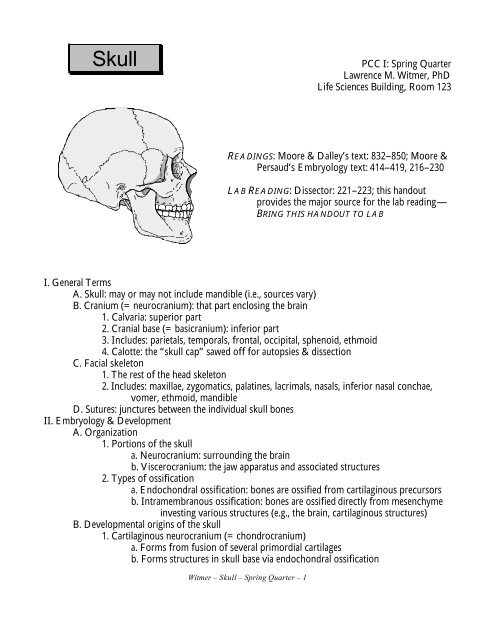
<strong>Skull</strong><br />
PCC I: Spring Quarter<br />
Lawrence M. Witmer, PhD<br />
Life Sciences Building, Room 123<br />
READINGS: Moore & Dalley’s text: 832–850; Moore &<br />
Persaud’s Embryology text: 414–419, 216–230<br />
LAB READING: Dissector: 221–223; this handout<br />
provides the major source for the lab reading—<br />
BRING THIS HANDOUT TO LAB<br />
I. General Terms<br />
A. <strong>Skull</strong>: may or may not include mandible (i.e., sources vary)<br />
B. Cranium (= neurocranium): that part enclosing the brain<br />
1. Calvaria: superior part<br />
2. Cranial base (= basicranium): inferior part<br />
3. Includes: parietals, temporals, frontal, occipital, sphenoid, ethmoid<br />
4. Calotte: the “skull cap” sawed off for autopsies & dissection<br />
C. Facial skeleton<br />
1. The rest of the head skeleton<br />
2. Includes: maxillae, zygomatics, palatines, lacrimals, nasals, inferior nasal conchae,<br />
vomer, ethmoid, mandible<br />
D. Sutures: junctures between the individual skull bones<br />
II. Embryology & Development<br />
A. Organization<br />
1. Portions of the skull<br />
a. Neurocranium: surrounding the brain<br />
b. Viscerocranium: the jaw apparatus and associated structures<br />
2. Types of ossification<br />
a. Endochondral ossification: bones are ossified from cartilaginous precursors<br />
b. Intramembranous ossification: bones are ossified directly from mesenchyme<br />
investing various structures (e.g., the brain, cartilaginous structures)<br />
B. Developmental origins of the skull<br />
1. Cartilaginous neurocranium (= chondrocranium)<br />
a. Forms from fusion of several primordial cartilages<br />
b. Forms structures in skull base via endochondral ossification<br />
Witmer – <strong>Skull</strong> – Spring Quarter – 1
c. Adult bones: occipital, petromastoid part of temporal bone, sphenoid,<br />
ethmoid<br />
2. Membranous neurocranium<br />
a. Forms bones around the brain via intramembranous ossification<br />
b. Adult bones: frontal, parietal<br />
3. Cartilaginous viscerocranium<br />
a. Forms from the cartilages of the first two branchial arches via endochondral<br />
ossification<br />
b. Adult first branchial arch bones: malleus and incus (middle ear ossicles)<br />
c. Adult second branchial arch bones: stapes (another middle ear ossicle), styloid<br />
process of temporal bone<br />
(note: the second branchial arch also contributes to the lesser horn and part of<br />
the body of the hyoid bone and the third branchial arch contributes the<br />
rest of the hyoid bone)<br />
4. Membranous viscerocranium<br />
a. Forms bones around the maxillary and mandibular prominences of the first<br />
branchial arch via intramembranous ossification<br />
b. Adult bones: premaxilla, maxilla, zygomatic, palatine, nasal, vomer, lacrimal,<br />
squamous part of temporal, mandible<br />
C. Many of the adult skull bones are compound bones representing fusions of more than one<br />
embryonic element<br />
1. Frontal: fusion of right and left elements<br />
2. Occipital: 4 ossification centers<br />
3. Temporal: 8 ossification centers<br />
4. Sphenoid: 14 ossification centers<br />
5. Ethmoid: 3 ossification centers<br />
6. Maxilla: 2 ossification centers<br />
D. Perinatal skull<br />
1. Proportions<br />
a. Large cranium and small face<br />
b. Face increases in relative size with growth<br />
(1). Eruption of teeth in the maxillae, premaxillae, & mandible<br />
(2). Development of paranasal sinuses in the maxilla, frontal, ethmoid,<br />
and sphenoid<br />
2. Fontanelles<br />
a. Allow molding of the head during parturition; molding also is facilitated by<br />
the flexibility of the bones (esp. the frontal)<br />
b. Six named fontanelles<br />
(1). Unpaired<br />
(a). Anterior fontanelle: juncture of frontals & parietals<br />
(b) Posterior fontanelle: juncture of parietals & occipital<br />
(2). Paired<br />
(a). Sphenoidal fontanelle: juncture of frontal, parietal, squamous<br />
part of temporal, & greater wing of sphenoid<br />
Witmer – <strong>Skull</strong> – Spring Quarter – 2
(b). Mastoid fontanelle: juncture of parietal, occipital, and<br />
petromastoid part of temporal<br />
E. Anomalies<br />
1. Cranioschisis: acrania (absence of the calvaria and often a large vertebral defect) +<br />
anencephaly (absence of most of the brain)<br />
2. Craniosynostosis: premature suture closure<br />
a. Scaphocephaly: sagittal suture closure: skull becomes long & narrow due to<br />
expansion in the frontal and occipital regions<br />
b. Acrocephaly (= oxycephaly, turricephaly, tower skull): coronal suture closure:<br />
skull becomes short and high<br />
c. Plagiocephaly: unilateral closure of lambdoid and coronal sutures: skull<br />
becomes asymmetrical<br />
III. Craniofacial Osteology: based on "views" rather than individual bones<br />
A. Anterior view (= Norma frontalis)<br />
1. Forehead: frontal bone<br />
2. Cheek prominence: zygomatic bone<br />
3. Nares (= anterior nasal aperture)<br />
a. Open posteriorly in the nasal cavity<br />
b. Bounded by nasals & maxillae (includes premaxillae, which fuse to maxillary<br />
bones early in ontogeny)<br />
4. Upper jaw (maxillae) and lower jaw (mandible): tooth-bearing elements<br />
5. Orbit<br />
a. Margin: frontal, maxilla, zygomatic<br />
b. Superior wall: orbital plate of frontal, sphenoid bones<br />
c. Medial wall: orbital lamina (lamina papyracea) of ethmoid bone, lacrimal,<br />
sphenoid, and frontal<br />
d. Inferior wall: mostly maxilla, also zygomatic & palatine<br />
e. Lateral wall: zygomatic bone and greater wing of sphenoid<br />
f. Openings within the orbit<br />
(1). Superior orbital fissure<br />
(a). Largely within sphenoid bone<br />
(b). Opens posteriorly into middle cranial fossa<br />
(c). Transmits: oculomotor n. (CN III), trochlear n. (CN IV),<br />
abducens n. (CN VI), branches of ophthalmic n. (CN V 1 ),<br />
superior ophthalmic v., sympathetics<br />
(2). Optic canal<br />
(a). Within sphenoid bone<br />
(b). Transmits optic n. (CN II), and ophthalmic a.<br />
(3). Supraorbital foramen (or notch)<br />
(a). Within frontal bone<br />
(b). Transmits: supraorbital vessels and nerve<br />
(4). Anterior & posterior ethmoidal foramina<br />
(a). Within or near suture between frontal & ethmoid bones<br />
(b). Transmits: anterior & posterior ethmoidal vessels & nerves<br />
Witmer – <strong>Skull</strong> – Spring Quarter – 3
(5). Nasolacrimal canal and groove<br />
(a). Between lacrimal and maxilla<br />
(b). Transmits: lacrimal sac and nasolacrimal duct<br />
(6). Inferior orbital fissure<br />
(a). Between greater wing of sphenoid, maxilla, and zygomatic,<br />
and a very small part of the palatine<br />
(b). Opens into the pterygopalatine fossa and thereby into the<br />
infratemporal and temporal regions—(i.e., the superior<br />
orbital fissure opens internally into the brain (cranial cavity)<br />
whereas the inferior orbital fissure opens externally into<br />
the temporal region)<br />
(c). Transmits: maxillary n. (CN V 2 ), infraorbital vessels, branches<br />
from the pterygopalatine ganglion<br />
6. Foramina for general somatic afferent (sensory) branches of trigeminal n. (CN V) and<br />
accompanying vessels: form a vertical line<br />
a. Supraorbital foramen (or notch)<br />
(1). Within frontal bone<br />
(2). Branches of ophthalmic division (CN V 1 )<br />
b. Infraorbital foramen<br />
(1). Within maxillary bone<br />
(2). Branches of maxillary division (CN V 2 )<br />
c. Mental foramen<br />
(1). Within mandible<br />
(2). Branches of mandibular division (CN V 3 )<br />
B. Posterior view (= Norma occipitalis)<br />
1. Cranial vault: formed by the parietal bones<br />
2. Occiput<br />
a. Formed mostly by occipital bone and a little of the temporal bone<br />
b. Area of muscle attachment<br />
(1). Highest (= supreme) nuchal line: attachment of epicranial<br />
aponeurosis (= galea aponeurotica), which is the aponeurosis<br />
between the frontal and occipital bellies of occipitofrontalis<br />
(2) Superior nuchal line: attachment of occipitofrontalis, trapezius,<br />
sternocleidomastoid, and splenius capitis<br />
(3). Between superior & inferior nuchal lines: semispinalis capitis and<br />
superior oblique (= obliquus capitis superior)<br />
(4). Inferior nuchal line: rectus major and rectus minor (= rectus capitis<br />
posterior major and minor)<br />
(5). External occipital protuberance: attachment of lig. nuchae<br />
(6). Mastoid process: attachment of sternocleidomastoid, splenius capitis,<br />
longissimus capitis, and posterior belly of digastric<br />
3. Sutures & landmarks<br />
a. Sagittal suture: between parietal bones<br />
b. Lambdoid suture: between occipital & parietals<br />
Witmer – <strong>Skull</strong> – Spring Quarter – 4
c. Lambda: point of intersection of sagittal & lambdoid sutures<br />
4. Sutural bones: presence is highly variable<br />
C. Superior view (= Norma verticalis)<br />
1. Dominated by cranial vault: frontal, parietals, occipital<br />
2. Sutures & landmarks<br />
a. Coronal suture: between frontal and parietals<br />
b. Sagittal suture: between parietal bones<br />
c. Lambdoid suture: between occipital & parietals<br />
d. Bregma: point of intersection of sagittal & coronal sutures<br />
e. Lambda: point of intersection of sagittal & lambdoid sutures<br />
3. Parietal foramina (= emissary v. foramina)<br />
a. Situated on either side of the sagittal suture<br />
b. Transmit emissary vv. from scalp to intracranial dural venous sinuses<br />
D. Ventral view (= Norma basalis)<br />
1. Hard palate<br />
a. Composed of palatine processes of maxillary bones and horizontal plates of<br />
palatine bones<br />
b. Teeth: 3 molars, 2 premolars, 1 canine, 2 incisors on each side<br />
c. Foramina<br />
(1). Within maxilla—incisive canal, fossa, and foramen: transmits<br />
nasopalatine nerve and sphenopalatine vessels<br />
(2). Within palatine<br />
(a). Greater palatine foramen: transmits greater palatine nerve and<br />
vessels<br />
(b). Lesser palatine foramen: transmits lesser palatine nerve and<br />
vessels<br />
d. Choana (= posterior nasal aperture)<br />
(1). Bounded by palatine, vomer, & sphenoid<br />
(2). Opening from nasal cavity into nasopharynx<br />
2. Cheekbone: zygomatic arch, formed by zygomatic bone & zygomatic part of<br />
temporal bone<br />
3. Jaw articulation: mandibular fossa<br />
4. Occiput (see above)<br />
5. Cranial base<br />
a. Composed of sphenoid, occipital, & temporal<br />
b. Occipital condyles: articulate with atlas vertebra (C1), forming<br />
atlantooccipital joint<br />
c. Site of attachment of numerous muscles that move the head on the neck, close<br />
& open the jaws, support the pharynx, & work the tongue<br />
(1). On pharyngeal tubercle of occipital bone: pharyngeal raphe (e.g.,<br />
superior constrictor)<br />
(2). On occipital bone anterior to occipital condyles: longus capitis,<br />
rectus capitis anterior<br />
(3). On occipital bone lateral to occipital condyles: rectus capitis lateralis<br />
Witmer – <strong>Skull</strong> – Spring Quarter – 5
(4). On temporal bone lateral to foramen lacerum: levator veli palatini<br />
(5). On sphenoid & temporal: tensor veli palatini<br />
(6). Between lateral & medial pterygoid plates of sphenoid: medial<br />
pterygoid<br />
(7). On external surface of lateral pterygoid plate of sphenoid: lateral<br />
pterygoid<br />
(8). Styloid process of the temporal: stylohyoid ligament, stylohyoid,<br />
styloglossus, stylopharyngeus<br />
d. Numerous foramina for nerves and vessels<br />
(1). Occipital bone<br />
(a). Foramen magnum: transmits spinal cord<br />
(b). Hypoglossal canal (= anterior condylar canal): within base of<br />
occipital condyle; transmits hypoglossal n. (CN XII)<br />
(c). Condylar fossa, canal, & foramen: variably present; transmits<br />
emissary v. from sigmoid sinus to vertebral vv.<br />
(2). Between occipital and petrous part of temporal: jugular fossa and<br />
foramen—transmits:<br />
(a). Superior bulb of internal jugular v., receiving sigmoid sinus<br />
(b). Glossopharyngeal n. (CN IX)<br />
(c). Vagus n. (CN X)<br />
(d). (Spinal) Accessory n. (CN XI)<br />
(e). Inferior petrosal sinus<br />
(3). Temporal bone<br />
(a). Carotid canal: internal carotid a. and carotid plexus<br />
(sympathetic nn.)<br />
(b). Stylomastoid foramen<br />
(i). Termination of facial canal for facial n. (CN VII)<br />
(ii). Located between styloid and mastoid processes<br />
(c). Some very small openings<br />
(i). Tympanic canaliculus: between carotid canal and<br />
jugular fossa; transmits tympanic branch of<br />
glossopharyngeal n. (CN IX)<br />
(ii). Mastoid canaliculus: within jugular fossa; transmits<br />
auricular branch of vagus n. (CN X)<br />
(iii). Petrotympanic fissure: transmits chorda tympani n.<br />
(4). Between petrous temporal and greater wing of sphenoid: bony<br />
portion of auditory tube<br />
(5). Sphenoid bone (greater wing)<br />
(a). Foramen ovale: transmits mandibular n. (CN V 3 )<br />
(b). Foramen spinosum: transmits middle meningeal vessels<br />
(branches of maxillary vessels)<br />
(6). Between occipital, sphenoid, and temporal—foramen lacerum:<br />
plugged with fibrocartilage in life such that nothing truly passes<br />
through it, the internal carotid a. passes over it superiorly<br />
Witmer – <strong>Skull</strong> – Spring Quarter – 6
E. Lateral view (= Norma lateralis)<br />
1. Cheekbone: processes from zygomatic and temporal<br />
2. Cranial vault: frontal, greater wing of sphenoid, temporal, parietal, occipital<br />
3. Ear opening: external acoustic meatus<br />
4. Mastoid and styloid processes<br />
5. Temporalis origin<br />
a. Superior temporal line: attachment of temporalis fascia<br />
b. Inferior temporal line: attachment of temporalis muscle<br />
6. Sutures & landmarks<br />
a. Coronal suture: between frontal and parietals<br />
b. Lambdoid suture: between occipital & parietals<br />
c. Squamosal suture: between squamous part of temporal & parietal<br />
d. Asterion: point of intersection of lambdoid & squamosal sutures<br />
e. Pterion<br />
(1). Point of intersection of frontal, parietal, sphenoid, & temporal<br />
(2). Clinically important because a branch of the middle meningeal a. runs<br />
in an internal calvarial groove that crosses pterion; fractures at<br />
pterion may tear the artery & result in extradural hematoma<br />
F. Internal surface of skullcap (= calotte)<br />
1. Sulcus for superior sagittal sinus (a dural venous sinus)<br />
2. Sulcus for middle meningeal vessels on parietal<br />
G. Cranial fossae<br />
1. General<br />
a. Internal, superior aspect of the skull<br />
b. Three fossae have a step-like arrangement such that each more posterior fossa<br />
is located more inferiorly<br />
c. Each supports a different portion of the brain<br />
(1). Anterior cranial fossa: frontal lobes<br />
(2). Middle cranial fossa: temporal lobes<br />
(3). Posterior cranial fossa: cerebellum, pons, medulla<br />
2. Anterior cranial fossa<br />
a. Formed mostly by frontal, also lesser wing of sphenoid and ethmoid<br />
b. Supports the frontal lobes of the cerebrum<br />
c. Brain markings on the bone form shallow, sinuous grooves; produced by<br />
convolutions (gyri) of the frontal lobe<br />
d. Ethmoid bone<br />
(1). Crista galli: a median plate of bone to which the falx cerebri (a<br />
median dural septum) attaches<br />
(2). Cribriform plate: overlain by olfactory bulb of brain<br />
e. Lesser wing of sphenoid bone<br />
(1). Overhangs the middle cranial fossa<br />
(2). Anterior clinoid processes: provides attachment to the tentorium<br />
cerebelli (a dural septum)<br />
f. Foramina<br />
Witmer – <strong>Skull</strong> – Spring Quarter – 7
(1). Foramina within cribriform plate: olfactory nerve fibers pierce<br />
foramina to enter olfactory bulb<br />
(2). Anterior & posterior ethmoidal foramina: for vessels and nerves of<br />
the same name<br />
(3). Foramen cecum: for emissary v. from nasal cavity to superior sagittal<br />
sinus; variable & relatively unimportant<br />
3. Middle cranial fossa<br />
a. Separated from:<br />
(1). Anterior cranial fossa by ridges from lesser wings<br />
(2). Posterior cranial fossa by dorsum sellae and superior border of<br />
petrous part of temporal<br />
b. Formed by sphenoid and temporal<br />
c. Supports temporal lobe of cerebrum<br />
d. Sella turcica<br />
(1). Saddle-shaped, central portion of sphenoid<br />
(2). Tuberculum sellae: anteriorly situated; divides hypophyseal area from<br />
optic nerve area<br />
(3). Hypophyseal fossa: for pituitary<br />
(4). Dorsum sellae: posteriorly situated<br />
(5). Posterior clinoid processes: at tips of dorsum sellae<br />
e. Foramina<br />
(1). Optic canals<br />
(a). Within sphenoid, just medial to anterior clinoid processes<br />
(b). Transmits: optic n., ophthalmic a.<br />
(2). Crescent of foramina within greater wing<br />
(a). Superior orbital fissure: four cranial nerves (CN III, IV, V 1 ,<br />
VI) & ophthalmic vv.<br />
(b). Foramen rotundum: maxillary n. (CN V 2 )<br />
(c). Foramen ovale: mandibular n. (CN V 3 ), accessory meningeal<br />
a.<br />
(d). Foramen spinosum: middle meningeal vessels; note: vessels<br />
enscribe a prominent groove within the middle cranial<br />
fossa<br />
(3). Foramen lacerum: located at apex of petrous temporal<br />
(a). Filled with fibrocartilage inferiorly<br />
(b). Superiorly, internal carotid a. and carotid plexus pass over<br />
(not through) the foramen<br />
(4). Greater petrosal hiatus and groove<br />
(a). Slit at posterolateral apex of foramen lacerum<br />
(b). Transmits greater petrosal n. & an artery (petrosal branch of<br />
middle meningeal a.)<br />
4. Posterior cranial fossa<br />
a. Formed mostly by occipital, also temporal and sphenoid<br />
b. Supports cerebellum, pons, and medulla<br />
Witmer – <strong>Skull</strong> – Spring Quarter – 8
c. Grooves for intracranial sinuses<br />
(1). Transverse sinus: right groove is usually larger because sagittal sinus<br />
dumps into it; tentorium cerebelli attaches on either side of the<br />
groove<br />
(2). Sigmoid sinus: from transverse sinus to jugular foramen<br />
d. Internal occipital crest: divides cerebellar fossae<br />
e. Internal occipital protuberance: attachment of dura<br />
f. Clivus: sloping portion of occipital leading to sphenoid: supports pons and<br />
medulla<br />
g. Foramina<br />
(1). Foramen magnum<br />
(a). Large, unpaired<br />
(b). Communication between cranial cavity & vertebral canal<br />
(c). Transmits: medulla, meninges, spinal roots of spinal accessory<br />
n. (CN XI), vertebral vessels, spinal vessels<br />
(2). Jugular foramen (see above, section III.D.5.d.(2))<br />
(3). Hypoglossal canal<br />
(a). Between jugular foramen and foramen magnum<br />
(b). Transmits hypoglossal n. (CN XII)<br />
(4). Condylar canal: variable; emissary v.<br />
(5). Internal acoustic meatus<br />
(a). Within petrous temporal<br />
(b). Transmits: facial n. (CN VII; including nervus intermedius),<br />
vestibulocochlear n (CN VIII), and vessels<br />
H. Mandible<br />
1. Ramus<br />
a. Condylar process (with head and neck): articulates with the mandibular fossa<br />
of the temporal bone<br />
b. Coronoid process: attachment of temporalis muscle<br />
c. Mandibular notch: separates condylar & coronoid<br />
d. Mandibular foramen<br />
(1). Entrance to mandibular canal<br />
(2). Transmits inferior alveolar nerves & vessels<br />
e. Lingula: attachment of sphenomandibular ligament<br />
f. Mylohyoid groove: for mylohyoid n. & vessels<br />
2. Body<br />
a. Alveolar process: tooth-bearing portion<br />
b. Mental foramen<br />
(1). Exit of mandibular canal<br />
(2). Transmits mental n. (CN V1) and vessels<br />
c. Mylohyoid line: attachment of mylohyoid muscle<br />
d. Mental symphysis, tubercles, and protuberance: chin<br />
e. Mental spine (= genial tubercles): attachment of genioglossus<br />
3. Angle: juncture of ramus & body; attachment of masseter & medial pterygoid<br />
Witmer – <strong>Skull</strong> – Spring Quarter – 9
SELF TEST<br />
1. Describe the differences between the cartilaginous and membranous neurocranium and<br />
viscerocranium. To which adult bones does each type contribute?<br />
2. Name the muscle “scars” on the occiput and provide at least two examples of muscle<br />
attachments for each.<br />
3. What are the sagittal, lambdoid, coronal, and squamosal sutures? Between which bones do<br />
they lie? What are the names of the landmarks that lie at their intersections?<br />
4. Why are fractures to the cranial base both so common and so serious?<br />
5. List the exits of all the cranial nerves and major blood vessels.<br />
SAMPLE QUESTION<br />
A helmet-less hockey player receives a hard blow to the temple from a slap-shot puck. In<br />
addition to a concussion, the individual shows signs of extradural hematoma. What is<br />
the most likely cause of this injury?<br />
A. The blow fractured the superior orbital fissure with resultant tearing of the superior<br />
ophthalmic veins<br />
B. The blow fractured the skull in the area of pterion with resultant tearing of the middle<br />
meningeal artery<br />
C. The blow fractured the cranial base with resultant tearing of the internal jugular vein<br />
D. The blow cracked the calvaria with resultant tearing of emissary veins<br />
E. The blow fractured the frontal bone with resultant tearing of the supraorbital vessels<br />
Witmer – <strong>Skull</strong> – Spring Quarter – 10
Witmer – <strong>Skull</strong> – Spring Quarter – 11
Witmer – <strong>Skull</strong> – Spring Quarter – 12
Witmer – <strong>Skull</strong> – Spring Quarter – 13
Witmer – <strong>Skull</strong> – Spring Quarter – 14
Witmer – <strong>Skull</strong> – Spring Quarter – 15
Witmer – <strong>Skull</strong> – Spring Quarter – 16
Witmer – <strong>Skull</strong> – Spring Quarter – 17
Witmer – <strong>Skull</strong> – Spring Quarter – 18
Witmer – <strong>Skull</strong> – Spring Quarter – 19
Witmer – <strong>Skull</strong> – Spring Quarter – 20


