- Page 1 and 2:
Th. Kahn, F. A. Jolesz, J. S. Lewin
- Page 3 and 4:
Welcome to the 8th Interventional M
- Page 5 and 6:
Symposium Chairman • Thomas Kahn,
- Page 7 and 8:
Session I Friday, September 24, 08:
- Page 9 and 10:
11:55 V-16 Visualization of ablatio
- Page 11 and 12:
Session V Saturday, September 25, 0
- Page 13 and 14:
12:00 V-44 Interventional MRI—vas
- Page 15 and 16:
04:25 V-59 Improved prostate-cancer
- Page 17 and 18:
P-10 Chemical shift-compensated hyb
- Page 19 and 20:
P-29 Passive navigation method for
- Page 21 and 22:
P-50 Comparison of MR imaging chara
- Page 23 and 24:
V-01 Anticipated highlights of the
- Page 25 and 26:
V-02 Interventional MRI systems rev
- Page 27 and 28:
MR-guided focused ultrasound system
- Page 29 and 30:
V-04 Optimal design for an intraope
- Page 31 and 32:
angiography suite with surgical cap
- Page 33 and 34:
V-06 Laser based laparoscopic liver
- Page 35 and 36:
V-07 Hybrid PRF-thermometry in the
- Page 37 and 38:
Figure 1. Magnitude images and temp
- Page 39 and 40:
≥1 signified tissue damage and th
- Page 41 and 42:
V-09 MRI guided cryoablation: in vi
- Page 43 and 44:
Figure 3. Expected iceball size and
- Page 45 and 46:
V-10 Continuous real-time MR thermo
- Page 47 and 48:
Figure 1. Pulse sequence diagram of
- Page 49 and 50:
References [1] M. Lepetit-Coiffé,
- Page 51 and 52:
direction. Rotation of the probe al
- Page 53 and 54:
V-13 MR guidance and thermal monito
- Page 55 and 56:
V-14 MR-guided radiofrequency ablat
- Page 57 and 58:
Figure 1. 3D-MR-Imaging during posi
- Page 59 and 60:
V-15 Clinical experience in univers
- Page 61 and 62:
Figure 3. RF ablation with real-tim
- Page 63 and 64:
Results The proposed DynCE analysis
- Page 65 and 66:
V-17 Cryoablation: rationale, techn
- Page 67 and 68:
V-18 MRI guided percutaneous cryoab
- Page 69 and 70:
V-19 MR image-guided percutaneous t
- Page 71 and 72:
Conclusion The use of a 1.5 T large
- Page 73 and 74:
Figure 1. Negative plain CT (a) as
- Page 75 and 76:
and most readily available method f
- Page 77 and 78:
V-22 Heart, cell therapy, and how t
- Page 79 and 80:
V-23 Intra-cerebral administration
- Page 81 and 82:
Figure 3. CED of a larger volume (~
- Page 83 and 84:
Results All PFOB-APA injection site
- Page 85 and 86:
The targeting efficiency depends pr
- Page 87 and 88:
Acknowledgement P. Vartholomeos wor
- Page 89 and 90:
tissue stimulation or can lead to b
- Page 91 and 92:
V-28 MR-guided high intensity focus
- Page 93 and 94:
V-29 In vivo MR acoustic radiation
- Page 95 and 96:
Conclusion High Intensity Focused U
- Page 97 and 98:
where ∠ 123 and ∠ 124 were the
- Page 99 and 100:
[4] Netter FH. Atlas of Human Anato
- Page 101 and 102:
Results The hybrid ARF-sensitive/PR
- Page 103 and 104:
V-32 Navigation techniques for MR-g
- Page 105 and 106:
V-33 Real-time scan plane selection
- Page 107 and 108:
V-34 Multi-touch enabled real time
- Page 109 and 110:
V-35 Near real-time MR imaging with
- Page 111 and 112:
Acknowledgement The authors would l
- Page 113 and 114:
Table 1 Puncture-to-target time (PT
- Page 115 and 116:
solution was then used to verify th
- Page 117 and 118:
Conclusion In conclusion, the new s
- Page 119 and 120:
(Juvenile OCD) or development of lo
- Page 121 and 122:
for calculation of the overall erro
- Page 123 and 124:
significance could only be demonstr
- Page 125 and 126:
V-42 Introduction of a new device f
- Page 127 and 128:
V-43 Robotics in MRI-guided therapy
- Page 129 and 130:
Haptics is a tactile feedback techn
- Page 131 and 132:
complications. For safe guidance of
- Page 133 and 134:
V-45 Developing guidelines for succ
- Page 135 and 136:
Figure 2. a: Reference GRASS axial
- Page 137 and 138:
A reference plate containing five C
- Page 139 and 140:
V-48 Initial evaluation of a novel
- Page 141 and 142:
Conclusion The use of a segmentable
- Page 143 and 144:
Results The nitinol-based self-expa
- Page 145 and 146:
V-50 TrueFISP based catheter tracki
- Page 147 and 148:
V-51 Real-time MRI at high spatial
- Page 149 and 150:
V-52 Real-time intravascular-coil b
- Page 151 and 152:
V-53 Augmented reality based MR ima
- Page 153 and 154:
Discussion Using this application,
- Page 155 and 156:
V-55 Simultaneous ultrasound/MRI mo
- Page 157 and 158:
Results During simultaneous US/MRI
- Page 159 and 160:
V-57 Role of 3D imaging with SPACE
- Page 161 and 162:
Figure 7. Coronal oblique view reco
- Page 163 and 164:
deformable registration. These prov
- Page 165 and 166:
V-59 Improved prostate-cancer stagi
- Page 167 and 168:
esult of imaging during severe moti
- Page 169 and 170:
V-60 MR-guided trans-perineal cryoa
- Page 171 and 172:
V-61 Preliminary experience with a
- Page 173 and 174:
The procedure has been successfully
- Page 175 and 176:
The accuracy of MRI alone in locali
- Page 177 and 178:
Poster Presentations 175
- Page 179 and 180:
Results In the all cases, the proce
- Page 181 and 182:
Figure 1. Image series obtained in
- Page 183 and 184:
phantom/cadaver, such that they cov
- Page 185 and 186:
P-04 Supine breast MRI: first steps
- Page 187 and 188:
The supine DCE images, which were a
- Page 189 and 190:
The temperature maps were obtained
- Page 191 and 192:
P-06 Fat temperature imaging based
- Page 193 and 194:
Discussion Separations of chemical
- Page 195 and 196:
Figure 2. Iterative approximation o
- Page 197 and 198:
Conclusion None of the mathematical
- Page 199 and 200:
capture all physically realistic va
- Page 201 and 202:
ablation gadolinium and T2 weighted
- Page 203 and 204:
Methods The extended hybrid method
- Page 205 and 206:
P-11 Chemically selective asymmetri
- Page 207 and 208:
phase images agreed within 0.15°C
- Page 209 and 210:
Figure 1. A 3D unspoiled gradient-r
- Page 211 and 212:
P-14 Evaluation of the thermal dose
- Page 213 and 214:
P-15 PRFS thermometry during radiof
- Page 215 and 216:
the ellipsoidal Gaussian source was
- Page 217 and 218:
P-16 Echo time optimization in mult
- Page 219 and 220:
20 20 20 40 a 60 40 b 60 40 c 60 20
- Page 221 and 222:
Results and Discussion For the refe
- Page 223 and 224:
P-18 First clinical experience with
- Page 225 and 226:
Conclusion The presented method all
- Page 227 and 228:
Figure 1. Non-interfered MR image o
- Page 229 and 230:
P-20 Laser-induced thermotherapy (L
- Page 231 and 232:
Figure 1. Series from left to right
- Page 233 and 234:
Trio a Tim system, Siemens AG, Germ
- Page 235 and 236:
P-23 Three-dimensional motion analy
- Page 237 and 238:
¦£ ¦¢ ¦¡ ¦¤ ¡¢£¢¡¤¥
- Page 239 and 240:
P-25 High-resolution MRI of RF lesi
- Page 241 and 242:
and fading away later, with fresh (
- Page 243 and 244:
where PATTERN_SIZE stands for the l
- Page 245 and 246:
P-27 Clinical experience with navig
- Page 247 and 248:
Conclusion The presented navigation
- Page 249 and 250:
traditionally has not been consider
- Page 251 and 252:
Figure 1. Custom-made navigation de
- Page 253 and 254:
P-31 Tailored interactive sequences
- Page 255 and 256:
without radiation as well as the ar
- Page 257 and 258: P-32 Imaging speed for MR guided pu
- Page 259 and 260: P-33 Localization accuracy and perf
- Page 261 and 262: References [1] M. Burl, G. A. Coutt
- Page 263 and 264: Results The radiologist was able to
- Page 265 and 266: P-35 Advanced scan-geometry plannin
- Page 267 and 268: fibrillation (AF) or flutter (AFL).
- Page 269 and 270: CNR Cement/Bone was 42 ± 4. By the
- Page 271 and 272: P-37 Online scan control using a st
- Page 273 and 274: P-38 Construction of a MR compatibl
- Page 275 and 276: P-39 Communication in intraoperativ
- Page 277 and 278: P-40 Development of a real-time int
- Page 279 and 280: P-41 Evaluation of accuracy and cli
- Page 281 and 282: Results All four needle insertions
- Page 283 and 284: Figure 2. Head-on (a) and lateral (
- Page 285 and 286: P-43 Esophageal imaging in vivo usi
- Page 287 and 288: Figure 2. Cross-sectional MR images
- Page 289 and 290: An MR-compatible 100pF-capacitor is
- Page 291 and 292: made up MR based on MR imaging para
- Page 293 and 294: P-46 Image-based correction of trac
- Page 295 and 296: Conclusion When the offset of track
- Page 297 and 298: segmentation tools. At the end of t
- Page 299 and 300: P-48 Automatic scan plane adjustmen
- Page 301 and 302: P-49 Physiological saline as a cont
- Page 303 and 304: deliveries. Nowadays in our practic
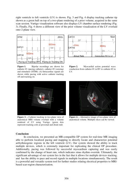
- Page 305 and 306: images (Figure 1). The mean signal
- Page 307: P-51 Integrated system for electrop
- Page 311 and 312: Fig. 1. Gold plated copper inductor
- Page 313 and 314: P-53 Coronary sinus extraction for
- Page 315 and 316: Results Our proposed method was eva
- Page 317 and 318: 1 2 3 4 5 5 4 3 2 1 Figure 1. 2D Fi
- Page 319 and 320: The HEFEWEIZEN sequence was segment
- Page 321 and 322: Figure 1. Left: 3T Pre-procedural T
- Page 323 and 324: image of the phantom, 5 target poin
- Page 325 and 326: catheter is connected to the extens
- Page 327 and 328: “billabong” leads with reversed
- Page 329 and 330: Fischbach, Frank Department of Radi
- Page 331 and 332: Kuroda, Kagayaki Graduate School of
- Page 333 and 334: Rump, Jens Department of Radiology
- Page 335 and 336: Weiss, Steffen Imaging Systems and
- Page 337 and 338: Flammang A V-09 Flask CA V-33 Foltz
- Page 339 and 340: R Razavi R V-47, V-48 Rempp H V-14