Transverse Nuclear Spin Relaxation in Phospholipid Bilayer ...
Transverse Nuclear Spin Relaxation in Phospholipid Bilayer ...
Transverse Nuclear Spin Relaxation in Phospholipid Bilayer ...

You also want an ePaper? Increase the reach of your titles
YUMPU automatically turns print PDFs into web optimized ePapers that Google loves.
2104 B IOC H E M IS T R Y<br />
ACCELERATED PUBLICATIONS<br />
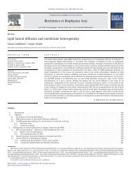
' t /<br />
?<br />
1400 -<br />
I- I of about 1.6 pm. In this section we mention briefly additional<br />
theoretical and experimental results which <strong>in</strong>dicate that lateral<br />
diffusion is, <strong>in</strong>deed, responsible for the long correlation times<br />
established by our experiments. These po<strong>in</strong>ts will be described<br />
more fully <strong>in</strong> a subsequent paper,<br />
We have developed a theory of transverse relaxation <strong>in</strong><br />
which the diffusion equation is explicitly solved for the lateral<br />
diffusion along a curved surface. Our calculation shows that<br />
the relaxation curve <strong>in</strong> the two-pulse qe experiment should be<br />
I I I I I I I I 1<br />
T2 (x10-8 s*c2)<br />
FIGURE 3: Initial slopes of the relaxation curves of Figure 2 vs. T ~ .<br />
The actual<br />
-<br />
values of T29mg are listed <strong>in</strong> Table I. The solid l<strong>in</strong>e is<br />
the result of a least-squares fit to eq 7, yield<strong>in</strong>g (1/Ti) = 1 /695 ps<br />
and T~ 100 ms.<br />
S<strong>in</strong>ce the sample has a distribution of values of R, AM2,<br />
and bilayer orientations, the nonexponential relaxations observed<br />
<strong>in</strong> Figure 2 are not unexpected. However, the <strong>in</strong>itial<br />
slopes of the relaxation curves of Figure 2 are exactly equal<br />
to ( 1 / T2*a), where (...) denotes the average over all of these<br />
parameters. A plot of these <strong>in</strong>itial slopes of the relaxation<br />
curves of Figure 2 vs. r2 is shown <strong>in</strong> Figure 3 to be l<strong>in</strong>ear as<br />
predicted by eq 5.<br />
S<strong>in</strong>ce the motions associated with the long correlation times<br />
are too slow to contribute to motional averag<strong>in</strong>g, they are only<br />
capable of gradually modulat<strong>in</strong>g the quadrupolar splitt<strong>in</strong>gs,<br />
which armnemmd <strong>in</strong> the 2H NMR spectrum. For spherical<br />
bilayers we may, therefore, identify ( AM2) with the "residual<br />
second moment", M2,, of the *H NMR spectrum (Bloom et<br />
al., 1978; Davis, 1979). Substitut<strong>in</strong>g eq 6 <strong>in</strong>to eq 5 and de-<br />
f<strong>in</strong><strong>in</strong>g an effective radius for diffusional relaxation, Reff, by<br />
RefC2 = (K2), we obta<strong>in</strong><br />
The l<strong>in</strong>ear plot <strong>in</strong> Figure 3 gives 2M2@/Reff2 = 93 X 10' s-~<br />
and ( 1 / Ti) = 1 /695 ps. For lipid bilayers hav<strong>in</strong>g a diffusion<br />
constant D i= 4 X 10-l2 m2 s-' (Bloom et al., 1978) and a value<br />
of M2r that we measured to be M2r = 3.0 X lo9 s-~, <strong>in</strong><br />
agreement with Davis (1979), we obta<strong>in</strong> Reff = 1.6 pm and,<br />
substitut<strong>in</strong>g <strong>in</strong>to eq 6, r2 = 100 ms. As noted by Hope et al.<br />
( 1986), multilamellar phospholipid dispersions prepared <strong>in</strong> the<br />
manner we have described "exhibit broad size distributions<br />
centered around diameters of a micron or more". An <strong>in</strong>dependent<br />
measurement of the average size of the dispersions<br />
<strong>in</strong> our DPPC sample by means of light scatter<strong>in</strong>g (Stern<strong>in</strong>,<br />
unpublished results) confirms this estimate.<br />
CONCLUSIONS<br />
We have demonstrated <strong>in</strong> this paper that 2H NMR transverse<br />
relaxation <strong>in</strong> DPPC bilayers is strongly <strong>in</strong>fluenced by<br />
molecular motions hav<strong>in</strong>g correlation times much longer than<br />
hundreds of microseconds. The most plausible candidate for<br />
such slow motions is diffusion of the phospholipid molecules<br />
along the curved membrane surfaces. This motion modulates<br />
the precession frequency <strong>in</strong> a random manner because the 2H<br />
NMR quadrupolar splitt<strong>in</strong>g is proportional to 3 cos2 8 - 1,<br />
where 8 is the angle between the local bilayer normal and the<br />
external magnetic field. Indeed, the theoretical results of<br />
Blicharski (1 986) fit the r2 dependence of the average CPMG<br />
relaxation rate for a plausible membrane radius of curvature<br />
I<br />
j<br />
nonexponential <strong>in</strong> analogy with the case of diffusion <strong>in</strong> an<br />
<strong>in</strong>homogeneous magnetic field (Abragam, 1961). It also<br />
confirms the use of eq 7 for the average relaxation rate, assum<strong>in</strong>g<br />
a s<strong>in</strong>gle correlation time model. This more general<br />
treatment predicts the dependence of the relaxation rate on<br />
bilayer orientation to be ( 1/T2q-Cpmg) c: s<strong>in</strong>2 8 cos2 8 at short<br />
times. Our unpublished results on specifically labeled lipid<br />
cha<strong>in</strong>s are <strong>in</strong> agreement with this prediction. We f<strong>in</strong>d that<br />
the <strong>in</strong>itial relaxation rate is slowest near the spectral edges of<br />
the 2H NMR powder spectra correspond<strong>in</strong>g to 8 = 90' and<br />
near the shoulders at 8 = 0'. Such an angular dependence<br />
has been observed empirically <strong>in</strong> 2tI-labeled lipids by Perly<br />
et al. (1985, see Figure 4). A similar behavior was observed<br />
for 2H NMR of heavy water, 2H20, <strong>in</strong> contact with membrane<br />
surfaces (Volke, 1984). In addition, our sample consists of<br />
31 2H nuclei per molecule, many of which have different<br />
quadrupolar splitt<strong>in</strong>gs; this results <strong>in</strong> an additional distribution<br />
of relaxation rates. A complicated superposition of nonexponential<br />
relaxation contributions leads <strong>in</strong> this case to the<br />
exponential behavior observed <strong>in</strong> the qe experiment.<br />
By means of the quadrupolar CPMG pulse tra<strong>in</strong> described<br />
here, it is now possible to separate the contributions of extremely<br />
long correlation times to transverse relaxation. Systematic<br />
study of the rema<strong>in</strong><strong>in</strong>g contribution, denoted by Ti<br />
<strong>in</strong> eq 5 and 7, will give <strong>in</strong>formation on the slowest of the<br />
conformational motions. This will be useful <strong>in</strong> the study of<br />
lipid-prote<strong>in</strong> <strong>in</strong>teractions. The methods used <strong>in</strong> this paper<br />
could also be applied to detect a wide class of slow motions<br />
<strong>in</strong> membranes, such as conformational changes <strong>in</strong> <strong>in</strong>tegral<br />
membrane prote<strong>in</strong>s or motions associated with lipids bound<br />
to such prote<strong>in</strong>s.<br />
ACKNOWLEDGMENTS<br />
We thank Prof. R. J. Cushley for provid<strong>in</strong>g the lipids used<br />
<strong>in</strong> this study, Dr. C. P. S. Tilcock for his help <strong>in</strong> prepar<strong>in</strong>g<br />
the samples, and U. Narger for her assistance with the light<br />
scatter<strong>in</strong>g measurements. We are grateful to J. C. Wallace<br />
for shar<strong>in</strong>g her data, and we thank her and Dr. A. L. MacKay<br />
for helpful discussions.<br />
REFERENCES<br />
Abragam, A. (1961) The Pr<strong>in</strong>ciples of <strong>Nuclear</strong> Magnetism,<br />
Oxford University Press, London.<br />
Bienvenue, A., Bloom, M., Davis, J. H., & Devaux, P. F.<br />
(1982) J. Biol. Chem. 257, 3032-3038.<br />
Blicharski, J. S. (1986) Can. J. Phys. 64, 733-735.<br />
Bloom, M., & Smith, I. C. P. (1985) <strong>in</strong> Progress <strong>in</strong> Prote<strong>in</strong>-<br />
Lipid Interactions (Watts, A., & DePont, J. J. H. H. M.,<br />
Eds.) pp 61-88, Elsevier, New York.<br />
Bloom, M., Burnell, E. E., MacKay, A. L., Nichol, C. P.,<br />
Valic, M. I., & Weeks, G. (1978) Biochemistry 17,<br />
5750-5 762.<br />
Brown, M. F. (1982) J. Chem. Phys. 77, 1576-1599.<br />
Brown, M. F. (1983) Proc. Natl. Acad. Sci. U.S.A. 80,<br />
4325-4329.<br />
Davis, J. H. (1979) Biophys. J. 27, 339-358.<br />
Davis, J. H. (1983) Biochim. Biophys. Acta 737, 117-171.