Basics of NMR spectroscopy
Basics of NMR spectroscopy
Basics of NMR spectroscopy

You also want an ePaper? Increase the reach of your titles
YUMPU automatically turns print PDFs into web optimized ePapers that Google loves.
<strong>Basics</strong> <strong>of</strong> <strong>NMR</strong> <strong>spectroscopy</strong><br />
Pr<strong>of</strong>. Klaus Ruhland<br />
N<br />
-<br />
+<br />
p<br />
p<br />
n<br />
+<br />
-<br />
S<br />
1
1. Introduction<br />
Nuclear magnetic resonance <strong>spectroscopy</strong> (<strong>NMR</strong> <strong>spectroscopy</strong>) is one <strong>of</strong> the most important<br />
characterisation methods in chemistry in particular for soluble low-molecular-weight<br />
compounds.<br />
History<br />
1940‘s first evidence for <strong>NMR</strong> signals (1945)<br />
1950‘s explanation <strong>of</strong> the influence on the chemical<br />
environment on the shift <strong>of</strong> <strong>NMR</strong> signals<br />
1952 Noble prize for Bloch/Purcell for developing<br />
<strong>NMR</strong> <strong>spectroscopy</strong><br />
1960‘s improvement <strong>of</strong> sensitivity by multiple<br />
measurements and Fourier-Transformation<br />
evaluation<br />
1970‘s improvement <strong>of</strong> resolution by application <strong>of</strong><br />
superconducting magnets yielding in higher field<br />
strengths<br />
1980‘s development <strong>of</strong> multidimensional methods<br />
1990‘s development <strong>of</strong> pulsed field gradient methods<br />
1991 Noble prize for R. Ernst for high-resolution <strong>NMR</strong><br />
2000‘s Coupling <strong>of</strong> <strong>NMR</strong> with chromatographic methods<br />
development <strong>of</strong> even higher fields <strong>of</strong> the magnets<br />
The detectable probe in this case is the magnetic dipole/multipole <strong>of</strong> the atomic nuclei, about<br />
the cause and existence <strong>of</strong> it we will talk first.<br />
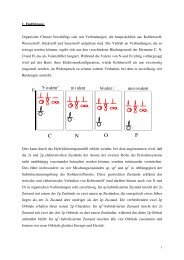
Atomic nuclei consist as is well known <strong>of</strong> protons and neutrons, from which one can at least<br />
as a model imagine, that they move around, held together by strong but short-ranging<br />
interactions and especially rotate (quantized) about their own axies. The accelerated<br />
movement <strong>of</strong> charges results in a magnetic field (Maxwell equations), so it will not come as a<br />
surprise, positively charged protons, when rotating about thier own axis (changes in<br />
orientation are also accelerations!) cause a magnetic field. In fact, both nucleons (protons and<br />
neutrons) contribute to the magnetic field <strong>of</strong> the atomic nucleus, which we are up to detect.<br />
This, in the case <strong>of</strong> the neutral neutrons, is not plausible in the first place, since no charge<br />
movement seems to be connected with the rotation about their own axis..<br />
2
N<br />
-<br />
+<br />
p<br />
p<br />
n<br />
+<br />
-<br />
S<br />
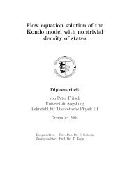
To better understand this, we must go a resolution step deeper and have a look, how protons<br />
and in particular neutrons themselves are composed <strong>of</strong>. Indeed, they consist each <strong>of</strong> 3 quarks (<br />
protons: up/up/down; neutrons: up/down/down). the up-quarks possess a charge <strong>of</strong> +⅔ and a<br />
spin <strong>of</strong> ½. The down-quarks possess a charge <strong>of</strong> -⅓ and also a spin <strong>of</strong> ½. If one assumes in a<br />
simplified manner, that the spins <strong>of</strong> u and d each act in opposite direction, on this basis one<br />
will predict for a proton (uud) the charge +1 and the spin ½. For the neutron (udd) one would<br />
expect a charge <strong>of</strong> 0 and a spin <strong>of</strong> ½. Both is in accordance with the experimental findings.<br />
Taking this into account we can easier accept, that also a neutron by rotation about its own<br />
axis contributes to the magnetic field, since the three charge carriers udd can be imagined to<br />
be anisotropically distributed in space, so that a self-rotation <strong>of</strong> neutrons with spin ½, if not<br />
connected with separated charges, is at least connected with the acceleration <strong>of</strong> electric<br />
dipoles.<br />
Particle Charge Spin Magnetic Moment<br />
(relative)<br />
Gyromagnetic<br />
constant γ<br />
10 -7 rad T -1 s -1<br />
Proton +1 ½ +1.6 26.8<br />
Neutron 0 ½ -1 -18.3<br />
Electron -1 ½ -1060 -17608<br />
The upper table compares the most decisive properties <strong>of</strong> the three „elemental“ particles <strong>of</strong><br />
matery. We realize, that the magnetic moment <strong>of</strong> neutrons and protons are approximately <strong>of</strong><br />
the same size and opposite in sign, whereas that <strong>of</strong> an electron is larger by a factor <strong>of</strong> 1000.<br />
The also listed gyromagnetic constant γ will be introduced and explained later in this text.<br />
3
It is <strong>of</strong> course not possible, to examine matery only consistent <strong>of</strong> nuclei. We always have to<br />
carry with us the electrons a neutralising kit. And concerning the magnetic properties, which<br />
we want to use to characterize the material, these obviously show a much more pronounced<br />
influence. In diamagnetic materials however the magnetic contributions <strong>of</strong> the electrons<br />
cancel out each other, since electrons with opposite spin each are present in the same amount<br />
in the sample, so in such samples we really can exclusively observe the (decisively smaller)<br />
magnetic behaviour <strong>of</strong> the nuclei. This means at the same time, that we have to expect<br />
complications for measurements <strong>of</strong> paramagnetic samples, which is indeed true.<br />
For the most important cases we can ignore the electron cloud (in first approximation; it will<br />
become important again later with a secondary effect, but not by its spin) and we can<br />
concentrate completely on the nucleus. In general several protons and neutrons will be sharing<br />
the nucleus. Which consequences will this have on the resulting magnetic moments <strong>of</strong> the<br />
nucleus?<br />
We will distinguish three different cases:<br />
1. Both the amount <strong>of</strong> protons and the amount <strong>of</strong> neutrons is even<br />
In this case the opposite spins <strong>of</strong> two neutrons and two protons each cancel out (Fermions ⇒<br />
Pauli-Prinziple). The complete spin <strong>of</strong> the nucleus I is zero, and there does not exist a<br />
resulting magnetic dipole/multipole. Nuclei, e. g. 12 C, for which this constellation is found,<br />
are called magic nuclei. For <strong>NMR</strong> <strong>spectroscopy</strong> they are inactive and therefor useless.<br />
2. Either the amount <strong>of</strong> protons or the amount <strong>of</strong> neutrons is uneven<br />
In this case the spins <strong>of</strong> one kind <strong>of</strong> particle cancel out each other, whereas for the other<br />
particle type with uneven amount at least one spin remains uncompensated. The interaction<br />
with the other existing spins however influences the complete nucleus spin. At least it can be<br />
predicted, that these nuclei will possess a half-numbered resulting nucleus spin I. If exactly<br />
one spin remains uncompensated, the nucleus spin is I=½. Nuclei <strong>of</strong> this kind are particularly<br />
attractive for <strong>NMR</strong> <strong>spectroscopy</strong>, because they do not possess a magnetic quadrupole<br />
moment, only a magnetic dipole moment (please just accept this as a matter <strong>of</strong> fact without<br />
explanation). Many nuclei important for organic materials, especially 1 H, 13 C, 19 F, 31 P belong<br />
to this group. Not at least because <strong>of</strong> this fact, <strong>NMR</strong> <strong>spectroscopy</strong> has gained its importance<br />
for chemists nowadays.<br />
3. Both the amount <strong>of</strong> protons and the amount <strong>of</strong> neutrons are uneven<br />
4
This constellation leads to whole-numbered complete nucleus spins I larger than zero. These<br />
will always possess a magnetic quadrupole moment (necessary and sufficient condition I>½),<br />
which makes the signal detection more difficult.<br />
5
2. Nuclei with spin I=½<br />
We will first concentrate on nuclei with I=½, because they are so important for <strong>NMR</strong><br />
<strong>spectroscopy</strong>. The following periodic system <strong>of</strong> the elements shows, which nuclei/isotopes we<br />
are talking about (some <strong>of</strong> the elements possess several isotopes with I=½. In the periodic<br />
system only that with the highest natural abundance is shown).<br />
Group<br />
1<br />
2<br />
3<br />
4<br />
5<br />
6<br />
7<br />
8<br />
9<br />
10<br />
11<br />
12<br />
13<br />
14<br />
15<br />
16<br />
17<br />
18<br />
Period<br />
The number below the element symbol represents the natural abundance <strong>of</strong> the Isotope with I=1/2 in %<br />
1<br />
1<br />
1 H<br />
100<br />
2<br />
He<br />
-<br />
2<br />
3<br />
Li<br />
-<br />
4<br />
Be<br />
-<br />
5<br />
B<br />
-<br />
6<br />
13 C<br />
1.1<br />
7<br />
15 N<br />
0.3<br />
8<br />
O<br />
-<br />
9<br />
19 F<br />
100<br />
10<br />
Ne<br />
-<br />
3<br />
11<br />
Na<br />
-<br />
12<br />
Mg<br />
-<br />
13<br />
Al<br />
-<br />
14<br />
29 Si<br />
4.7<br />
15<br />
31 P<br />
100<br />
16<br />
S<br />
-<br />
17<br />
Cl<br />
-<br />
18<br />
Ar<br />
-<br />
4<br />
19<br />
K<br />
-<br />
20<br />
Ca<br />
-<br />
21<br />
Sc<br />
-<br />
22<br />
Ti<br />
-<br />
23<br />
V<br />
-<br />
24<br />
Cr<br />
-<br />
25<br />
Mn<br />
-<br />
26<br />
57 Fe<br />
2.2<br />
27<br />
Co<br />
-<br />
28<br />
Ni<br />
-<br />
29<br />
Cu<br />
-<br />
30<br />
Zn<br />
-<br />
31<br />
Ga<br />
-<br />
32<br />
Ge<br />
33<br />
As<br />
-<br />
34<br />
Se<br />
-<br />
35<br />
Br<br />
-<br />
36<br />
Kr<br />
-<br />
5<br />
37<br />
Rb<br />
-<br />
38<br />
Sr<br />
-<br />
39<br />
89 Y<br />
100<br />
40<br />
Zr<br />
-<br />
41<br />
Nb<br />
-<br />
42<br />
Mo<br />
-<br />
43<br />
Tc<br />
-<br />
44<br />
Ru<br />
-<br />
45<br />
103 Rh<br />
100<br />
46<br />
Pd<br />
-<br />
47<br />
107 Ag<br />
51.8<br />
48<br />
111 Cd<br />
12.8<br />
49<br />
In<br />
-<br />
50<br />
119 Sn<br />
8.6<br />
51<br />
Sb<br />
-<br />
52<br />
125 Te<br />
7.0<br />
53<br />
I<br />
-<br />
54<br />
129 Xe<br />
26.4<br />
6<br />
55<br />
Cs<br />
-<br />
56<br />
Ba<br />
-<br />
*<br />
71<br />
Lu<br />
-<br />
72<br />
Hf<br />
-<br />
73<br />
Ta<br />
-<br />
74<br />
183 W<br />
14.4<br />
75<br />
Re<br />
-<br />
76<br />
187 Os<br />
1.6<br />
77<br />
Ir<br />
-<br />
78<br />
195 Pt<br />
33.8<br />
79<br />
Au<br />
-<br />
80<br />
Hg<br />
-<br />
81<br />
205 Tl<br />
70.5<br />
82<br />
207 Pb<br />
22.6<br />
83<br />
Bi<br />
-<br />
84<br />
Po<br />
-<br />
85<br />
At<br />
-<br />
86<br />
Rn<br />
-<br />
7<br />
87<br />
Fr<br />
-<br />
88<br />
Ra<br />
-<br />
*<br />
*<br />
103<br />
Lr<br />
-<br />
104<br />
Rf<br />
-<br />
105<br />
Db<br />
-<br />
106<br />
Sg<br />
-<br />
107<br />
Bh<br />
-<br />
108<br />
Hs<br />
-<br />
109<br />
Mt<br />
-<br />
110<br />
Ds<br />
-<br />
111<br />
Rg<br />
-<br />
112<br />
Uub<br />
-<br />
113<br />
Uut<br />
-<br />
114<br />
Uuq<br />
-<br />
115<br />
Uup<br />
-<br />
116<br />
Uuh<br />
-<br />
117<br />
Uus<br />
-<br />
118<br />
Uuo<br />
-<br />
*Lanthanoids<br />
*<br />
57<br />
La<br />
-<br />
58<br />
Ce<br />
-<br />
59<br />
Pr<br />
-<br />
60<br />
Nd<br />
-<br />
61<br />
Pm<br />
-<br />
62<br />
Sm<br />
-<br />
63<br />
Eu<br />
-<br />
64<br />
Gd<br />
-<br />
65<br />
Tb<br />
-<br />
66<br />
Dy<br />
-<br />
67<br />
Ho<br />
-<br />
68<br />
Er<br />
-<br />
69<br />
169 T<br />
m<br />
100<br />
70<br />
173 Yb<br />
14.3<br />
**Actinoids<br />
*<br />
*<br />
89<br />
Ac<br />
-<br />
90<br />
Th<br />
-<br />
91<br />
Pa<br />
-<br />
92<br />
U<br />
-<br />
93<br />
Np<br />
-<br />
94<br />
Pu<br />
-<br />
95<br />
Am<br />
-<br />
96<br />
Cm<br />
-<br />
97<br />
Bk<br />
-<br />
98<br />
Cf<br />
-<br />
99<br />
Es<br />
-<br />
100<br />
Fm<br />
-<br />
101<br />
Md<br />
-<br />
102<br />
No<br />
-<br />
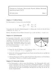
What happens, if an external magnetic field is applied to such kind <strong>of</strong> nucleus?<br />
For a nucleus with spin I=½ there exist only two allowed quantized states ±½ (in general:<br />
amount <strong>of</strong> allowed states = 2 I + 1). Without an external magnetic field these two states are<br />
degenerated, but the stronger the external applied magnetic field becomes, the more decisive<br />
6
the degeneration will be lost and an energy difference proportional to the external magnetic<br />
field will results between the two states.<br />
Energy<br />
Magn. field strength<br />
How strong the energy splitting between the two states proportional to the external magnetic<br />
field will be, depends on the proportionality constant γ, which is individual for each isotope:<br />
∆E = γ ħ B 0 = h ν<br />
This proportionality constant is the aforementioned gyromagnetic constant. The larger it is the<br />
more sensitive is the isotope for <strong>NMR</strong> spectroscopic examinations.<br />
7
The upper table summarizes the most decisive properties concerning <strong>NMR</strong> <strong>spectroscopy</strong> <strong>of</strong><br />
the most important isotopes.<br />
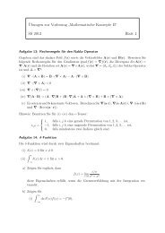
In <strong>NMR</strong> <strong>spectroscopy</strong> will be measured the transition between the allowed states (in case <strong>of</strong><br />
I=½ between the two states ±½), resulting from the external magnetic energy irradiation,<br />
which is transformed in an energy-dependent signal. This can be done in two ways. On the<br />
one hand the external magnetic field can be systematically changed on constant irradiation <strong>of</strong><br />
an amount <strong>of</strong> energy h ν and on the other hand the irradiated energy can be systematically<br />
changed at constant external magnetic field strength. In both cases the energy absorption by<br />
the sample results in a signal.<br />
Energy<br />
Energyabsorption<br />
Magn. field strength<br />
Energyabsorption<br />
Magn. field strength<br />
Energyabsorption<br />
Magn. field strength<br />
8
Energyabsorption<br />
Magn. field strength<br />
Energyabsorption<br />
Magn. field strength<br />
Because the external magnetic field should be as constant and homogeneous as possible the<br />
more appropriate method is, to keep the magnetic field unchanged and instead vary the<br />
irradiated energy.<br />
Frequency ν<br />
Energy<br />
Energyabsorption<br />
Magn. field strength<br />
9
Frequency ν<br />
Energy<br />
Energyabsorption<br />
Magn. field strength<br />
Frequency ν<br />
Energy<br />
Energyabsorption<br />
Magn. field strength<br />
Frequency ν<br />
Energy<br />
Energyabsorption<br />
Magn. field strength<br />
10
Frequency ν<br />
Energy<br />
Energyabsorption<br />
Magn. field strength<br />
The measurement is even more simplified nowadays by the fact, that the frequencies <strong>of</strong> the<br />
irradiated energy (brought about by a AC current device) are not stepwise applied, but all at<br />
once a broad band <strong>of</strong> frequencies are used. This is done by irradiation <strong>of</strong> an as short as<br />
possible current signal (the shorter the more frequencies) in an as long as possible periodic<br />
time distance (the longer the time distance the larger the frequency window, that is irradiated<br />
at once). From the temporal response <strong>of</strong> this at-once-irradiation <strong>of</strong> a broad frequency band the<br />
intensity <strong>of</strong> absorption as a function <strong>of</strong> the irradiating frequency can be received by Fouriertransform<br />
analysis (which we only mention here without further detail). This is then the <strong>NMR</strong><br />
spectrum.<br />
1 Frequenz gleichzeitig<br />
5 Frequenzen gleichzeitig<br />
1,5<br />
1<br />
0,5<br />
0<br />
0 1 2 3 4 5 6 7<br />
-0,5<br />
-1<br />
-1,5<br />
10<br />
8<br />
6<br />
4<br />
2<br />
0<br />
0 1 2 3 4 5 6 7<br />
-2<br />
-4<br />
-6<br />
-8<br />
-10<br />
10 Frequenzen gleichzeitig<br />
50 Frequenzen gleichzeitig<br />
25<br />
20<br />
15<br />
10<br />
5<br />
0<br />
0 1 2 3 4 5 6 7<br />
-5<br />
-10<br />
-15<br />
-20<br />
25<br />
20<br />
15<br />
10<br />
5<br />
0<br />
0 1 2 3 4 5 6 7<br />
-5<br />
-10<br />
-15<br />
-25<br />
-20<br />
11
100 Frequenzen gleichzeitig<br />
250 Frequenzen gleichzeitig<br />
80<br />
70<br />
60<br />
50<br />
40<br />
30<br />
20<br />
10<br />
0<br />
0<br />
-10<br />
1 2 3 4 5 6 7<br />
-20<br />
-30<br />
100<br />
80<br />
60<br />
40<br />
20<br />
0<br />
0 1 2 3 4 5 6 7<br />
-20<br />
-40<br />
The pulse length is in the range <strong>of</strong> µs.<br />
Let us discuss for a short while the energy window <strong>of</strong> <strong>NMR</strong> <strong>spectroscopy</strong>:<br />
Energy differencies in <strong>NMR</strong> <strong>spectroscopy</strong> (intensity <strong>of</strong> signals)<br />
Magnetic Energy<br />
∆E = h ν = ħγ B ˜ 4 10 -25 J<br />
Thermal Energy<br />
E = k T ˜ 4 10 -21 J<br />
∆E<br />
m=½<br />
∆E = h ν<br />
B<br />
n m=½<br />
- ∆E<br />
k T<br />
n =e ≈ 0.9999<br />
m=-½<br />
m=-½<br />
In fact the energy difference between the nucleus spin states is much smaller than thermal<br />
energy at room temperature. A Boltzmann distribution assumed, this means, that the<br />
distribution difference between the two states (I=½), which is the most important parameter<br />
for the sensitivity <strong>of</strong> the measurement, is very small. This explains the search for as strong as<br />
possible external magnetic fields, because the energy difference between the spin states grows<br />
proportional to this external magnetic field. At the moment the maximum is 900 MHz (21.15<br />
Tesla) and it is determined by the superconducting material <strong>of</strong> the coil, which provides the<br />
magnetic field, and its critical magnetic field strength, above which the superconducting<br />
properties are lost.<br />
Concerning the range <strong>of</strong> frequencies (E = h ν) <strong>NMR</strong> <strong>spectroscopy</strong> is a fairly „slow“<br />
spectroscopic method.<br />
12
Comparison <strong>of</strong> energies in <strong>spectroscopy</strong><br />
Electromagnetic Wave length Frequency Properties to be examined Spectroscopic method<br />
irradiation<br />
γ rays 100 pm – 1 pm 3 10 18 – 3 10 20 Hz Change <strong>of</strong> nuclear states γ-Spectroscopy, Mößbauer-<br />
Spectroscopy<br />
x-rays 10 nm – 100 pm 3 10 16 – 3 10 18 Hz Change <strong>of</strong> state <strong>of</strong> inner electron x-ray Spectroscopy<br />
core state<br />
UV light, visible light 1 µm – 10 nm 3 10 14 – 3 10 16 Hz Change <strong>of</strong> state <strong>of</strong> avlence UV Spectroscopy<br />
electrons<br />
Infrared rays 100 µm – 1 µm 3 10 12 – 3 10 14 Hz Change <strong>of</strong> vibrational<br />
IR/Raman Spectroscopy<br />
states<br />
Microwaves 1 cm – 100 µm 30 GHz – 3 10 12 Hz Change <strong>of</strong> rotational<br />
Microwave Spectroscopy<br />
states<br />
Microwaves 1m – 1 cm 300 MHz – 30 GHz Change <strong>of</strong> electron spin Electron spin resonance (ESR)<br />
states<br />
Radiowaves 100 m – 1 m 3 MHz – 300 MHz Change <strong>of</strong> nuclear spin<br />
states<br />
Nuclear spin resonance (<strong>NMR</strong>)<br />
This means, that on doing <strong>NMR</strong> spectroscopic examinations in several cases only a timeaveraged<br />
picture <strong>of</strong> the behaviour <strong>of</strong> matery in comparison with „faster“ methods as for<br />
instance IR <strong>spectroscopy</strong> with higher time resolution is detected.<br />
IR <strong>spectroscopy</strong><br />
13<br />
C-<strong>NMR</strong><br />
13
In the upper example for instance only one type <strong>of</strong> symmetry-equivalent carbons can be<br />
detected by <strong>NMR</strong> <strong>spectroscopy</strong> for the compound Co 2 (CO) 9 . The IR spectrum <strong>of</strong> the very<br />
same compound however shows, that this is only true time-averaged and in fact is based on an<br />
overlay <strong>of</strong> different isomers/conformers.<br />
Below is shown the schematic composition <strong>of</strong> an <strong>NMR</strong> machine.<br />
Let us have a look step by step, what happens during the measurement.<br />
A typical <strong>NMR</strong> sample tube for solvent <strong>NMR</strong> <strong>spectroscopy</strong> is shown in the upper right. This<br />
will be lifted, attached to a probe holder, by release <strong>of</strong> air pressure along the probe channel<br />
into the strong constant external magnetic field. The probe holder enables (as well by a stream<br />
<strong>of</strong> air), that the <strong>NMR</strong> sample tube rotates about its long axis. This contributes decisively to the<br />
fact, that the complete sample time-averaged experiences an as homogeneous magnetic field<br />
as possible. The rotation must be faster than the desired resolution in the spectrum. In general<br />
14
it is spinned at 20 Hz. As the next step one optimizes the emitter coil (which generates a<br />
magnetic field perpendicular to the external strong magnetic field, if a current pulse is<br />
applied) for the measurement. Two actions are implemented to homogenize the emitter<br />
magnetic field concerning time and space. The first action is called „Locking“. For it the<br />
deuterium signal <strong>of</strong> the <strong>NMR</strong> solvent is used as leading signal (that is the main reason why<br />
the <strong>NMR</strong> solvent is deuterated; also <strong>of</strong> course to hold free the 1 H window from disturbing<br />
signals), to guarantee the time-averaged constancy <strong>of</strong> the emitter magnetic field. The second<br />
action is called „Shimming“. On doing this the optimal orientation <strong>of</strong> the emitter coil is<br />
arranged. It is done by maximising the lock signal (again using the deuterium channel).<br />
The measurement can be started now!<br />
Perpendicular to the external strong magnetic field an emitter magnetic field is applied via DC<br />
coil to the sample.<br />
N<br />
B<br />
B'<br />
M<br />
emitter<br />
coil<br />
S<br />
receiver<br />
coil M=0<br />
N<br />
B'<br />
M<br />
emitter<br />
coil<br />
S<br />
rotating angle<br />
is dependent on<br />
the irradiation time<br />
and irradiation power<br />
receiver<br />
coil M>0<br />
N<br />
B'<br />
M<br />
emitter<br />
coil<br />
S<br />
The magnetisations <strong>of</strong> the sample that are in resonance with the irradiation <strong>of</strong> the emitter are<br />
rotated the more the longer the emitter coil is applied and the higher its power is. Ideal fort he<br />
sensitivity is a 90°-Puls, because in that case the magnetisation is exactly parallel to the<br />
receiver coil and thus induces the strongest magnetic field as signal.<br />
15
What happens as soon as the emitter pulse stops? In that case the system is no longer at its<br />
equilibrium state, because the magnetisation <strong>of</strong> the sample is perpendicular to the external<br />
magnetic field, without another magnetic field being operative (the emitter is <strong>of</strong>f). The<br />
equilibrium state a parrallel orientation <strong>of</strong> the sample magnetisation relative to the external<br />
magnetic field. The equilibrium state however cannot be reached instantaneously. A physical<br />
process, at which an equilibrium state is reached with time delay, is called relaxation. In<br />
<strong>NMR</strong> <strong>spectroscopy</strong> there exist two relaxation mechanisms:<br />
A. Longitudinale or Spin-Lattice-Relaxation<br />
Relaxation<br />
A. spin-lattice relaxation T 1 :<br />
receiver<br />
static magnetic field<br />
transmitter<br />
B z<br />
receiver<br />
T 1<br />
B y<br />
transmitter<br />
static magnetic field<br />
B z<br />
receiver<br />
B. spin-spin relaxation T 2 :<br />
transmitter<br />
B z<br />
B z<br />
T 2<br />
B z<br />
∆E ∆t ≥ h<br />
B y<br />
B y<br />
This mechanism describes the loss in sample magnetisation via interaction <strong>of</strong> the magnetic<br />
mlti(di)poles <strong>of</strong> the nucleus with other molecular magnetic fields in ist proximity (solvent<br />
molecules, other nclei (↑ towards →)).<br />
B. Transversale or Spin-Spin-Relaxation<br />
This mechanism explains the loss in sample magnetisation by a „Spin-Flipping“-Process (e.<br />
g.: ↑↓ → ↓↑), through which the preferred orientation <strong>of</strong> the spins along the receiver coil<br />
decreases, until no preferred orientation exists any more.<br />
The next diagram shows the time scale <strong>of</strong> <strong>NMR</strong> <strong>spectroscopy</strong> (spectral resolution) in relation<br />
to chemical and physical processes.<br />
16
Molecular<br />
vibration<br />
Dynamic<br />
Process<br />
Chemical<br />
exchange processes<br />
Molecular<br />
Rotation<br />
Macroscopic<br />
transport processes<br />
slow<br />
s ms µs ns ps fs<br />
fast<br />
Spectroscopic<br />
Finding<br />
slow<br />
exchange<br />
Longitudinal<br />
Magnetization exchange<br />
Line wideperturbation<br />
fast<br />
exchange<br />
The <strong>NMR</strong> time scale<br />
Relaxation<br />
time scale<br />
Spectral<br />
time scale<br />
Larmortime<br />
scale<br />
The <strong>NMR</strong> time scale which overlays conveniently with that <strong>of</strong> chemical exchange processes<br />
is another reason, why this characterization method is so important for chemists.<br />
17
3. The chemical shift<br />
Until now we understand, that, if a particular nucleus (e. g. H or C or P) is present in the<br />
material, we can detect it via <strong>NMR</strong> <strong>spectroscopy</strong> (as far as it is <strong>NMR</strong>-aktive).<br />
In fact the diagnostic information content <strong>of</strong> <strong>NMR</strong> <strong>spectroscopy</strong> is decisively higher, because<br />
the molecular electronic environment <strong>of</strong> the nucleus influences the resonance frequency as<br />
well. This influence is called chemical shift. Nuclei <strong>of</strong> one and the same isotope will show a<br />
magnetic transition at a very similar resonance frequency between the spin states, but within<br />
this window the direct electronic environment <strong>of</strong> the nucleus will lead to small changes in the<br />
resonance frequency. The chemical (electronic) environment acts thus as a perturbation <strong>of</strong> the<br />
resonance frequency <strong>of</strong> the bare nucleus. The physical reason is the magnetic Lorentz effect.<br />
Electron density near the nucleus is influenced by the external magnetic field in a way, that it<br />
generates a magnetic field that counteracts the external field. This screens the nucleus against<br />
the external field.<br />
Resonance<br />
frequency<br />
ν Α,Ref.<br />
bare nucleus A<br />
nucleus A<br />
with electron density<br />
e<br />
nucleus A<br />
with more electron density<br />
δ<br />
e<br />
chemical<br />
shift<br />
ν Β,Ref.<br />
bare nucleus B nucleus B<br />
with electron density<br />
e<br />
δ<br />
nucleus B<br />
with electron density<br />
e<br />
A nucleus, surrounded by large electron density, will be exited at a (slightly) higher resonance<br />
frequency excited in comparison to a nucleus <strong>of</strong> the same isotope, surrounded by a smaller<br />
electron density.<br />
The diagram below shows the different reasons, that contribute to the chemical shift.<br />
Intermolecular influences are (in contrast to solid-state-<strong>NMR</strong> <strong>spectroscopy</strong>) in case <strong>of</strong><br />
solution <strong>NMR</strong> <strong>spectroscopy</strong> <strong>of</strong> second order, because they cancel out in time via free rotation<br />
<strong>of</strong> the single molecules (which is faster than the spectral resolution).<br />
18
A quite special influence on the chemical shift is the ring-current effect, which is found in<br />
cyclic conjugated (aromatic) compounds.<br />
The protons in the periphery <strong>of</strong> the benzene ring are strongly deshielded by the ring-current<br />
effect (resonance frequency decisively smaller than for other protons, e. g. those bound to a<br />
non-cyclic conjugated double bond). The chemical shift is measured in ppm according to the<br />
equation<br />
δ = (ν A, Ref. – ν A ) / ν A,Ref.<br />
The ppm (parts per million) scale stems from the fact, that the difference between ν A and<br />
ν A,Ref . is in the range <strong>of</strong> Hz, while ν A,Ref. itself has a value <strong>of</strong> MHz.<br />
19
The upper two diagrams show typical chemical shifts in 1 H (left) and 13 C (right) <strong>NMR</strong><br />
<strong>spectroscopy</strong> for well-established organic functionalities.<br />
4. Scalar Spin-Spin coupling<br />
Let us go one step further, since <strong>NMR</strong> <strong>spectroscopy</strong> contains even more information than<br />
mentioned so far. Not only the type <strong>of</strong> nucleus and its electronic environment, but also the<br />
neighboring nucleus spins influence the appearance <strong>of</strong> the <strong>NMR</strong> spectrum, the latter by the<br />
multiplicity <strong>of</strong> the signal.<br />
In contrast to solid-state-<strong>NMR</strong> <strong>spectroscopy</strong> in case <strong>of</strong> solution <strong>NMR</strong> <strong>spectroscopy</strong> only those<br />
nucleus spin interactions contribute, that are mediated by a chemical bond (shared electron<br />
density). Magnetically dipolar couplings through spatial proximity time-average to zero in<br />
solution.<br />
Let us have a look on the situation in formaldehyde H 2 C=O. 16 O and 12 C, the most abundant<br />
isotopes <strong>of</strong> these two elements, are magic nuclei with spin 0 and thus <strong>NMR</strong>-inactive. We can<br />
thus concentrate in the following only on the two protons. So let us have a look, how this<br />
constellation will develop in the 1 H-<strong>NMR</strong> spectrum.<br />
20
Spectrum:<br />
ν 0 ν 1 ν 1<br />
β(β)<br />
H<br />
β<br />
β<br />
β(α)<br />
C<br />
O<br />
H<br />
ν 0 ν 1<br />
ν 1<br />
ν 1<br />
α<br />
α<br />
α(β)<br />
α(α)<br />
bare nucleus<br />
Chemical<br />
shift<br />
by surrounding<br />
electrons<br />
Chemical<br />
shift<br />
additional by<br />
neighboring spins<br />
The protons experience an electronic screening through the neighboring electrons <strong>of</strong> the COgroup<br />
(chemical shift). Additionally the spins feel each other, which also leads to shielding or<br />
deshielding. Starting from the two energy states for a pare proton now four energy states<br />
result. Only transitions with ∆I=1 are allowed. This selection rule stems from the fact , that on<br />
a transition a photon is absorbed. Photons possess a spin <strong>of</strong> 1; this means, that on absorption<br />
the moment <strong>of</strong> inerta <strong>of</strong> the system changes by –1. Since the moment <strong>of</strong> inerta must be<br />
retained, the loss must be compensated for an allowed transition; in our case by a change <strong>of</strong><br />
the spin nucleus <strong>of</strong> +1.<br />
It might seem so, that for formaldehyde one should expect two signals in the 1 H <strong>NMR</strong><br />
spectrum instead <strong>of</strong> only one. Butt his is not the case, because the two allowed transitions are<br />
brought about by the same energy h ν and thus appear at the same position in the spectrum.<br />
This is only the case, because the two protons are symmetry-equivalent (remember:<br />
symmetry-equivalent nuclei do not couple with each other!). The situation changes already in<br />
Aceto-chlorimin H 2 C=NCl. Now the symmetry-inequivalent nuclei couple with each other<br />
additionally to the chemical shift. This coupling splits <strong>of</strong> the signals fort he two protons each<br />
to a doublet. The scalar coupling is transferred via bonding electrons. The size <strong>of</strong> the coupling<br />
is therefore indicative for the electronic situation <strong>of</strong> the bond. For example one can deduce the<br />
degree <strong>of</strong> hybridization <strong>of</strong> a C-H bond by means <strong>of</strong> the 1 J 1H/13C -coupling constant.<br />
21
Spectrum:<br />
ν 0<br />
ν 1 ν 2 ν 1 ν 2 ν 1a/b ν 2a/b<br />
β<br />
β(β)<br />
β(α)<br />
β@β<br />
β@α<br />
H<br />
Cl<br />
β<br />
β<br />
β(β)<br />
β(α)<br />
β@β<br />
β@α<br />
C<br />
N<br />
H<br />
ν<br />
ν 2b<br />
ν 2<br />
ν 2 ν 0 ν 1 ν 1b<br />
1 ν 2a<br />
ν 2 ν 1 ν 1a<br />
α<br />
α<br />
α(β)<br />
α(α)<br />
α@β<br />
α@α<br />
bare nucleus<br />
α<br />
chemical<br />
shift<br />
by surrounding<br />
electrons<br />
α(β)<br />
α(α)<br />
chemical<br />
shift<br />
by neighboring spins<br />
α@β<br />
α@α<br />
additional active coupling<br />
between<br />
neighboring sins<br />
If two nuclei interact with each other by magnetic coupling, the visible splitting <strong>of</strong> the signals<br />
must bet he same for both nuclei (J ab = J ba ). The following diagram shows the continuous<br />
transition starting from an A 2 -system (both nuclei symmetry-equivalent) to an AX-System<br />
(active coupling between the two nuclei).<br />
In general it holds:<br />
3 J > 1 J >> 2 J<br />
The higher number expresses, how many bonds there are between the actively coupling<br />
nuclei.<br />
22
The splitting pattern in organic compounds <strong>of</strong> a proton CH-X, where X ist he group as shown<br />
below, shows in dependence <strong>of</strong> the number <strong>of</strong> bound protons k to the neighboring C-atom a<br />
characteristic behaviour (multiplicity: (k + 1); try to make this plausible to you yourself!):<br />
The splitting follows concerning the multiplicity and intensity <strong>of</strong> the single multiplett-signals<br />
a Pascal’s triangle.<br />
For the 3 J-coupling the Karplus-equation holds:<br />
3 J = A cos 2 (θ) + b cos(θ) + c<br />
θ is the torsional angle between bond one and three.<br />
23
5. Line broadening<br />
We have by now collected quite a bit <strong>of</strong> information power, connected with <strong>NMR</strong><br />
<strong>spectroscopy</strong>, but still there is more to come. Additionally to the position <strong>of</strong> the peak in the<br />
spectrum (electronic environment), the intensity <strong>of</strong> the peak (amount <strong>of</strong> the nucleus in a<br />
specific environment) and the multiplicity <strong>of</strong> the peak (spin environment <strong>of</strong> the nucleus) the<br />
line-broadening <strong>of</strong> the signal is another valuable diagnostic criterion, since it tells us<br />
something about the life-time <strong>of</strong> a state.<br />
Is in particular a chemical exchange between two positions in the spectral time scale <strong>of</strong> <strong>NMR</strong><br />
<strong>spectroscopy</strong> (1-1000 Hz), this will be detectable by line-broadening, from which (also<br />
quantitatively) the rate <strong>of</strong> this exchange can be determined. In general at modern machines<br />
there is the possibility, to cool and heat the probe head (from about -110°C, caused by the<br />
melting point <strong>of</strong> the well-established <strong>NMR</strong> solvents up to 110°C, caused by the stability limit<br />
<strong>of</strong> the probe head material (tubing etc.)), so that by temperature-dependent measurement <strong>of</strong><br />
the line-broadening very conveniently activation parameters can be determined.<br />
Line broadening (Chemical exchange)<br />
The upper example demonstrates, how the line-broadening <strong>of</strong> the CH 2 -group with temperature<br />
changes. At low temperatures only two doublets are visible (why?), which at higher<br />
temperatures step by step develop into a singlet as time-averaged signal, because at elevated<br />
temperatures the back- and for-vibration <strong>of</strong> the CH 2 -groups becomes so fast, That on timeaverage<br />
the appear equivalent.<br />
25
In summary the last diagram shows once again, which conclusions can be drawn from a 1-<br />
dimensional solution <strong>NMR</strong> spectrum.<br />
Information content <strong>of</strong> a 1D <strong>NMR</strong> spectrum<br />
Line width <strong>of</strong> the signal:<br />
life time <strong>of</strong> the spin state<br />
in the specific environment<br />
Intensity <strong>of</strong> the signal:<br />
amount <strong>of</strong> nuclei with<br />
specific environment<br />
Fine structure <strong>of</strong> the signal:<br />
neighboring nuclear spins<br />
Position <strong>of</strong> the signal:<br />
electronic environment <strong>of</strong> the nucleus<br />
26
Which compound might this be?<br />
27