

Principles of MRI - Department of Radiology
Principles of MRI - Department of Radiology
Principles of MRI - Department of Radiology

Create successful ePaper yourself
Turn your PDF publications into a flip-book with our unique Google optimized e-Paper software.
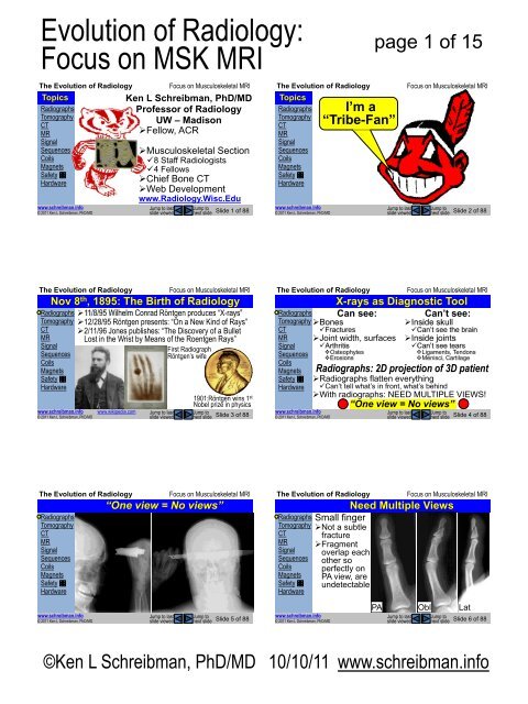
Evolution <strong>of</strong> <strong>Radiology</strong>:<br />
Focus on MSK <strong>MRI</strong><br />
The Evolution <strong>of</strong> <strong>Radiology</strong><br />
Topics<br />
Radiographs<br />
Tomography<br />
CT<br />
MR<br />
Signal<br />
Sequences<br />
Coils<br />
Magnets<br />
Safety<br />
Hardware<br />
www.schreibman.info<br />
© 2011 Ken L Schreibman, PhD/MD<br />
Jump to last<br />
slide viewed<br />
Focus on Musculoskeletal <strong>MRI</strong><br />
Ken L Schreibman, PhD/MD<br />
Pr<strong>of</strong>essor <strong>of</strong> <strong>Radiology</strong><br />
UW – Madison<br />
‣Fellow, ACR<br />
‣Musculoskeletal Section<br />
8 Staff Radiologists<br />
4 Fellows<br />
‣Chief Bone CT<br />
‣Web Development<br />
www.<strong>Radiology</strong>.Wisc.Edu<br />
Jump to<br />
next slide<br />
Slide 1 <strong>of</strong> 88<br />
The Evolution <strong>of</strong> <strong>Radiology</strong><br />
Topics<br />
Radiographs<br />
Tomography<br />
CT<br />
MR<br />
Signal<br />
Sequences<br />
Coils<br />
Magnets<br />
Safety<br />
Hardware<br />
www.schreibman.info<br />
© 2011 Ken L Schreibman, PhD/MD<br />
I’m a<br />
Schreibman<br />
“Tribe-Fan”<br />
page 1 <strong>of</strong> 15<br />
Jump to last<br />
slide viewed<br />
Focus on Musculoskeletal <strong>MRI</strong><br />
Jump to<br />
next slide<br />
Slide 2 <strong>of</strong> 88<br />
The Evolution <strong>of</strong> <strong>Radiology</strong><br />
Radiographs ‣11/8/95 Wilhelm Conrad Röntgen produces “X-rays”<br />
Tomography ‣12/28/95 Röntgen presents: “On a New Kind <strong>of</strong> Rays”<br />
CT ‣2/11/96 Jones publishes: “The Discovery <strong>of</strong> a Bullet<br />
MR<br />
Lost in the Wrist by Means <strong>of</strong> the Roentgen Rays”<br />
Signal<br />
First Radiograph<br />
Sequences<br />
Röntgen’s wife<br />
Coils<br />
Magnets<br />
Safety<br />
Hardware<br />
www.schreibman.info<br />
© 2011 Ken L Schreibman, PhD/MD<br />
Jump to last<br />
slide viewed<br />
Focus on Musculoskeletal <strong>MRI</strong><br />
Nov 8 th , 1895: The Birth <strong>of</strong> <strong>Radiology</strong><br />
www.wikipedia.com<br />
1901:Röntgen wins 1 st<br />
Nobel prize in physics<br />
Jump to<br />
next slide Slide 3 <strong>of</strong> 88<br />
The Evolution <strong>of</strong> <strong>Radiology</strong><br />
Radiographs<br />
Tomography<br />
CT<br />
MR<br />
Signal<br />
Sequences<br />
Coils<br />
Magnets<br />
Safety<br />
Hardware<br />
www.schreibman.info<br />
© 2011 Ken L Schreibman, PhD/MD<br />
Jump to last<br />
slide viewed<br />
Focus on Musculoskeletal <strong>MRI</strong><br />
X-rays as Diagnostic Tool<br />
Can see: Can’t see:<br />
‣Inside skull<br />
Can’t see the brain<br />
‣Inside joints<br />
Can’t see tears<br />
Ligaments, Tendons<br />
Menisci, Cartilage<br />
‣Bones<br />
Fractures<br />
‣Joint width, surfaces<br />
Arthritis<br />
Osteophytes<br />
Erosions<br />
Radiographs: 2D projection <strong>of</strong> 3D patient<br />
‣Radiographs flatten everything<br />
Can’t tell what’s in front, what’s behind<br />
‣With radiographs: NEED MULTIPLE VIEWS!<br />
“One view = No views”<br />
Jump to<br />
next slide<br />
Slide 4 <strong>of</strong> 88<br />
The Evolution <strong>of</strong> <strong>Radiology</strong><br />
Radiographs<br />
Tomography<br />
CT<br />
MR<br />
Signal<br />
Sequences<br />
Coils<br />
Magnets<br />
Safety<br />
Hardware<br />
Focus on Musculoskeletal <strong>MRI</strong><br />
“One view = No views”<br />
The Evolution <strong>of</strong> <strong>Radiology</strong><br />
Radiographs<br />
Tomography<br />
CT<br />
MR<br />
Signal<br />
Sequences<br />
Coils<br />
Magnets<br />
Safety<br />
Hardware<br />
Focus on Musculoskeletal <strong>MRI</strong><br />
Need Multiple Views<br />
Small finger<br />
‣Not a subtle<br />
fracture<br />
‣Fragment<br />
overlap each<br />
other so<br />
perfectly on<br />
PA view, are<br />
undetectable<br />
www.schreibman.info<br />
© 2011 Ken L Schreibman, PhD/MD<br />
Jump to last<br />
slide viewed<br />
Jump to<br />
next slide<br />
Slide 5 <strong>of</strong> 88<br />
www.schreibman.info<br />
© 2011 Ken L Schreibman, PhD/MD<br />
PA Obl Lat<br />
Jump to last<br />
slide viewed<br />
Jump to<br />
next slide<br />
Slide 6 <strong>of</strong> 88<br />
©Ken L Schreibman, PhD/MD 10/10/11 www.schreibman.info
1995<br />
1996<br />
1997<br />
1998<br />
1999<br />
2000<br />
2001<br />
2002<br />
2003<br />
2004<br />
2005<br />
2006<br />
2007<br />
2008<br />
2009<br />
2010<br />
2011<br />
2012<br />
Evolution <strong>of</strong> <strong>Radiology</strong>:<br />
Focus on MSK <strong>MRI</strong><br />
The Evolution <strong>of</strong> <strong>Radiology</strong><br />
Radiographs<br />
Tomography<br />
CT<br />
MR<br />
Signal<br />
Sequences<br />
Coils<br />
Magnets<br />
Safety<br />
Hardware<br />
PA<br />
www.schreibman.info<br />
© 2011 Ken L Schreibman, PhD/MD<br />
Jump to last<br />
slide viewed<br />
Focus on Musculoskeletal <strong>MRI</strong><br />
For Joints: Need 3 Views!<br />
PIP<br />
Obl<br />
PIP<br />
Lat<br />
Jump to<br />
next slide<br />
PIP<br />
Slide 7 <strong>of</strong> 88<br />
The Evolution <strong>of</strong> <strong>Radiology</strong><br />
Radiographs<br />
Tomography<br />
CT<br />
MR<br />
Signal<br />
Sequences<br />
Coils<br />
Magnets<br />
Safety<br />
Hardware<br />
www.schreibman.info British Journal <strong>of</strong> <strong>Radiology</strong><br />
© 2011 Ken L Schreibman, PhD/MD<br />
8:733-751,1935<br />
page 2 <strong>of</strong> 15<br />
Jump to last<br />
slide viewed<br />
Focus on Musculoskeletal <strong>MRI</strong><br />
Tomography: Small Step Forward<br />
To overcome<br />
flat 2D nature <strong>of</strong><br />
radiographs…<br />
‣ Structures in the<br />
Focal Plane <br />
are in focus.<br />
‣ Structures out <strong>of</strong><br />
focal plane are<br />
blurred out.<br />
‣ At best, we got<br />
blurry pictures.<br />
‣ Long exposures<br />
= high radiation.<br />
1935<br />
Grossmann<br />
Tomograph<br />
Can’t use<br />
this to see<br />
the brain <br />
Focal Plane<br />
Film<br />
Jump to<br />
next slide<br />
Slide 8 <strong>of</strong> 88<br />
The Evolution <strong>of</strong> <strong>Radiology</strong><br />
Radiographs<br />
Tomography<br />
CT<br />
MR<br />
Signal<br />
Sequences<br />
Coils<br />
Magnets<br />
Safety<br />
Hardware<br />
Focus on Musculoskeletal <strong>MRI</strong><br />
CT: Giant Leap Forward<br />
CT: Computed Tomography (Tomo [Gr]: part, slice)<br />
CAT: Computed Axial Tomography<br />
1917 Johann Radon, Austrian mathematician, proved<br />
image <strong>of</strong> a 3D object could be reconstructed from an<br />
infinite number <strong>of</strong> 2D projection images <strong>of</strong> the object.<br />
‣ Had to await the advent <strong>of</strong> mainframe<br />
computers in the 1970’s.<br />
The Evolution <strong>of</strong> <strong>Radiology</strong><br />
Focus on Musculoskeletal <strong>MRI</strong><br />
Hounsfield & EMI Brain Scanner<br />
Radiographs 1972: Godfrey Hounsfield, a British electrical engineer<br />
Tomography at EMI Laboratories, developed EMI Brain Scanner.<br />
CT<br />
‣Finally, could see through the skull into the brain!<br />
Awarded Nobel Prize for Medicine 1979; Knighted 1981.<br />
MR<br />
“Hounsfield Units” is the scale we use to measure CT density.<br />
Signal ‣ EMI: “Electric and Musical Industries”<br />
Sequences<br />
Coils<br />
Magnets<br />
Safety<br />
Hardware<br />
www.schreibman.info<br />
© 2011 Ken L Schreibman, PhD/MD<br />
www.wikipedia.com<br />
Jump to last<br />
slide viewed<br />
Jump to<br />
next slide<br />
Slide 9 <strong>of</strong> 88<br />
www.schreibman.info www.sciencemuseum.org.uk<br />
© 2011 Ken L Schreibman, PhD/MD<br />
Jump to last<br />
slide viewed<br />
Jump to<br />
next slide<br />
Slide 10 <strong>of</strong> 88<br />
The Evolution <strong>of</strong> <strong>Radiology</strong><br />
Radiographs<br />
Tomography<br />
CT<br />
MR<br />
Signal<br />
Sequences<br />
Coils<br />
Magnets<br />
Safety<br />
Hardware<br />
www.schreibman.info<br />
© 2011 Ken L Schreibman, PhD/MD<br />
Jump to last<br />
slide viewed<br />
Focus on Musculoskeletal <strong>MRI</strong><br />
Why CT is So Great<br />
Can see the brain<br />
‣Strokes, bleeds, tumors<br />
Can see organs (lungs, liver, bowel)<br />
‣Tumors, trauma, acute/chronic diseases<br />
Can see fractures otherwise missed<br />
‣Cervical spine, pelvis<br />
And now with ultra-fast, multi-slice…<br />
‣Can scan the heart in a single beat!<br />
Can see coronary arteries, pulmonary emboli<br />
Hospitals have CT scanners in the ER<br />
Jump to<br />
next slide<br />
Slide 11 <strong>of</strong> 88<br />
The Evolution <strong>of</strong> <strong>Radiology</strong><br />
Radiographs<br />
Tomography<br />
CT<br />
MR<br />
Signal<br />
Sequences<br />
Coils<br />
Magnets<br />
Safety<br />
Hardware<br />
25%<br />
20%<br />
15%<br />
10%<br />
5%<br />
0%<br />
www.schreibman.info<br />
© 2011 Ken L Schreibman, PhD/MD<br />
Jump to last<br />
slide viewed<br />
Focus on Musculoskeletal <strong>MRI</strong><br />
CT Usage Increasing in ERs<br />
Percentage <strong>of</strong> patients seen in US ERs who get CT<br />
30%<br />
26%<br />
National Trends in CT Use in the<br />
22%<br />
Emergency <strong>Department</strong>: 1995-2007<br />
20%<br />
presented at RSNA 11/29/2010 17%<br />
15%<br />
David B. Larson, MD, MBA et. al.<br />
Extrapolated<br />
3% 3% 3% 4% 5% 5% 6% 7% 8% 9% 10% 11% 13% Data<br />
Sample Data<br />
radiology.rsna.org<br />
Jump to<br />
next slide<br />
Slide 12 <strong>of</strong> 88<br />
©Ken L Schreibman, PhD/MD 10/10/11 www.schreibman.info
Evolution <strong>of</strong> <strong>Radiology</strong>:<br />
Focus on MSK <strong>MRI</strong><br />
The Evolution <strong>of</strong> <strong>Radiology</strong><br />
Radiographs<br />
Tomography<br />
CT<br />
MR<br />
Signal<br />
Sequences<br />
Coils<br />
Magnets<br />
Safety<br />
Hardware<br />
www.schreibman.info<br />
© 2011 Ken L Schreibman, PhD/MD<br />
Jump to last<br />
slide viewed<br />
Focus on Musculoskeletal <strong>MRI</strong><br />
Biggest Problem with CT<br />
High radiation dose<br />
We are exposed to low levels <strong>of</strong> radiation<br />
every day, “Background Radiation”<br />
‣Earth: naturally occurring radionuclides<br />
Uranium-238, potassium-40<br />
‣Atmosphere: Radon-222 (from U-238)<br />
2 nd leading cause <strong>of</strong> lung cancer after smoking<br />
‣Space: cosmic rays<br />
Airline crews, who spend a lot <strong>of</strong> time in the upper<br />
atmosphere, receive 2x typical background dose.<br />
Ave background dose ≈ 2.4mSv/year<br />
www.wikipedia.com<br />
Jump to<br />
next slide<br />
Slide 13 <strong>of</strong> 88<br />
The Evolution <strong>of</strong> <strong>Radiology</strong><br />
Radiographs<br />
Tomography<br />
CT<br />
MR<br />
Signal<br />
Sequences<br />
Coils<br />
Magnets<br />
Safety<br />
Hardware<br />
www.schreibman.info<br />
© 2011 Ken L Schreibman, PhD/MD<br />
page 3 <strong>of</strong> 15<br />
Jump to last<br />
slide viewed<br />
Focus on Musculoskeletal <strong>MRI</strong><br />
Radiation from Diagnostic Imaging<br />
Ave background dose ≈ 2.4mSv/year<br />
Chest Radiograph ≈ 0.06mSv<br />
‣≈1 week <strong>of</strong> background radiation<br />
Chest CT ≈ 7.0mSv<br />
‣≈3 YEARS <strong>of</strong> background radiation<br />
How much radiation is too much?<br />
Who the heck knows…<br />
Health Physics Society<br />
Jump to<br />
next slide<br />
Slide 14 <strong>of</strong> 88<br />
The Evolution <strong>of</strong> <strong>Radiology</strong><br />
Radiographs<br />
Tomography<br />
CT<br />
MR<br />
Signal<br />
Sequences<br />
Coils<br />
Magnets<br />
Safety<br />
Hardware<br />
www.schreibman.info<br />
© 2011 Ken L Schreibman, PhD/MD<br />
Jump to last<br />
slide viewed<br />
Focus on Musculoskeletal <strong>MRI</strong><br />
Other Problems with CT<br />
Usually requires IV contrast<br />
‣1% patients are allergic to CT contrast<br />
‣Can affect renal function<br />
Costs more than radiographs<br />
‣Knee radiographs (4 views): $154<br />
‣Knee CT (no contrast): $1,200<br />
Can’t see structures inside joints<br />
‣Knee: Menisci, Ligaments, Cartilage<br />
‣Shoulder: Rotator Cuff, Labrum<br />
‣Spine: Disks, Spinal Cord<br />
Jump to<br />
next slide<br />
Slide 15 <strong>of</strong> 88<br />
The Evolution <strong>of</strong> <strong>Radiology</strong><br />
Radiographs<br />
Tomography<br />
CT<br />
MR<br />
Signal<br />
Sequences<br />
Coils<br />
Magnets<br />
Safety<br />
Hardware<br />
www.schreibman.info<br />
© 2011 Ken L Schreibman, PhD/MD<br />
Jump to last<br />
slide viewed<br />
Focus on Musculoskeletal <strong>MRI</strong><br />
<strong>MRI</strong>: Giant Leap Sideways<br />
<strong>MRI</strong> doesn’t rely on X-rays to see<br />
projected shadows <strong>of</strong> patients<br />
‣Unlike radiographs, tomography, CT -<br />
<strong>MRI</strong> sees tissues based upon<br />
sub-atomic characteristics<br />
‣Proton nucleus <strong>of</strong> Hydrogen<br />
“NMR”<br />
‣“Nuclear Magnetic Resonance”<br />
‣“No More Radiologists”<br />
<strong>MRI</strong><br />
‣“Magnetic Resonance Imaging”<br />
Jump to<br />
next slide<br />
+<br />
Slide 16 <strong>of</strong> 88<br />
The Evolution <strong>of</strong> <strong>Radiology</strong><br />
Radiographs<br />
Tomography<br />
CT<br />
MR<br />
Signal<br />
Sequences<br />
Coils<br />
Magnets<br />
Safety<br />
Hardware<br />
Focus on Musculoskeletal <strong>MRI</strong><br />
<strong>MRI</strong> Scanner: 2 Components<br />
THE THE MAGNET COIL<br />
N<br />
S<br />
The Evolution <strong>of</strong> <strong>Radiology</strong><br />
Radiographs<br />
Tomography<br />
CT<br />
MR<br />
Signal<br />
Sequences<br />
Coils<br />
Magnets<br />
Safety<br />
Hardware<br />
Focus on Musculoskeletal <strong>MRI</strong><br />
<strong>MRI</strong> Scanner: 2 Components<br />
THE THE MAGNET COIL : Jobs<br />
Radio<br />
Frequency<br />
Transmitter<br />
Radio<br />
Frequency<br />
Receiver<br />
www.schreibman.info<br />
© 2011 Ken L Schreibman, PhD/MD<br />
Jump to last<br />
slide viewed<br />
Jump to<br />
next slide<br />
Slide 17 <strong>of</strong> 88<br />
www.schreibman.info<br />
© 2011 Ken L Schreibman, PhD/MD<br />
Jump to last<br />
slide viewed<br />
Jump to<br />
next slide<br />
Slide 18 <strong>of</strong> 88<br />
©Ken L Schreibman, PhD/MD 10/10/11 www.schreibman.info
Evolution <strong>of</strong> <strong>Radiology</strong>:<br />
Focus on MSK <strong>MRI</strong><br />
The Evolution <strong>of</strong> <strong>Radiology</strong><br />
Radiographs<br />
Tomography<br />
CT<br />
MR<br />
Signal<br />
Sequences<br />
Coils<br />
Magnets<br />
Safety<br />
Hardware<br />
www.schreibman.info<br />
© 2011 Ken L Schreibman, PhD/MD<br />
Jump to last<br />
slide viewed<br />
Focus on Musculoskeletal <strong>MRI</strong><br />
How MR Scanner Works<br />
Magnet<br />
‣Aligns spins <strong>of</strong> protons in hydrogen nuclei<br />
Align in direction <strong>of</strong> magnetic field, B 0<br />
Coil<br />
1)Sends RF pulse to flip spinning protons<br />
After RF pulse is <strong>of</strong>f, protons realign to B 0<br />
As protons realign, resonate RF energy<br />
2)Measures strength <strong>of</strong> resonant RF echo<br />
At a specific time, T E , “Echo Time”<br />
Steps 1&2 repeated many times / image slice<br />
At a specific “Repetition Time”, T R<br />
Jump to<br />
next slide<br />
Slide 19 <strong>of</strong> 88<br />
The Evolution <strong>of</strong> <strong>Radiology</strong><br />
page 4 <strong>of</strong> 15<br />
Radiographs<br />
Specific tissue types<br />
Tomography have specific resonant echoes (T1, T2)<br />
CT<br />
depending upon specified T R & T E<br />
MR ‣ Fluid (Hydrogen protons in H 2 O)<br />
Signal Cysts<br />
Sequences Joint effusions<br />
Edema (in s<strong>of</strong>t tissues, in bone marrow)<br />
Coils<br />
‣ Fat (Hydrogen protons in fat)<br />
Magnets Sub-cutaneous fat<br />
Safety Fatty yellow bone marrow<br />
Hardware ‣ Dense Stuff (with few Hydrogen protons)<br />
Cortical bone<br />
Ligaments, tendons<br />
Menisci<br />
www.schreibman.info<br />
© 2011 Ken L Schreibman, PhD/MD<br />
<br />
Key to <strong>MRI</strong><br />
Jump to last<br />
slide viewed<br />
Focus on Musculoskeletal <strong>MRI</strong><br />
Jump to<br />
next slide<br />
Slide 20 <strong>of</strong> 88<br />
The Evolution <strong>of</strong> <strong>Radiology</strong><br />
Radiographs<br />
High<br />
Tomography<br />
CT<br />
MR<br />
Signal<br />
Sequences Signal<br />
Coils<br />
Magnets<br />
Safety<br />
Hardware<br />
Low<br />
www.schreibman.info<br />
© 2011 Ken L Schreibman, PhD/MD<br />
Jump to last<br />
slide viewed<br />
Focus on Musculoskeletal <strong>MRI</strong><br />
T1 Recovery Curve (T R ~500ms)<br />
Fat<br />
10ms<br />
Fluid<br />
Dense<br />
Stuff<br />
T1-weighted image<br />
(T R : short, T E : short)<br />
‣Fat: High<br />
‣Fluid: Low<br />
‣Dense Stuff: Low<br />
Time to Echo T E (ms)<br />
Jump to<br />
next slide Slide 21 <strong>of</strong> 88<br />
The Evolution <strong>of</strong> <strong>Radiology</strong><br />
Radiographs<br />
Tomography<br />
CT<br />
MR<br />
Signal<br />
Sequences<br />
Coils<br />
Magnets<br />
Safety<br />
Hardware<br />
High<br />
Signal<br />
Low<br />
www.schreibman.info<br />
© 2011 Ken L Schreibman, PhD/MD<br />
Dense<br />
Stuff<br />
80ms<br />
Jump to last<br />
slide viewed<br />
Focus on Musculoskeletal <strong>MRI</strong><br />
T2 Decay Curve (T R ~2,000ms)<br />
T2-weighted image<br />
(T R : long, T E : long)<br />
‣Fluid: Intermed.<br />
‣Fat: Intermediate<br />
‣Dense Stuff: Low<br />
Fluid To increase separation <strong>of</strong><br />
fluid from fat, can apply<br />
“Fat-Suppression”<br />
(“Fat-Saturation”)<br />
Fat<br />
Time to Echo T E (ms)<br />
Jump to<br />
next slide Slide 22 <strong>of</strong> 88<br />
The Evolution <strong>of</strong> <strong>Radiology</strong><br />
Radiographs<br />
High<br />
Tomography<br />
CT<br />
MR<br />
Signal<br />
Sequences Signal<br />
Coils<br />
Magnets<br />
Safety<br />
Hardware<br />
Low<br />
www.schreibman.info<br />
© 2011 Ken L Schreibman, PhD/MD<br />
Dense<br />
Stuff<br />
80ms<br />
Jump to last<br />
slide viewed<br />
Focus on Musculoskeletal <strong>MRI</strong><br />
Fat-Sat T2 Decay Curve (T R ~2,000ms)<br />
It is always preferable to<br />
suppress the fat on T2<br />
to increase fluid<br />
conspicuity.<br />
Inversion Recovery<br />
(IR, STIR) is<br />
equivalent<br />
T2-weighted image<br />
(T R : long, T E : long)<br />
‣Fluid: Intermed.<br />
‣Fat: Low (suppressed)<br />
‣Dense Stuff: Low<br />
Fluid To increase separation <strong>of</strong><br />
fluid from fat, can apply<br />
“Fat-Suppression”<br />
(“Fat-Saturation”)<br />
Fat<br />
Time to Echo T E (ms)<br />
Jump to<br />
next slide Slide 23 <strong>of</strong> 88<br />
The Evolution <strong>of</strong> <strong>Radiology</strong><br />
Radiographs<br />
Tomography<br />
CT<br />
MR<br />
Signal<br />
Sequences<br />
Coils<br />
Magnets<br />
Safety<br />
Hardware<br />
www.schreibman.info<br />
© 2011 Ken L Schreibman, PhD/MD<br />
Dense<br />
Stuff<br />
Fluid<br />
80ms<br />
Jump to last<br />
slide viewed<br />
Focus on Musculoskeletal <strong>MRI</strong><br />
Fat-Sat T2 Decay Curve (T R ~2,000ms)<br />
Compress<br />
signal<br />
scale<br />
<br />
<br />
<br />
<br />
<br />
High<br />
Signal<br />
Low<br />
T2-weighted image<br />
(T R : long, T E : long)<br />
‣Fluid: High (relatively)<br />
‣Fat: Low (suppressed)<br />
‣Dense Stuff: Low<br />
Fat<br />
Time to Echo T E (ms)<br />
Jump to<br />
next slide Slide 24 <strong>of</strong> 88<br />
©Ken L Schreibman, PhD/MD 10/10/11 www.schreibman.info
Evolution <strong>of</strong> <strong>Radiology</strong>:<br />
Focus on MSK <strong>MRI</strong><br />
The Evolution <strong>of</strong> <strong>Radiology</strong><br />
Radiographs<br />
Tomography<br />
CT<br />
MR<br />
Signal<br />
Sequences<br />
Coils<br />
Magnets<br />
Safety<br />
Hardware<br />
www.schreibman.info<br />
© 2011 Ken L Schreibman, PhD/MD<br />
Jump to last<br />
slide viewed<br />
Focus on Musculoskeletal <strong>MRI</strong><br />
How We Make MR Images<br />
Magnetic field divides body into slices<br />
Each slice is divided into “voxels”<br />
‣voxel: 3D pixel<br />
‣voxel size = 2D pixel size X slice thickness<br />
Coil measures signal in each voxel<br />
Computer maps this onto 2D slices<br />
‣High signal: White (“Bright”)<br />
‣Intermediate signal: Gray (“Iso-intense”)<br />
‣Low signal: Black (“Dark”)<br />
Jump to<br />
next slide<br />
Slide 25 <strong>of</strong> 88<br />
Radiographs<br />
Tomography<br />
CT<br />
MR<br />
Signal<br />
Sequences<br />
Coils<br />
Magnets<br />
Safety<br />
Hardware<br />
Sub<br />
Q<br />
www.schreibman.info<br />
© 2011 Ken L Schreibman, PhD/MD<br />
page 5 <strong>of</strong> 15<br />
The Evolution <strong>of</strong> <strong>Radiology</strong><br />
Focus on Musculoskeletal <strong>MRI</strong><br />
Knee, same<br />
Comparing Sequences mid-sagittal slice<br />
T1 T2 T2fs<br />
T R =400<br />
T R =2500<br />
T R =2500<br />
T E =10ms<br />
T E =80ms<br />
T E =80ms<br />
Cyst<br />
Bone<br />
Marrow<br />
Fat<br />
Pad<br />
Fluid:<br />
Low<br />
Fat:<br />
High<br />
Cyst<br />
Bone<br />
Marrow<br />
Fat<br />
Pad<br />
Sub<br />
Q<br />
Fluid:<br />
High<br />
Fat:<br />
Inter<br />
Jump to last<br />
slide viewed<br />
Cyst<br />
Jump to<br />
next slide<br />
Bone<br />
Marrow<br />
Fat<br />
Pad<br />
Sub<br />
Q<br />
Fluid:<br />
High<br />
Fat:<br />
Low<br />
Slide 26 <strong>of</strong> 88<br />
The Evolution <strong>of</strong> <strong>Radiology</strong><br />
Radiographs<br />
High<br />
Tomography<br />
CT<br />
MR<br />
Signal<br />
Sequences Signal<br />
Coils<br />
Magnets<br />
Safety<br />
Hardware<br />
Low<br />
www.schreibman.info<br />
© 2011 Ken L Schreibman, PhD/MD<br />
Dense<br />
Stuff<br />
80ms<br />
Jump to last<br />
slide viewed<br />
Focus on Musculoskeletal <strong>MRI</strong><br />
T2 Decay Curve (T R ~2,000ms)<br />
“Proton<br />
Density”<br />
Poor separation<br />
fat from fluid<br />
20ms<br />
Fluid<br />
PD-weighted image<br />
(T R : long, T E : short)<br />
‣Fluid: Intermed.<br />
‣Fat: Intermediate<br />
‣Dense Stuff: Low<br />
Great separation <strong>of</strong><br />
the Dense Stuff from<br />
fluid & fat<br />
Fat<br />
Time to Echo T E (ms)<br />
Jump to<br />
next slide Slide 27 <strong>of</strong> 88<br />
The Evolution <strong>of</strong> <strong>Radiology</strong><br />
Focus on Musculoskeletal <strong>MRI</strong><br />
Knee, same<br />
Comparing Sequences mid-sagittal slice<br />
T1 PD T2fs<br />
T R =400<br />
T R =2500<br />
T R =2500<br />
T E =10ms<br />
T E =20ms<br />
T E =80ms<br />
Radiographs<br />
Tomography<br />
CT<br />
MR<br />
Signal<br />
Sequences<br />
Coils<br />
Magnets<br />
Safety<br />
Hardware<br />
Cyst<br />
Fluid:<br />
Low<br />
www.schreibman.info<br />
© 2011 Ken L Schreibman, PhD/MD<br />
Dense<br />
Stuff:<br />
Low<br />
Menisci<br />
Fat:<br />
High<br />
Cyst<br />
Fluid:<br />
Inter<br />
Dense<br />
Stuff:<br />
Low<br />
Menisci<br />
Fat:<br />
Inter<br />
Jump to last<br />
slide viewed<br />
Cyst<br />
Fluid:<br />
High<br />
Jump to<br />
next slide<br />
Dense<br />
Stuff:<br />
Low<br />
Menisci<br />
Fat:<br />
Low<br />
Slide 28 <strong>of</strong> 88<br />
The Evolution <strong>of</strong> <strong>Radiology</strong><br />
Radiographs<br />
Tomography<br />
CT<br />
MR<br />
Signal<br />
Sequences<br />
Coils<br />
Magnets<br />
Safety<br />
Hardware<br />
www.schreibman.info<br />
© 2011 Ken L Schreibman, PhD/MD<br />
Jump to last<br />
slide viewed<br />
Focus on Musculoskeletal <strong>MRI</strong><br />
<strong>MRI</strong>: Need Multiple Sequences<br />
T1 shows Fat best<br />
‣Most normal anatomy surrounded by fat<br />
‣In essence, T1 shows anatomy best<br />
T2 shows Fluid best<br />
‣Most pathology contains fluid (edema)<br />
‣In essence, T2 shows pathology best<br />
Fat-suppression makes fluid more conspicuous<br />
PD shows Dense Stuff best<br />
‣Good for meniscal and tendon tears<br />
‣Used mostly for <strong>MRI</strong> <strong>of</strong> joint pain<br />
Jump to<br />
next slide<br />
Slide 29 <strong>of</strong> 88<br />
The Evolution <strong>of</strong> <strong>Radiology</strong><br />
Radiographs<br />
Tomography<br />
CT<br />
MR<br />
Signal<br />
Sequences<br />
Coils<br />
Magnets<br />
Safety<br />
Hardware<br />
www.schreibman.info<br />
© 2011 Ken L Schreibman, PhD/MD<br />
Jump to last<br />
slide viewed<br />
Focus on Musculoskeletal <strong>MRI</strong><br />
Limitations <strong>of</strong> <strong>MRI</strong><br />
Limited Field <strong>of</strong> View (FOV)<br />
Image resolution related to voxel size<br />
‣Smaller FOV = smaller voxels<br />
‣Smaller voxels = higher resolution<br />
‣To maximize resolution, try to limit FOV<br />
Can only image inside the coil<br />
‣Requires an assortment <strong>of</strong><br />
different coils for different body parts<br />
Jump to<br />
next slide<br />
Slide 30 <strong>of</strong> 88<br />
©Ken L Schreibman, PhD/MD 10/10/11 www.schreibman.info
Evolution <strong>of</strong> <strong>Radiology</strong>:<br />
Focus on MSK <strong>MRI</strong><br />
The Evolution <strong>of</strong> <strong>Radiology</strong><br />
Radiographs<br />
Tomography<br />
CT<br />
MR<br />
Signal<br />
Sequences<br />
Coils<br />
Magnets<br />
Safety<br />
Hardware<br />
Knee Coil<br />
Focus on Musculoskeletal <strong>MRI</strong><br />
The Evolution <strong>of</strong> <strong>Radiology</strong><br />
Radiographs<br />
Tomography<br />
CT<br />
MR<br />
Signal<br />
Sequences<br />
Coils<br />
Magnets<br />
Safety<br />
Hardware<br />
page 6 <strong>of</strong> 15<br />
Focus on Musculoskeletal <strong>MRI</strong><br />
Knee Coil for the Ankle<br />
www.schreibman.info<br />
© 2011 Ken L Schreibman, PhD/MD<br />
Jump to last<br />
slide viewed<br />
Jump to<br />
next slide<br />
Slide 31 <strong>of</strong> 88<br />
www.schreibman.info<br />
© 2011 Ken L Schreibman, PhD/MD<br />
Jump to last<br />
slide viewed<br />
Jump to<br />
next slide<br />
Slide 32 <strong>of</strong> 88<br />
The Evolution <strong>of</strong> <strong>Radiology</strong><br />
Radiographs<br />
Tomography<br />
CT<br />
MR<br />
Signal<br />
Sequences<br />
Coils<br />
Magnets<br />
Safety<br />
Hardware<br />
Foot Coil<br />
Focus on Musculoskeletal <strong>MRI</strong><br />
The Evolution <strong>of</strong> <strong>Radiology</strong><br />
Radiographs<br />
Tomography<br />
CT<br />
MR<br />
Signal<br />
Sequences<br />
Coils<br />
Magnets<br />
Safety<br />
Hardware<br />
Elbow Coil<br />
Focus on Musculoskeletal <strong>MRI</strong><br />
www.schreibman.info<br />
© 2011 Ken L Schreibman, PhD/MD<br />
Jump to last<br />
slide viewed<br />
Jump to<br />
next slide<br />
Slide 33 <strong>of</strong> 88<br />
www.schreibman.info<br />
© 2011 Ken L Schreibman, PhD/MD<br />
Jump to last<br />
slide viewed<br />
Jump to<br />
next slide<br />
Slide 34 <strong>of</strong> 88<br />
The Evolution <strong>of</strong> <strong>Radiology</strong><br />
Radiographs<br />
Tomography<br />
CT<br />
MR<br />
Signal<br />
Sequences<br />
Coils<br />
Magnets<br />
Safety<br />
Hardware<br />
Wrist Coil<br />
Focus on Musculoskeletal <strong>MRI</strong><br />
The Evolution <strong>of</strong> <strong>Radiology</strong><br />
Radiographs<br />
Tomography<br />
CT<br />
MR<br />
Signal<br />
Sequences<br />
Coils<br />
Magnets<br />
Safety<br />
Hardware<br />
2 Part Torso Coil<br />
Focus on Musculoskeletal <strong>MRI</strong><br />
www.schreibman.info<br />
© 2011 Ken L Schreibman, PhD/MD<br />
Jump to last<br />
slide viewed<br />
Jump to<br />
next slide<br />
Slide 35 <strong>of</strong> 88<br />
www.schreibman.info<br />
© 2011 Ken L Schreibman, PhD/MD<br />
Jump to last<br />
slide viewed<br />
Jump to<br />
next slide<br />
Slide 36 <strong>of</strong> 88<br />
©Ken L Schreibman, PhD/MD 10/10/11 www.schreibman.info
Evolution <strong>of</strong> <strong>Radiology</strong>:<br />
Focus on MSK <strong>MRI</strong><br />
The Evolution <strong>of</strong> <strong>Radiology</strong><br />
Radiographs<br />
Tomography<br />
CT<br />
MR<br />
Signal<br />
Sequences<br />
Coils<br />
Magnets<br />
Safety<br />
Hardware<br />
www.schreibman.info<br />
© 2011 Ken L Schreibman, PhD/MD<br />
Jump to last<br />
slide viewed<br />
Focus on Musculoskeletal <strong>MRI</strong><br />
Many Coils are Needed<br />
Jump to<br />
next slide<br />
Slide 37 <strong>of</strong> 88<br />
The Evolution <strong>of</strong> <strong>Radiology</strong><br />
Radiographs<br />
Tomography<br />
CT<br />
MR<br />
Signal<br />
Sequences<br />
Coils<br />
Magnets<br />
Safety<br />
Hardware<br />
www.schreibman.info<br />
© 2011 Ken L Schreibman, PhD/MD<br />
page 7 <strong>of</strong> 15<br />
Jump to last<br />
slide viewed<br />
Focus on Musculoskeletal <strong>MRI</strong><br />
<strong>MRI</strong> Scans are Expensive<br />
‣Coils are expensive: >$25,000 EACH!<br />
‣Scanners are expensive: >$2,000,000<br />
‣Specialty trained technologists are expensive<br />
‣MR scans take 30-60 minutes<br />
Run several sequences in several planes<br />
Can scan only a limited number <strong>of</strong> patients per day<br />
Have to charge a lot per scan<br />
Knee Radiographs (4 views): $154<br />
Knee CT (no contrast): $1,200<br />
Knee MR (no contrast): $2,400<br />
Don’t order MSK MR before getting Radiographs!<br />
Jump to<br />
next slide<br />
Slide 38 <strong>of</strong> 88<br />
The Evolution <strong>of</strong> <strong>Radiology</strong><br />
Radiographs<br />
Tomography<br />
CT<br />
MR<br />
Signal<br />
Sequences<br />
Coils<br />
Magnets<br />
Safety<br />
Hardware<br />
www.schreibman.info<br />
© 2011 Ken L Schreibman, PhD/MD<br />
Jump to last<br />
slide viewed<br />
Focus on Musculoskeletal <strong>MRI</strong><br />
MR Scans are Long<br />
MR scans take 30-60 min<br />
‣Patient’s need to lie still…<br />
like a statue…<br />
for the entire time.<br />
‣If the patient is ill the day<br />
<strong>of</strong> the scan and can’t stop<br />
coughing or sneezing,<br />
should reschedule.<br />
‣Patients who can’t lie flat,<br />
severe heart failure (CHF),<br />
can’t get <strong>MRI</strong>.<br />
Jump to<br />
next slide<br />
Slide 39 <strong>of</strong> 88<br />
The Evolution <strong>of</strong> <strong>Radiology</strong><br />
Radiographs<br />
Tomography<br />
CT<br />
MR<br />
Signal<br />
Sequences<br />
Coils<br />
Magnets<br />
Safety<br />
Hardware<br />
www.schreibman.info<br />
© 2011 Ken L Schreibman, PhD/MD<br />
Scanners<br />
A CT scanner…<br />
is a doughnut<br />
Jump to last<br />
slide viewed<br />
Focus on Musculoskeletal <strong>MRI</strong><br />
Jump to<br />
next slide<br />
Slide 40 <strong>of</strong> 88<br />
The Evolution <strong>of</strong> <strong>Radiology</strong><br />
Radiographs<br />
Tomography<br />
CT<br />
MR<br />
Signal<br />
Sequences<br />
Coils<br />
Magnets<br />
Safety<br />
Hardware<br />
A CT scanner…<br />
is a doughnut<br />
Scanners<br />
Focus on Musculoskeletal <strong>MRI</strong><br />
An MR scanner…<br />
is a cannoli<br />
The Evolution <strong>of</strong> <strong>Radiology</strong><br />
Radiographs<br />
Tomography<br />
CT<br />
MR<br />
Signal<br />
Sequences<br />
Coils<br />
Magnets<br />
Safety<br />
Hardware<br />
Focus on Musculoskeletal <strong>MRI</strong><br />
MR Scanner is a Tube<br />
They don’t build tubes to torture patients.<br />
Tubular design is needed to achieve the<br />
high magnetic fields inherent to <strong>MRI</strong>.<br />
This is a 1.5 T magnet<br />
www.schreibman.info<br />
© 2011 Ken L Schreibman, PhD/MD<br />
Jump to last<br />
slide viewed<br />
Jump to<br />
next slide<br />
Slide 41 <strong>of</strong> 88<br />
www.schreibman.info<br />
© 2011 Ken L Schreibman, PhD/MD<br />
Jump to last<br />
slide viewed<br />
Jump to<br />
next slide<br />
Slide 42 <strong>of</strong> 88<br />
©Ken L Schreibman, PhD/MD 10/10/11 www.schreibman.info
Evolution <strong>of</strong> <strong>Radiology</strong>:<br />
Focus on MSK <strong>MRI</strong><br />
The Evolution <strong>of</strong> <strong>Radiology</strong><br />
Radiographs<br />
Tomography<br />
CT<br />
MR<br />
Signal<br />
Sequences<br />
Coils<br />
Magnets<br />
Safety<br />
Hardware<br />
www.schreibman.info<br />
© 2011 Ken L Schreibman, PhD/MD<br />
Jump to last<br />
slide viewed<br />
Focus on Musculoskeletal <strong>MRI</strong><br />
Tesla: Measure Magnetic Field Strength<br />
Earth's magnetic field:<br />
‣30 µT (3×10 −5 T)<br />
Typical refrigerator magnet:<br />
‣3 mT (3×10 −3 T)<br />
High Field <strong>MRI</strong> scanner:<br />
‣1.5 – 3 T<br />
‣1,000 times the strength refrigerator magnet<br />
‣100,000 times the Earth’s magnetic field<br />
Jump to<br />
next slide<br />
Slide 43 <strong>of</strong> 88<br />
The Evolution <strong>of</strong> <strong>Radiology</strong><br />
Radiographs<br />
Tomography<br />
CT<br />
MR<br />
Signal<br />
Sequences<br />
Coils<br />
Magnets<br />
Safety<br />
Hardware<br />
www.schreibman.info<br />
© 2011 Ken L Schreibman, PhD/MD<br />
page 8 <strong>of</strong> 15<br />
Jump to last<br />
slide viewed<br />
Focus on Musculoskeletal <strong>MRI</strong><br />
“Open” <strong>MRI</strong> = Low Field<br />
Favored by commercial stand-alone <strong>MRI</strong> sites<br />
Our <strong>MRI</strong> scanner is open on all four sides; that’s a major advantage for large<br />
people who find a tunnel too confining, for children who might become<br />
frightened inside a tunnel, and for anyone with a touch <strong>of</strong> claustrophobia.<br />
open-mri-inc.com<br />
Jump to<br />
next slide<br />
Slide 44 <strong>of</strong> 88<br />
The Evolution <strong>of</strong> <strong>Radiology</strong><br />
Radiographs<br />
Tomography<br />
CT<br />
MR<br />
Signal<br />
Sequences<br />
Coils<br />
Magnets<br />
Safety<br />
Hardware<br />
www.schreibman.info<br />
© 2011 Ken L Schreibman, PhD/MD<br />
Jump to last<br />
slide viewed<br />
Focus on Musculoskeletal <strong>MRI</strong><br />
“Open” <strong>MRI</strong> = Low Field<br />
Favored by commercial stand-alone <strong>MRI</strong> sites<br />
‣ Typical<br />
open MR:<br />
0.1-0.3T<br />
‣ 1/10 th<br />
strength <strong>of</strong><br />
a high field<br />
scanner…<br />
‣ 1/10 th image resolution <strong>of</strong> a high field scanner.<br />
‣ Costs 1/10 th the price to buy low field scanner…<br />
‣ They charge the same price as a high field scan.<br />
Diagnostic value <strong>of</strong> low field MR is<br />
inferior to that <strong>of</strong> high field MR.<br />
Jump to<br />
next slide<br />
Slide 45 <strong>of</strong> 88<br />
The Evolution <strong>of</strong> <strong>Radiology</strong><br />
Radiographs<br />
Tomography<br />
CT<br />
MR<br />
Signal<br />
Sequences<br />
Coils<br />
Magnets<br />
Safety<br />
Hardware<br />
www.schreibman.info<br />
© 2011 Ken L Schreibman, PhD/MD<br />
Jump to last<br />
slide viewed<br />
Focus on Musculoskeletal <strong>MRI</strong><br />
UW Experience with Open MR<br />
0.7 T “Mid Field”<br />
‣This is highest field<br />
open scanner made<br />
Our accuracy: Knee<br />
‣In 1.5 T MR: ≈ 95% <br />
‣In this scanner: 75% <br />
Same UW radiologists<br />
Same UW protocols<br />
Diagnostic value <strong>of</strong><br />
low field MR is inferior<br />
to that <strong>of</strong> high field MR.<br />
Jump to<br />
next slide<br />
Slide 46 <strong>of</strong> 88<br />
The Evolution <strong>of</strong> <strong>Radiology</strong><br />
Radiographs<br />
Tomography<br />
CT<br />
MR<br />
Signal<br />
Sequences<br />
Coils<br />
Magnets<br />
Safety<br />
Hardware<br />
Focus on Musculoskeletal <strong>MRI</strong><br />
UW Experience with Open MR<br />
Our surgeons refused<br />
to schedule patients in<br />
our open scanner.<br />
‣Ran it only 2 days/week<br />
‣Primarily: Obese patients<br />
‣As bad as this scanner<br />
was, it did a particularly<br />
poor job with…<br />
obese patients.<br />
‣Got rid <strong>of</strong> it for a 3 T !<br />
The Evolution <strong>of</strong> <strong>Radiology</strong><br />
Radiographs<br />
Tomography<br />
CT<br />
MR<br />
Signal<br />
Sequences<br />
Coils<br />
Magnets<br />
Safety<br />
Hardware<br />
Focus on Musculoskeletal <strong>MRI</strong><br />
My Recommendations<br />
For yourself or your patients:<br />
‣Don’t use open low field scanners<br />
‣Always want to use at least a 1.5 T scanner<br />
‣Go to a 3 T if available!<br />
What about obese patients?<br />
‣Patients who don’t fit in the standard 1.5 T?<br />
‣We now have an alternative to low field<br />
open scanners for the “Wisconsin-sized”<br />
patient…<br />
www.schreibman.info<br />
© 2011 Ken L Schreibman, PhD/MD<br />
Jump to last<br />
slide viewed<br />
Jump to<br />
next slide<br />
Slide 47 <strong>of</strong> 88<br />
www.schreibman.info<br />
© 2011 Ken L Schreibman, PhD/MD<br />
Jump to last<br />
slide viewed<br />
Jump to<br />
next slide<br />
Slide 48 <strong>of</strong> 88<br />
©Ken L Schreibman, PhD/MD 10/10/11 www.schreibman.info
Evolution <strong>of</strong> <strong>Radiology</strong>:<br />
Focus on MSK <strong>MRI</strong><br />
The Evolution <strong>of</strong> <strong>Radiology</strong><br />
Radiographs<br />
Tomography<br />
CT<br />
MR<br />
Signal<br />
Sequences<br />
Coils<br />
Magnets<br />
Safety<br />
Hardware<br />
Focus on Musculoskeletal <strong>MRI</strong><br />
New Wide Bore 1.5T<br />
It’s still a tube…<br />
But it’s a much wider tube<br />
Same size opening as a CT scanner<br />
Table can hold up to 500 lbs!<br />
The Evolution <strong>of</strong> <strong>Radiology</strong><br />
Radiographs<br />
Tomography<br />
CT<br />
MR<br />
Signal<br />
Sequences<br />
Coils<br />
Magnets<br />
Safety<br />
Hardware<br />
page 9 <strong>of</strong> 15<br />
Focus on Musculoskeletal <strong>MRI</strong><br />
Wide Bore 1.5T, also Short Bore<br />
Wide bore + short bore<br />
= less “closed in” feeling<br />
www.schreibman.info<br />
© 2011 Ken L Schreibman, PhD/MD<br />
Jump to last<br />
slide viewed<br />
Jump to<br />
next slide<br />
Slide 49 <strong>of</strong> 88<br />
www.schreibman.info<br />
© 2011 Ken L Schreibman, PhD/MD<br />
Jump to last<br />
slide viewed<br />
Jump to<br />
next slide<br />
Slide 50 <strong>of</strong> 88<br />
The Evolution <strong>of</strong> <strong>Radiology</strong><br />
Radiographs<br />
Tomography<br />
CT<br />
MR<br />
Signal<br />
Sequences<br />
Coils<br />
Magnets<br />
Safety<br />
Hardware<br />
www.schreibman.info<br />
© 2011 Ken L Schreibman, PhD/MD<br />
Jump to last<br />
slide viewed<br />
Focus on Musculoskeletal <strong>MRI</strong><br />
MR scanner is a tube<br />
Claustrophobia<br />
Don’t make patients claustrophobic<br />
‣Things I’ve seen clinicians write:<br />
I told my patient how traumatic an MR scan is<br />
I told my patient it’s like laying inside a COFFIN<br />
I told my patient it’s like laying in a SEWER PIPE<br />
Jump to<br />
next slide<br />
Slide 51 <strong>of</strong> 88<br />
The Evolution <strong>of</strong> <strong>Radiology</strong><br />
Radiographs<br />
Tomography<br />
CT<br />
MR<br />
Signal<br />
Sequences<br />
Coils<br />
Magnets<br />
Safety<br />
Hardware<br />
www.schreibman.info<br />
© 2011 Ken L Schreibman, PhD/MD<br />
Jump to last<br />
slide viewed<br />
Focus on Musculoskeletal <strong>MRI</strong><br />
MR scanner is just a tube<br />
Nothing happens inside the tube<br />
‣Nothing moves<br />
‣Nothing crushes<br />
‣Open at both ends<br />
‣Plenty <strong>of</strong> air<br />
‣No radiation<br />
‣No X-rays<br />
‣No flashing lights<br />
If it didn’t make any noise you wouldn’t<br />
even know anything was happening.<br />
Jump to<br />
next slide<br />
Slide 52 <strong>of</strong> 88<br />
The Evolution <strong>of</strong> <strong>Radiology</strong><br />
Radiographs<br />
Tomography<br />
CT<br />
MR<br />
Signal<br />
Sequences<br />
Coils<br />
Magnets<br />
Safety<br />
Hardware<br />
www.schreibman.info<br />
© 2011 Ken L Schreibman, PhD/MD<br />
Jump to last<br />
slide viewed<br />
Focus on Musculoskeletal <strong>MRI</strong><br />
MR scanners make lots <strong>of</strong> noise<br />
We protect the patient’s ears<br />
‣Ear plugs<br />
‣Headphones<br />
Can play radio station<br />
or CD<br />
or patient’s iPod<br />
Our goal is to make patient relaxed<br />
‣We get our best pictures <strong>of</strong> people sleeping<br />
Jump to<br />
next slide<br />
Slide 53 <strong>of</strong> 88<br />
The Evolution <strong>of</strong> <strong>Radiology</strong><br />
Radiographs Can take something mild as an outpatient<br />
Tomography ‣ Valium (Diazepam)<br />
CT ‣ Ativan (Lorazepam)<br />
MR ‣ Cocktail? (not all 3)<br />
Signal ‣ Patient should not drive!<br />
Sequences ‣ NOT Haldol (Haloperidol)<br />
Coils If patient is really problematic<br />
Magnets ‣ We can provide conscience sedation at hospital<br />
Safety Not at outpatient facility<br />
Hardware If patient is really really problematic<br />
‣ General anesthesia can be arranged<br />
(It rarely comes to that)<br />
www.schreibman.info<br />
© 2011 Ken L Schreibman, PhD/MD<br />
Jump to last<br />
slide viewed<br />
Focus on Musculoskeletal <strong>MRI</strong><br />
If your patient is still anxious<br />
Jump to<br />
next slide<br />
Slide 54 <strong>of</strong> 88<br />
©Ken L Schreibman, PhD/MD 10/10/11 www.schreibman.info
Evolution <strong>of</strong> <strong>Radiology</strong>:<br />
Focus on MSK <strong>MRI</strong><br />
The Evolution <strong>of</strong> <strong>Radiology</strong><br />
Radiographs<br />
Tomography<br />
CT<br />
MR<br />
Signal<br />
Sequences<br />
Coils<br />
Magnets<br />
Safety<br />
Hardware<br />
www.schreibman.info<br />
© 2011 Ken L Schreibman, PhD/MD<br />
Jump to last<br />
slide viewed<br />
Focus on Musculoskeletal <strong>MRI</strong><br />
The Big Problem with <strong>MRI</strong><br />
It’s a Big Magnet<br />
It’s a Big Magnet<br />
It’s Always On<br />
Jump to<br />
next slide<br />
Slide 55 <strong>of</strong> 88<br />
The Evolution <strong>of</strong> <strong>Radiology</strong><br />
Radiographs<br />
Tomography<br />
CT<br />
MR<br />
Signal<br />
Sequences<br />
Coils<br />
Magnets<br />
Safety<br />
Hardware<br />
www.schreibman.info<br />
© 2011 Ken L Schreibman, PhD/MD<br />
page 10 <strong>of</strong> 15<br />
Jump to last<br />
slide viewed<br />
Focus on Musculoskeletal <strong>MRI</strong><br />
Why is it Always On?<br />
Isn’t it an electromagnet?<br />
‣Can’t we just flick a switch and turn it <strong>of</strong>f?<br />
‣It’s not that simple…<br />
‣Yes, it’s an electromagnet.<br />
‣Yes, it works by passing current through wire<br />
To achieve 1.5T, need to pass A LOT<br />
<strong>of</strong> current through wire<br />
‣Requires low resistance wire…<br />
‣…super-conducting wire<br />
‣Super-conducting materials operate at<br />
CRYOGENIC TEMPERATURES!<br />
‣Can’t turn <strong>of</strong>f magnet with venting cryogens.<br />
Jump to<br />
next slide<br />
Slide 56 <strong>of</strong> 88<br />
The Evolution <strong>of</strong> <strong>Radiology</strong><br />
Radiographs<br />
Tomography<br />
CT<br />
MR<br />
Signal<br />
Sequences<br />
Coils<br />
Magnets<br />
Safety<br />
Hardware<br />
www.schreibman.info<br />
© 2011 Ken L Schreibman, PhD/MD<br />
Jump to last<br />
slide viewed<br />
Focus on Musculoskeletal <strong>MRI</strong><br />
Occasionally Replenish Cryogens<br />
Jump to<br />
next slide<br />
Slide 57 <strong>of</strong> 88<br />
The Evolution <strong>of</strong> <strong>Radiology</strong><br />
Radiographs<br />
Tomography<br />
CT<br />
MR<br />
Signal<br />
Sequences<br />
Coils<br />
Magnets<br />
Safety<br />
Hardware<br />
www.schreibman.info<br />
© 2011 Ken L Schreibman, PhD/MD<br />
<strong>MRI</strong> Safety<br />
Everyone in the entire<br />
medical center needs<br />
to respect <strong>MRI</strong> safety<br />
Can’t bring into the<br />
scanner room<br />
anything that is:<br />
‣Ferromagnetic<br />
‣Electronic<br />
that is not certified<br />
<strong>MRI</strong> compatible.<br />
Jump to last<br />
slide viewed<br />
Focus on Musculoskeletal <strong>MRI</strong><br />
Jump to<br />
next slide<br />
Slide 58 <strong>of</strong> 88<br />
The Evolution <strong>of</strong> <strong>Radiology</strong><br />
Radiographs<br />
Tomography<br />
CT<br />
MR<br />
Signal<br />
Sequences<br />
Coils<br />
Magnets<br />
Safety<br />
Hardware<br />
Safety Videos<br />
Focus on Musculoskeletal <strong>MRI</strong><br />
The Evolution <strong>of</strong> <strong>Radiology</strong><br />
Radiographs<br />
Tomography<br />
CT<br />
MR<br />
Signal<br />
Sequences<br />
Coils<br />
Magnets<br />
Safety<br />
Hardware<br />
Focus on Musculoskeletal <strong>MRI</strong><br />
Things Stuck in Magnets: Floor Buffer<br />
www.schreibman.info www.patiencys.com/mri-safety<br />
© 2011 Ken L Schreibman, PhD/MD<br />
Jump to last<br />
slide viewed<br />
Jump to<br />
next slide<br />
Slide 59 <strong>of</strong> 88<br />
www.schreibman.info www.<strong>MRI</strong>metalDetector.com<br />
© 2011 Ken L Schreibman, PhD/MD<br />
Jump to last<br />
slide viewed<br />
Jump to<br />
next slide<br />
Slide 60 <strong>of</strong> 88<br />
©Ken L Schreibman, PhD/MD 10/10/11 www.schreibman.info
Evolution <strong>of</strong> <strong>Radiology</strong>:<br />
Focus on MSK <strong>MRI</strong><br />
The Evolution <strong>of</strong> <strong>Radiology</strong><br />
Radiographs<br />
Tomography<br />
CT<br />
MR<br />
Signal<br />
Sequences<br />
Coils<br />
Magnets<br />
Safety<br />
Hardware<br />
Focus on Musculoskeletal <strong>MRI</strong><br />
Things Stuck in Magnets: Gas Tank<br />
The Evolution <strong>of</strong> <strong>Radiology</strong><br />
Radiographs<br />
Tomography<br />
CT<br />
MR<br />
Signal<br />
Sequences<br />
Coils<br />
Magnets<br />
Safety<br />
Hardware<br />
page 11 <strong>of</strong> 15<br />
Focus on Musculoskeletal <strong>MRI</strong><br />
Things Stuck in Magnets: ICU Bed<br />
www.schreibman.info www.simplyphysics.com<br />
© 2011 Ken L Schreibman, PhD/MD<br />
Jump to last<br />
slide viewed<br />
Jump to<br />
next slide<br />
Slide 61 <strong>of</strong> 88<br />
www.schreibman.info www.<strong>MRI</strong>metalDetector.com<br />
© 2011 Ken L Schreibman, PhD/MD<br />
Jump to last<br />
slide viewed<br />
Jump to<br />
next slide<br />
Slide 62 <strong>of</strong> 88<br />
The Evolution <strong>of</strong> <strong>Radiology</strong><br />
Things Stuck in Magnets: Chair<br />
Radiographs<br />
Tomography<br />
CT<br />
MR<br />
Signal<br />
Sequences<br />
Coils<br />
Magnets<br />
Safety<br />
Hardware<br />
Focus on Musculoskeletal <strong>MRI</strong><br />
The Evolution <strong>of</strong> <strong>Radiology</strong><br />
Radiographs<br />
Tomography<br />
CT<br />
MR<br />
Signal<br />
Sequences<br />
Coils<br />
Magnets<br />
Safety<br />
Hardware<br />
Focus on Musculoskeletal <strong>MRI</strong><br />
Things Stuck in Magnets: Drug Cart<br />
www.schreibman.info www.simplyphysics.com<br />
© 2011 Ken L Schreibman, PhD/MD<br />
Jump to last<br />
slide viewed<br />
Jump to<br />
next slide<br />
Slide 63 <strong>of</strong> 88<br />
www.schreibman.info<br />
© 2011 Ken L Schreibman, PhD/MD<br />
Jump to last<br />
slide viewed<br />
Jump to<br />
next slide<br />
Slide 64 <strong>of</strong> 88<br />
The Evolution <strong>of</strong> <strong>Radiology</strong><br />
Radiographs<br />
Tomography<br />
CT<br />
MR<br />
Signal<br />
Sequences<br />
Coils<br />
Magnets<br />
Safety<br />
Hardware<br />
Warning Signs<br />
Focus on Musculoskeletal <strong>MRI</strong><br />
The Evolution <strong>of</strong> <strong>Radiology</strong><br />
Radiographs<br />
Tomography<br />
CT<br />
MR<br />
Signal<br />
Sequences<br />
Coils<br />
Magnets<br />
Safety<br />
Hardware<br />
Warning Signs<br />
Focus on Musculoskeletal <strong>MRI</strong><br />
www.schreibman.info<br />
© 2011 Ken L Schreibman, PhD/MD<br />
Jump to last<br />
slide viewed<br />
Jump to<br />
next slide<br />
Slide 65 <strong>of</strong> 88<br />
www.schreibman.info<br />
© 2011 Ken L Schreibman, PhD/MD<br />
Jump to last<br />
slide viewed<br />
Jump to<br />
next slide<br />
Slide 66 <strong>of</strong> 88<br />
©Ken L Schreibman, PhD/MD 10/10/11 www.schreibman.info
Evolution <strong>of</strong> <strong>Radiology</strong>:<br />
Focus on MSK <strong>MRI</strong><br />
The Evolution <strong>of</strong> <strong>Radiology</strong><br />
Radiographs<br />
Tomography<br />
CT<br />
MR<br />
Signal<br />
Sequences<br />
Coils<br />
Magnets<br />
Safety<br />
Hardware<br />
Focus on Musculoskeletal <strong>MRI</strong><br />
Metal Objects May Become Airborne<br />
The Evolution <strong>of</strong> <strong>Radiology</strong><br />
Radiographs<br />
Tomography<br />
CT<br />
MR<br />
Signal<br />
Sequences<br />
Coils<br />
Magnets<br />
Safety<br />
Hardware<br />
page 12 <strong>of</strong> 15<br />
Focus on Musculoskeletal <strong>MRI</strong><br />
<strong>MRI</strong> Safety in China<br />
www.schreibman.info<br />
© 2011 Ken L Schreibman, PhD/MD<br />
Jump to last<br />
slide viewed<br />
Jump to<br />
next slide<br />
Slide 67 <strong>of</strong> 88<br />
www.schreibman.info<br />
© 2011 Ken L Schreibman, PhD/MD<br />
Jump to last<br />
slide viewed<br />
Jump to<br />
next slide<br />
Slide 68 <strong>of</strong> 88<br />
The Evolution <strong>of</strong> <strong>Radiology</strong><br />
Radiographs<br />
Tomography<br />
CT<br />
MR<br />
Signal<br />
Sequences<br />
Coils<br />
Magnets<br />
Safety<br />
Hardware<br />
Focus on Musculoskeletal <strong>MRI</strong><br />
<strong>MRI</strong> Safety in China<br />
The Evolution <strong>of</strong> <strong>Radiology</strong><br />
Radiographs<br />
Tomography<br />
CT<br />
MR<br />
Signal<br />
Sequences<br />
Coils<br />
Magnets<br />
Safety<br />
Hardware<br />
Focus on Musculoskeletal <strong>MRI</strong><br />
Limit Access to MR Suite<br />
www.schreibman.info<br />
© 2011 Ken L Schreibman, PhD/MD<br />
Jump to last<br />
slide viewed<br />
Jump to<br />
next slide<br />
Slide 69 <strong>of</strong> 88<br />
www.schreibman.info<br />
© 2011 Ken L Schreibman, PhD/MD<br />
Jump to last<br />
slide viewed<br />
Jump to<br />
next slide<br />
Slide 70 <strong>of</strong> 88<br />
The Evolution <strong>of</strong> <strong>Radiology</strong><br />
Radiographs New York Daily News Online Tuesday, July 31, 2001<br />
Tomography Freak <strong>MRI</strong> Accident Kills<br />
CT<br />
MR<br />
Westchester Boy<br />
Signal Magnet send canister flying into him<br />
Sequences<br />
Coils<br />
Magnets 6-year-old boy undergoing an <strong>MRI</strong> exam<br />
Safety at a Westchester hospital died after the<br />
Hardware machine’s powerful 10-ton magnet turned<br />
an oxygen canister into a missile that<br />
www.schreibman.info<br />
© 2011 Ken L Schreibman, PhD/MD<br />
A True Tragedy<br />
smashed his skull, <strong>of</strong>ficials said yesterday.<br />
mrimetaldetector.com<br />
Jump to last<br />
slide viewed<br />
Focus on Musculoskeletal <strong>MRI</strong><br />
Michael Colombini<br />
Jump to<br />
next slide Slide 71 <strong>of</strong> 88<br />
The Evolution <strong>of</strong> <strong>Radiology</strong><br />
Radiographs<br />
Tomography<br />
CT<br />
MR<br />
Signal<br />
Sequences<br />
Coils<br />
Magnets<br />
Safety<br />
Hardware<br />
www.schreibman.info<br />
© 2011 Ken L Schreibman, PhD/MD<br />
<strong>MRI</strong> Safety<br />
Everyone in the entire<br />
medical center needs<br />
to respect <strong>MRI</strong> safety<br />
Can’t bring into the<br />
scanner room<br />
anything that is:<br />
‣Ferromagnetic<br />
‣Electronic<br />
that is not certified<br />
<strong>MRI</strong> compatible.<br />
Jump to last<br />
slide viewed<br />
Focus on Musculoskeletal <strong>MRI</strong><br />
Jump to<br />
next slide<br />
Slide 72 <strong>of</strong> 88<br />
©Ken L Schreibman, PhD/MD 10/10/11 www.schreibman.info
Evolution <strong>of</strong> <strong>Radiology</strong>:<br />
Focus on MSK <strong>MRI</strong><br />
The Evolution <strong>of</strong> <strong>Radiology</strong><br />
Radiographs<br />
Tomography<br />
CT<br />
MR<br />
Signal<br />
Sequences<br />
Coils<br />
Magnets<br />
Safety<br />
Hardware<br />
www.schreibman.info<br />
© 2011 Ken L Schreibman, PhD/MD<br />
Jump to last<br />
slide viewed<br />
Focus on Musculoskeletal <strong>MRI</strong><br />
No Implanted Electronics<br />
No pacemakers<br />
‣Magnet won’t suck<br />
pacer out <strong>of</strong> chest<br />
‣But magnet may…<br />
Drain the battery<br />
Make pacer fire<br />
erratically<br />
Scramble<br />
electronics<br />
May even<br />
reprogram pacer<br />
www.dotmed.com<br />
Jump to<br />
next slide<br />
Slide 73 <strong>of</strong> 88<br />
The Evolution <strong>of</strong> <strong>Radiology</strong><br />
Radiographs<br />
Tomography<br />
CT<br />
MR<br />
Signal<br />
Sequences<br />
Coils<br />
Magnets<br />
Safety<br />
Hardware<br />
www.schreibman.info www.advancedbionics.com<br />
© 2011 Ken L Schreibman, PhD/MD<br />
page 13 <strong>of</strong> 15<br />
Jump to last<br />
slide viewed<br />
Focus on Musculoskeletal <strong>MRI</strong><br />
No Implanted Electronics<br />
No pacemakers<br />
No cochlea<br />
implants<br />
Jump to<br />
next slide<br />
Slide 74 <strong>of</strong> 88<br />
The Evolution <strong>of</strong> <strong>Radiology</strong><br />
Radiographs<br />
Tomography<br />
CT<br />
MR<br />
Signal<br />
Sequences<br />
Coils<br />
Magnets<br />
Safety<br />
Hardware<br />
www.schreibman.info<br />
© 2011 Ken L Schreibman, PhD/MD<br />
Jump to last<br />
slide viewed<br />
Focus on Musculoskeletal <strong>MRI</strong><br />
No Implanted Electronics<br />
No pacemakers<br />
No cochlea<br />
implants<br />
No neurostimulators<br />
memory-alpha.org<br />
Jump to<br />
next slide<br />
Slide 75 <strong>of</strong> 88<br />
The Evolution <strong>of</strong> <strong>Radiology</strong><br />
Radiographs<br />
Tomography ‣Metal that can’t move<br />
CT is not a safety issue<br />
MR Fillings in the teeth<br />
Signal Orthopedic hardware<br />
Sequences ‣Need to worry about<br />
Coils metal that CAN move<br />
Magnets Metal in/around eyes<br />
Safety Welding equipment<br />
Grinding equipment<br />
Hardware Fire guns w/o protection<br />
People who’ve been shot<br />
Old aneurysm clips<br />
www.schreibman.info<br />
© 2011 Ken L Schreibman, PhD/MD<br />
Jump to last<br />
slide viewed<br />
Focus on Musculoskeletal <strong>MRI</strong><br />
Metal Inside Patients<br />
Safety Issues Imaging Issues<br />
from patient blog<br />
Jump to<br />
next slide<br />
Slide 76 <strong>of</strong> 88<br />
The Evolution <strong>of</strong> <strong>Radiology</strong><br />
Radiographs<br />
Tomography<br />
CT<br />
MR<br />
Signal<br />
Sequences<br />
Coils<br />
Magnets<br />
Safety<br />
Hardware<br />
www.schreibman.info<br />
© 2011 Ken L Schreibman, PhD/MD<br />
Jump to last<br />
slide viewed<br />
Focus on Musculoskeletal <strong>MRI</strong><br />
New UW Screening Sheet<br />
Jump to<br />
next slide<br />
Slide 77 <strong>of</strong> 88<br />
The Evolution <strong>of</strong> <strong>Radiology</strong><br />
Radiographs<br />
Tomography<br />
CT<br />
MR<br />
Signal<br />
Sequences<br />
Coils<br />
Magnets<br />
Safety<br />
Hardware<br />
www.schreibman.info<br />
© 2011 Ken L Schreibman, PhD/MD<br />
An actual case…<br />
We’re screening the patient to<br />
see if he’s MR compatible.<br />
We ask the patient if he has any<br />
metal in his body.<br />
He replies, “… yeah… I think I<br />
was shot in the head once.”<br />
Is this patient MR compatible?<br />
Maybe yes, maybe no.<br />
We get a skull radiograph…<br />
What do you say now?<br />
“One view = no views”<br />
Jump to last<br />
slide viewed<br />
Focus on Musculoskeletal <strong>MRI</strong><br />
Jump to<br />
next slide<br />
Slide 78 <strong>of</strong> 88<br />
©Ken L Schreibman, PhD/MD 10/10/11 www.schreibman.info
Evolution <strong>of</strong> <strong>Radiology</strong>:<br />
Focus on MSK <strong>MRI</strong><br />
The Evolution <strong>of</strong> <strong>Radiology</strong><br />
Radiographs<br />
Tomography<br />
CT<br />
What’s<br />
the<br />
MR<br />
answer?<br />
Signal<br />
Sequences<br />
Coils<br />
Magnets<br />
Safety<br />
Hardware<br />
Focus on Musculoskeletal <strong>MRI</strong><br />
Need to have Multiple Views<br />
AP View Waters View<br />
Bullet nowhere<br />
near the eye<br />
<br />
Bullet projecting<br />
next to the orbit<br />
The Evolution <strong>of</strong> <strong>Radiology</strong><br />
Radiographs Here’s the<br />
Tomography answer on<br />
CT the lateral<br />
MR view!<br />
Signal On the Waters<br />
Sequences view the bullet<br />
Coils just happened<br />
Magnets to project over<br />
Safety the eye.<br />
Hardware<br />
page 14 <strong>of</strong> 15<br />
Focus on Musculoskeletal <strong>MRI</strong><br />
Need to have Multiple Views<br />
Waters View<br />
Lateral View<br />
Bullet embedded in<br />
back <strong>of</strong> calvarium!<br />
AP View<br />
This patient IS<br />
MR Compatible<br />
www.schreibman.info<br />
© 2011 Ken L Schreibman, PhD/MD<br />
Jump to last<br />
slide viewed<br />
Jump to<br />
next slide<br />
Slide 79 <strong>of</strong> 88<br />
www.schreibman.info<br />
© 2011 Ken L Schreibman, PhD/MD<br />
Jump to last<br />
slide viewed<br />
Jump to<br />
next slide<br />
Slide 80 <strong>of</strong> 88<br />
The Evolution <strong>of</strong> <strong>Radiology</strong><br />
Radiographs<br />
Tomography<br />
CT<br />
MR<br />
Signal<br />
Sequences<br />
Coils<br />
Magnets<br />
Safety<br />
Hardware<br />
www.schreibman.info<br />
© 2011 Ken L Schreibman, PhD/MD<br />
Jump to last<br />
slide viewed<br />
Focus on Musculoskeletal <strong>MRI</strong><br />
This Patient is NOT MR Compatible<br />
Don’t want this<br />
knife blade to<br />
move from its<br />
current position.<br />
History?<br />
“Stabbing<br />
Chest<br />
Pain”<br />
Jump to<br />
next slide<br />
Slide 81 <strong>of</strong> 88<br />
The Evolution <strong>of</strong> <strong>Radiology</strong><br />
Radiographs<br />
Tomography No implanted electronics<br />
CT No metal that can move<br />
MR<br />
Signal OK: Orthopedic hardware<br />
Sequences OK: Modern aneurysm<br />
Coils<br />
clips<br />
Magnets OK: Modern heart valves<br />
Safety<br />
OK: Vascular stents<br />
Hardware<br />
OK: IVC filters<br />
www.schreibman.info<br />
© 2011 Ken L Schreibman, PhD/MD<br />
Jump to last<br />
slide viewed<br />
Focus on Musculoskeletal <strong>MRI</strong><br />
Metal Inside Patients<br />
Safety Issues<br />
Imaging Issues<br />
‣Metal can affect the<br />
magnetic field<br />
“Susceptibility artifact”<br />
‣May limit diagnostic<br />
value <strong>of</strong> the scan…<br />
‣But <strong>of</strong>ten the scans<br />
come out just fine.<br />
As long as the patient is<br />
MR safe, we’re willing to try.<br />
If we can’t get useful<br />
images, cancel all charges<br />
Jump to<br />
next slide<br />
Slide 82 <strong>of</strong> 88<br />
The Evolution <strong>of</strong> <strong>Radiology</strong><br />
Radiographs<br />
Tomography<br />
CT<br />
MR<br />
Signal<br />
Sequences<br />
Coils<br />
Magnets<br />
Safety<br />
Hardware<br />
www.schreibman.info<br />
© 2011 Ken L Schreibman, PhD/MD<br />
Jump to last<br />
slide viewed<br />
Focus on Musculoskeletal <strong>MRI</strong><br />
Metal Example: Femoral Rod<br />
Patient with T1 Rod causes<br />
lots <strong>of</strong> metal slight artifact<br />
Is it unsafe to<br />
put this patient<br />
in the magnet?<br />
Of course not! Fracture!<br />
Patient has<br />
unexplained<br />
knee pain.<br />
Even in retrospect<br />
this fracture cannot<br />
be seen on the<br />
radiograph.<br />
Jump to<br />
next slide<br />
Slide 83 <strong>of</strong> 88<br />
The Evolution <strong>of</strong> <strong>Radiology</strong><br />
Radiographs<br />
Tomography<br />
CT<br />
MR<br />
Signal<br />
Sequences<br />
Coils<br />
Magnets<br />
Safety<br />
Hardware<br />
www.schreibman.info<br />
© 2011 Ken L Schreibman, PhD/MD<br />
Jump to last<br />
slide viewed<br />
Focus on Musculoskeletal <strong>MRI</strong><br />
Metal Example: Interference Screws<br />
T2fs Coronal<br />
artifact<br />
artifact<br />
T2fs Sagittal<br />
ACL graft intact<br />
Jump to<br />
next slide<br />
Slide 84 <strong>of</strong> 88<br />
©Ken L Schreibman, PhD/MD 10/10/11 www.schreibman.info
Evolution <strong>of</strong> <strong>Radiology</strong>:<br />
Focus on MSK <strong>MRI</strong><br />
The Evolution <strong>of</strong> <strong>Radiology</strong><br />
Radiographs<br />
Tomography<br />
CT<br />
MR<br />
Signal<br />
Sequences<br />
Coils<br />
Magnets<br />
Safety<br />
Hardware<br />
www.schreibman.info<br />
© 2011 Ken L Schreibman, PhD/MD<br />
Jump to last<br />
slide viewed<br />
Focus on Musculoskeletal <strong>MRI</strong><br />
Metal Example: Interference Screws<br />
PD Sagittal<br />
Medial<br />
Tear Posterior Horn<br />
Medial Medicus<br />
PD Sagittal<br />
T2fs Sagittal<br />
ACL graft intact<br />
Jump to<br />
next slide<br />
Slide 85 <strong>of</strong> 88<br />
The Evolution <strong>of</strong> <strong>Radiology</strong><br />
Radiographs<br />
Tomography<br />
CT<br />
MR<br />
Signal<br />
Sequences<br />
Coils<br />
Magnets<br />
Safety<br />
Hardware<br />
www.schreibman.info<br />
© 2011 Ken L Schreibman, PhD/MD<br />
page 15 <strong>of</strong> 15<br />
Jump to last<br />
slide viewed<br />
Focus on Musculoskeletal <strong>MRI</strong><br />
What to Order When (WOW)<br />
Should always start with radiographs<br />
‣Least expensive study<br />
‣May show the answer<br />
‣Needed for planning other studies<br />
Tomography<br />
‣Ancient technology<br />
CT (MSK)<br />
‣Used in ER for fracture detection (spine)<br />
‣Used for surgical planning <strong>of</strong> known fractures<br />
<strong>MRI</strong><br />
‣Used for tears, occult fractures, infections, …<br />
Jump to<br />
next slide<br />
Slide 86 <strong>of</strong> 88<br />
The Evolution <strong>of</strong> <strong>Radiology</strong><br />
Radiographs<br />
Tomography<br />
CT<br />
MR<br />
Signal<br />
Sequences<br />
Coils<br />
Magnets<br />
Safety<br />
Hardware<br />
Focus on Musculoskeletal <strong>MRI</strong><br />
Any questions about anything?<br />
www.schreibman.info<br />
© 2011 Ken L Schreibman, PhD/MD<br />
Jump to last<br />
slide viewed<br />
Jump to<br />
next slide<br />
Slide 87 <strong>of</strong> 88<br />
©Ken L Schreibman, PhD/MD 10/10/11 www.schreibman.info


