Demonstration of the course of the posterior intercostal artery on CT ...
Demonstration of the course of the posterior intercostal artery on CT ...
Demonstration of the course of the posterior intercostal artery on CT ...

You also want an ePaper? Increase the reach of your titles
YUMPU automatically turns print PDFs into web optimized ePapers that Google loves.
study group c<strong>on</strong>sisted <str<strong>on</strong>g>of</str<strong>on</strong>g> 50 patients<br />
(male:female, 23:27) with a mean age<br />
<str<strong>on</strong>g>of</str<strong>on</strong>g> 63.7±18.77 years (range, 21–96<br />
years). Thirty-two patients underwent<br />
CPTA and 18 patients underwent<br />
<strong>CT</strong>A. Data were collected by a radiology<br />
resident (C.D.) and two board-certified<br />
radiologists (K.O.R. and M.M.),<br />
all <str<strong>on</strong>g>of</str<strong>on</strong>g> whom reported and logged relevant<br />
data in c<strong>on</strong>sensus.<br />
The 4th to <str<strong>on</strong>g>the</str<strong>on</strong>g> 8th IS were used for<br />
measurements as <str<strong>on</strong>g>the</str<strong>on</strong>g>y are <str<strong>on</strong>g>the</str<strong>on</strong>g> most<br />
comm<strong>on</strong> positi<strong>on</strong>s for chest interventi<strong>on</strong>al<br />
radiology procedures; <str<strong>on</strong>g>the</str<strong>on</strong>g>y<br />
were visualized and present in all 50<br />
patients and are included in <str<strong>on</strong>g>the</str<strong>on</strong>g> scan<br />
plane for both <strong>CT</strong>PA and <strong>CT</strong>A. A total<br />
<str<strong>on</strong>g>of</str<strong>on</strong>g> 428 out <str<strong>on</strong>g>of</str<strong>on</strong>g> a possible 500 PIA in <str<strong>on</strong>g>the</str<strong>on</strong>g><br />
IS were evaluated; some <str<strong>on</strong>g>of</str<strong>on</strong>g> <str<strong>on</strong>g>the</str<strong>on</strong>g> arteries<br />
were not clearly visualized in <str<strong>on</strong>g>the</str<strong>on</strong>g> lower<br />
spaces, and this was sec<strong>on</strong>dary to an ei<str<strong>on</strong>g>the</str<strong>on</strong>g>r<br />
poor c<strong>on</strong>trast bolus and opacificati<strong>on</strong><br />
<str<strong>on</strong>g>of</str<strong>on</strong>g> <str<strong>on</strong>g>the</str<strong>on</strong>g> vessel or not being able to<br />
fully evaluate <str<strong>on</strong>g>the</str<strong>on</strong>g> vessel entirely al<strong>on</strong>g<br />
its length.<br />
<strong>CT</strong>A was performed <strong>on</strong> our 4-row<br />
helical scanner (Aquili<strong>on</strong> MD<strong>CT</strong>,<br />
Toshiba America Medical Systems,<br />
Tustin, California, USA) using a standard<br />
c<strong>on</strong>trast-enhanced protocol for<br />
<strong>CT</strong>A or <strong>CT</strong>PA. <strong>CT</strong> images were obtained<br />
during patient breath-holding<br />
using <str<strong>on</strong>g>the</str<strong>on</strong>g> following parameters: 120<br />
kVp, 200–400 mA (depending <strong>on</strong> patient<br />
size), with a secti<strong>on</strong> thickness <str<strong>on</strong>g>of</str<strong>on</strong>g><br />
2.5 mm and an axial rec<strong>on</strong>structi<strong>on</strong><br />
interval <str<strong>on</strong>g>of</str<strong>on</strong>g> 2.5×2.5 mm for <strong>CT</strong>A, and<br />
a slice thickness <str<strong>on</strong>g>of</str<strong>on</strong>g> 5 mm and axial rec<strong>on</strong>structi<strong>on</strong><br />
interval <str<strong>on</strong>g>of</str<strong>on</strong>g> 5×5 mm for<br />
<strong>CT</strong>PA. A 120-mL dose <str<strong>on</strong>g>of</str<strong>on</strong>g> n<strong>on</strong>i<strong>on</strong>ic intravenous<br />
c<strong>on</strong>trast material (Iohexol<br />
300, Omnipaque, GE Healthcare,<br />
Milwaukee, Wisc<strong>on</strong>sin, USA) was administered<br />
with a power injector at a<br />
rate <str<strong>on</strong>g>of</str<strong>on</strong>g> 4.0 mL/s (or slower if mandated<br />
due to suboptimal venous access). All<br />
images were reviewed and rec<strong>on</strong>structed<br />
<strong>on</strong> a Vitrea Workstati<strong>on</strong> (Vital<br />
Images Inc., Minnet<strong>on</strong>ka, Minnesota,<br />
USA).<br />
Multiplanar rec<strong>on</strong>structi<strong>on</strong>s were<br />
performed <strong>on</strong> a Vitrea Workstati<strong>on</strong><br />
(Vital Images Inc.). Images were rec<strong>on</strong>structed<br />
in <str<strong>on</strong>g>the</str<strong>on</strong>g> cor<strong>on</strong>al plane using<br />
volume-rendered maximum intensity<br />
projecti<strong>on</strong>s (MIP) with a slice thickness<br />
<str<strong>on</strong>g>of</str<strong>on</strong>g> 7.00 mm and a window level<br />
<str<strong>on</strong>g>of</str<strong>on</strong>g> 1200(width)×300(length). Readers<br />
were aware that patients were being<br />
evaluated for PIA measurements, but<br />
<str<strong>on</strong>g>the</str<strong>on</strong>g>y were blinded to all o<str<strong>on</strong>g>the</str<strong>on</strong>g>r clinical,<br />
pathological, and imaging findings.<br />
ii • Diagnostic and Interventi<strong>on</strong>al Radiology<br />
Prior to image interpretati<strong>on</strong>, <str<strong>on</strong>g>the</str<strong>on</strong>g> readers<br />
met and agreed <strong>on</strong> <str<strong>on</strong>g>the</str<strong>on</strong>g> <strong>CT</strong> features<br />
to be used to measure <str<strong>on</strong>g>the</str<strong>on</strong>g> PIA and designed<br />
a data collecti<strong>on</strong> form.<br />
Image analysis<br />
The distance <str<strong>on</strong>g>of</str<strong>on</strong>g> PIA from <str<strong>on</strong>g>the</str<strong>on</strong>g> undersurface<br />
<str<strong>on</strong>g>of</str<strong>on</strong>g> <str<strong>on</strong>g>the</str<strong>on</strong>g> upper rib was measured<br />
at three clinically relevant points: <str<strong>on</strong>g>the</str<strong>on</strong>g><br />
<str<strong>on</strong>g>posterior</str<strong>on</strong>g> paravertebral level (PPV), angle<br />
<str<strong>on</strong>g>of</str<strong>on</strong>g> <str<strong>on</strong>g>the</str<strong>on</strong>g> rib (AR), and 25 mm lateral<br />
to <str<strong>on</strong>g>the</str<strong>on</strong>g> angle <str<strong>on</strong>g>of</str<strong>on</strong>g> <str<strong>on</strong>g>the</str<strong>on</strong>g> rib (LAR). The LAR<br />
point was chosen as this most accurately<br />
defined <str<strong>on</strong>g>the</str<strong>on</strong>g> mid-axillary level.<br />
The maximum height <str<strong>on</strong>g>of</str<strong>on</strong>g> <str<strong>on</strong>g>the</str<strong>on</strong>g> IS was<br />
also calculated at <str<strong>on</strong>g>the</str<strong>on</strong>g> same positi<strong>on</strong>s.<br />
The results were recorded <strong>on</strong> both sides<br />
from <str<strong>on</strong>g>the</str<strong>on</strong>g> 4th to <str<strong>on</strong>g>the</str<strong>on</strong>g> 8th IS.<br />
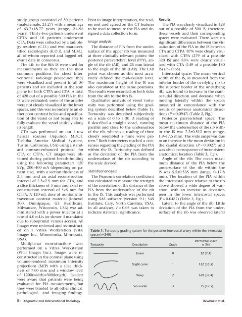
Qualitative analysis <str<strong>on</strong>g>of</str<strong>on</strong>g> vessel tortuosity<br />
was performed using <str<strong>on</strong>g>the</str<strong>on</strong>g> grading<br />
system described below (Table 1).<br />
Tortuosity was described subjectively<br />
<strong>on</strong> a scale <str<strong>on</strong>g>of</str<strong>on</strong>g> 0 to 3 (8). A reading <str<strong>on</strong>g>of</str<strong>on</strong>g><br />
0 described a straight vessel, running<br />
parallel and close to <str<strong>on</strong>g>the</str<strong>on</strong>g> undersurface<br />
<str<strong>on</strong>g>of</str<strong>on</strong>g> <str<strong>on</strong>g>the</str<strong>on</strong>g> rib, whereas a reading <str<strong>on</strong>g>of</str<strong>on</strong>g> three<br />
closely resembled a “sine wave pattern.”<br />
Both interpreters reached a c<strong>on</strong>sensus<br />
regarding <str<strong>on</strong>g>the</str<strong>on</strong>g> grading <str<strong>on</strong>g>of</str<strong>on</strong>g> <str<strong>on</strong>g>the</str<strong>on</strong>g> PIA<br />
within <str<strong>on</strong>g>the</str<strong>on</strong>g> IS. Tortuosity was defined<br />
as <str<strong>on</strong>g>the</str<strong>on</strong>g> deviati<strong>on</strong> <str<strong>on</strong>g>of</str<strong>on</strong>g> <str<strong>on</strong>g>the</str<strong>on</strong>g> PIA from <str<strong>on</strong>g>the</str<strong>on</strong>g><br />
undersurface <str<strong>on</strong>g>of</str<strong>on</strong>g> <str<strong>on</strong>g>the</str<strong>on</strong>g> rib according to<br />
<str<strong>on</strong>g>the</str<strong>on</strong>g> scale devised.<br />
Statistical analysis<br />
The Pears<strong>on</strong>’s correlati<strong>on</strong> coefficient<br />
was calculated to measure <str<strong>on</strong>g>the</str<strong>on</strong>g> strength<br />
<str<strong>on</strong>g>of</str<strong>on</strong>g> <str<strong>on</strong>g>the</str<strong>on</strong>g> correlati<strong>on</strong> <str<strong>on</strong>g>of</str<strong>on</strong>g> <str<strong>on</strong>g>the</str<strong>on</strong>g> distance <str<strong>on</strong>g>of</str<strong>on</strong>g> <str<strong>on</strong>g>the</str<strong>on</strong>g><br />
PIA from <str<strong>on</strong>g>the</str<strong>on</strong>g> undersurface <str<strong>on</strong>g>of</str<strong>on</strong>g> <str<strong>on</strong>g>the</str<strong>on</strong>g> rib<br />
in <str<strong>on</strong>g>the</str<strong>on</strong>g> IS. This analysis was performed<br />
using SAS s<str<strong>on</strong>g>of</str<strong>on</strong>g>tware (versi<strong>on</strong> 9.1; SAS<br />
Institute, Cary, North Carolina, USA).<br />
In all analyses, P < 0.05 was taken to<br />
indicate statistical significance.<br />
Results<br />
The PIA was clearly visualized in 428<br />
out <str<strong>on</strong>g>of</str<strong>on</strong>g> a possible <str<strong>on</strong>g>of</str<strong>on</strong>g> 500 IS; <str<strong>on</strong>g>the</str<strong>on</strong>g>refore,<br />
<str<strong>on</strong>g>the</str<strong>on</strong>g>se vessels and <str<strong>on</strong>g>the</str<strong>on</strong>g>ir corresp<strong>on</strong>ding<br />
spaces were evaluated. There were no<br />
significant differences between <str<strong>on</strong>g>the</str<strong>on</strong>g> visualizati<strong>on</strong><br />
<str<strong>on</strong>g>of</str<strong>on</strong>g> <str<strong>on</strong>g>the</str<strong>on</strong>g> PIA in <str<strong>on</strong>g>the</str<strong>on</strong>g> IS between<br />
<strong>CT</strong>A and <strong>CT</strong>PA: 87% were clearly visualized<br />
with <strong>CT</strong>PA (279 <str<strong>on</strong>g>of</str<strong>on</strong>g> a possible<br />
320 IS) and 83% were clearly visualized<br />
with <strong>CT</strong>A (149 <str<strong>on</strong>g>of</str<strong>on</strong>g> a possible 180<br />
IS) (P = 0.65).<br />
Intercostal space: The mean vertical<br />
width <str<strong>on</strong>g>of</str<strong>on</strong>g> <str<strong>on</strong>g>the</str<strong>on</strong>g> IS, as measured from <str<strong>on</strong>g>the</str<strong>on</strong>g><br />
inferior border <str<strong>on</strong>g>of</str<strong>on</strong>g> <str<strong>on</strong>g>the</str<strong>on</strong>g> overlying rib to<br />
<str<strong>on</strong>g>the</str<strong>on</strong>g> superior border <str<strong>on</strong>g>of</str<strong>on</strong>g> <str<strong>on</strong>g>the</str<strong>on</strong>g> underlying<br />
rib, was found to increase in <str<strong>on</strong>g>the</str<strong>on</strong>g> craniocaudal<br />
directi<strong>on</strong> and decrease when<br />
moving laterally within <str<strong>on</strong>g>the</str<strong>on</strong>g> spaces<br />
measured in c<strong>on</strong>cordance with <str<strong>on</strong>g>the</str<strong>on</strong>g><br />
results <str<strong>on</strong>g>of</str<strong>on</strong>g> previous anatomical dissecti<strong>on</strong>s<br />
(P = 0.0947) (Table 2, Fig.).<br />
Posterior paravertebral space: The<br />
mean maximum distance <str<strong>on</strong>g>of</str<strong>on</strong>g> <str<strong>on</strong>g>the</str<strong>on</strong>g> PIA<br />
below <str<strong>on</strong>g>the</str<strong>on</strong>g> undersurface <str<strong>on</strong>g>of</str<strong>on</strong>g> <str<strong>on</strong>g>the</str<strong>on</strong>g> rib within<br />
<str<strong>on</strong>g>the</str<strong>on</strong>g> IS was 7.2±0.512 mm (range,<br />
1.9–17.5 mm). The wide range was due<br />
to an increase in <str<strong>on</strong>g>the</str<strong>on</strong>g> mean deviati<strong>on</strong> in<br />
<str<strong>on</strong>g>the</str<strong>on</strong>g> caudal directi<strong>on</strong> (P = 0.0027) and<br />
was also a c<strong>on</strong>sequence <str<strong>on</strong>g>of</str<strong>on</strong>g> inc<strong>on</strong>sistent<br />
anatomical locati<strong>on</strong> (Table 3, Fig.).<br />
Angle <str<strong>on</strong>g>of</str<strong>on</strong>g> <str<strong>on</strong>g>the</str<strong>on</strong>g> rib: The mean maximum<br />
distance <str<strong>on</strong>g>of</str<strong>on</strong>g> <str<strong>on</strong>g>the</str<strong>on</strong>g> PIA below <str<strong>on</strong>g>the</str<strong>on</strong>g><br />
undersurface <str<strong>on</strong>g>of</str<strong>on</strong>g> <str<strong>on</strong>g>the</str<strong>on</strong>g> rib within <str<strong>on</strong>g>the</str<strong>on</strong>g><br />
IS was 5.5±0.535 mm (range, 0–17.8<br />
mm). The locati<strong>on</strong> <str<strong>on</strong>g>of</str<strong>on</strong>g> <str<strong>on</strong>g>the</str<strong>on</strong>g> PIA within<br />
<str<strong>on</strong>g>the</str<strong>on</strong>g> <str<strong>on</strong>g>intercostal</str<strong>on</strong>g> space relative to <str<strong>on</strong>g>the</str<strong>on</strong>g> rib<br />
above showed a wide degree <str<strong>on</strong>g>of</str<strong>on</strong>g> variati<strong>on</strong>,<br />
with an increase in deviati<strong>on</strong><br />
seen in <str<strong>on</strong>g>the</str<strong>on</strong>g> lower <str<strong>on</strong>g>intercostal</str<strong>on</strong>g> spaces<br />
(P = 0.0487) (Table 3, Fig.).<br />
Lateral to <str<strong>on</strong>g>the</str<strong>on</strong>g> angle <str<strong>on</strong>g>of</str<strong>on</strong>g> <str<strong>on</strong>g>the</str<strong>on</strong>g> rib: Little<br />
deviati<strong>on</strong> <str<strong>on</strong>g>of</str<strong>on</strong>g> <str<strong>on</strong>g>the</str<strong>on</strong>g> PIA from <str<strong>on</strong>g>the</str<strong>on</strong>g> undersurface<br />
<str<strong>on</strong>g>of</str<strong>on</strong>g> <str<strong>on</strong>g>the</str<strong>on</strong>g> rib was observed lateral<br />
Table 1. Tortuosity grading system for <str<strong>on</strong>g>the</str<strong>on</strong>g> <str<strong>on</strong>g>posterior</str<strong>on</strong>g> <str<strong>on</strong>g>intercostal</str<strong>on</strong>g> <str<strong>on</strong>g>artery</str<strong>on</strong>g> within <str<strong>on</strong>g>the</str<strong>on</strong>g> <str<strong>on</strong>g>intercostal</str<strong>on</strong>g><br />
space (n=248)<br />
Tortuosity Descripti<strong>on</strong> Grade<br />
Intercostal space<br />
n (%)<br />
Linear 0 32 (7.4)<br />
Slight curve 1 152 (35.5)<br />
Wavy 2 169 (39.4)<br />
Sinusoidal 3 75 (17.5)<br />
Dewhurst et al.