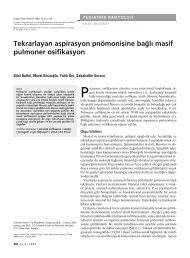
Demonstration of the course of the posterior intercostal artery on CT ...
Demonstration of the course of the posterior intercostal artery on CT ...
Demonstration of the course of the posterior intercostal artery on CT ...

You also want an ePaper? Increase the reach of your titles
YUMPU automatically turns print PDFs into web optimized ePapers that Google loves.
ib <str<strong>on</strong>g>of</str<strong>on</strong>g>fers more protecti<strong>on</strong> to <str<strong>on</strong>g>the</str<strong>on</strong>g> PIA<br />
laterally than medially, thus favoring<br />
<str<strong>on</strong>g>the</str<strong>on</strong>g> mid-axillary line approach for transthoracic<br />
interventi<strong>on</strong>s and avoidance<br />
<str<strong>on</strong>g>of</str<strong>on</strong>g> <str<strong>on</strong>g>the</str<strong>on</strong>g> paravertebral space (14).<br />
In a caudal directi<strong>on</strong>, from <str<strong>on</strong>g>the</str<strong>on</strong>g> 4th<br />
to <str<strong>on</strong>g>the</str<strong>on</strong>g> 8th <str<strong>on</strong>g>intercostal</str<strong>on</strong>g> space, <str<strong>on</strong>g>the</str<strong>on</strong>g>re is a<br />
significant increase in <str<strong>on</strong>g>the</str<strong>on</strong>g> mean width<br />
<str<strong>on</strong>g>of</str<strong>on</strong>g> <str<strong>on</strong>g>the</str<strong>on</strong>g> IS allowing <str<strong>on</strong>g>the</str<strong>on</strong>g> PIA to dip more<br />
significantly into <str<strong>on</strong>g>the</str<strong>on</strong>g> IS at <str<strong>on</strong>g>the</str<strong>on</strong>g> lower<br />
levels. C<strong>on</strong>versely, <str<strong>on</strong>g>the</str<strong>on</strong>g>re is a significant<br />
progressive decrease in <str<strong>on</strong>g>the</str<strong>on</strong>g> mean width<br />
<str<strong>on</strong>g>of</str<strong>on</strong>g> <str<strong>on</strong>g>the</str<strong>on</strong>g> IS when traveling laterally, with<br />
a maximal width at PPV. This results in<br />
a decrease in <str<strong>on</strong>g>the</str<strong>on</strong>g> lateral vulnerability <str<strong>on</strong>g>of</str<strong>on</strong>g><br />
<str<strong>on</strong>g>the</str<strong>on</strong>g> PIA as it dips less into <str<strong>on</strong>g>the</str<strong>on</strong>g> IS.<br />
Vessel tortuosity grades indicated<br />
that this was also unpredictable, independent<br />
<str<strong>on</strong>g>of</str<strong>on</strong>g> both age and gender, and<br />
could be ei<str<strong>on</strong>g>the</str<strong>on</strong>g>r straight or extremely<br />
complex in shape and locati<strong>on</strong>. This<br />
differs from <str<strong>on</strong>g>the</str<strong>on</strong>g> results <str<strong>on</strong>g>of</str<strong>on</strong>g> a previous<br />
study by Choi et al. (15) who examined<br />
<strong>on</strong>ly 160 IS, but c<strong>on</strong>cluded that <str<strong>on</strong>g>the</str<strong>on</strong>g>re<br />
was increased tortuosity <str<strong>on</strong>g>of</str<strong>on</strong>g> <str<strong>on</strong>g>the</str<strong>on</strong>g> vessel<br />
with increasing age; this was not observed<br />
in <str<strong>on</strong>g>the</str<strong>on</strong>g> present study (P = 0.79)<br />
and <str<strong>on</strong>g>the</str<strong>on</strong>g>re were no differences between<br />
<str<strong>on</strong>g>the</str<strong>on</strong>g> right and left sides.<br />
Thus, while cadaveric specimens<br />
have shown variability <str<strong>on</strong>g>of</str<strong>on</strong>g> <str<strong>on</strong>g>the</str<strong>on</strong>g> PIA within<br />
<str<strong>on</strong>g>the</str<strong>on</strong>g> IS, <str<strong>on</strong>g>the</str<strong>on</strong>g> present study defined its<br />
positi<strong>on</strong> in vivo using <strong>CT</strong>A. While this<br />
study was limited by <str<strong>on</strong>g>the</str<strong>on</strong>g> small number<br />
<str<strong>on</strong>g>of</str<strong>on</strong>g> patients, our data support <str<strong>on</strong>g>the</str<strong>on</strong>g> c<strong>on</strong>clusi<strong>on</strong><br />
that <str<strong>on</strong>g>the</str<strong>on</strong>g> PIA is variable in positi<strong>on</strong><br />
and tortuosity, particularly at <str<strong>on</strong>g>the</str<strong>on</strong>g><br />
PPV juncti<strong>on</strong>. In additi<strong>on</strong>, our study<br />
was limited to a 4-slice MD<strong>CT</strong>, and<br />
with <str<strong>on</strong>g>the</str<strong>on</strong>g> advent <str<strong>on</strong>g>of</str<strong>on</strong>g> 64-slice imaging,<br />
image quality and vascular anatomy<br />
can now be much better defined.<br />
In c<strong>on</strong>clusi<strong>on</strong>, our data suggest <str<strong>on</strong>g>the</str<strong>on</strong>g><br />
presence <str<strong>on</strong>g>of</str<strong>on</strong>g> high variability and tortuosity<br />
<str<strong>on</strong>g>of</str<strong>on</strong>g> <str<strong>on</strong>g>the</str<strong>on</strong>g> PIA, particularly at <str<strong>on</strong>g>the</str<strong>on</strong>g> PPV<br />
and AR. While <str<strong>on</strong>g>the</str<strong>on</strong>g> principle <str<strong>on</strong>g>of</str<strong>on</strong>g> “choosing<br />
a site above <str<strong>on</strong>g>the</str<strong>on</strong>g> rib below” is endorsed<br />
in <str<strong>on</strong>g>the</str<strong>on</strong>g> c<strong>on</strong>text <str<strong>on</strong>g>of</str<strong>on</strong>g> percutaneous<br />
transthoracic interventi<strong>on</strong>, we would<br />
advocate <str<strong>on</strong>g>the</str<strong>on</strong>g> additi<strong>on</strong> <str<strong>on</strong>g>of</str<strong>on</strong>g> increased cauti<strong>on</strong><br />
to avoid <str<strong>on</strong>g>the</str<strong>on</strong>g> <str<strong>on</strong>g>posterior</str<strong>on</strong>g> paravertebral<br />
space to this dictum, as <str<strong>on</strong>g>the</str<strong>on</strong>g>re is a<br />
potentially increased risk <str<strong>on</strong>g>of</str<strong>on</strong>g> inadvertent<br />
vessel injury in this area.<br />
C<strong>on</strong>flict <str<strong>on</strong>g>of</str<strong>on</strong>g> interest disclosure<br />
The authors declared no c<strong>on</strong>flicts <str<strong>on</strong>g>of</str<strong>on</strong>g> interest.<br />
References<br />
1. Kwan S, Bhargavan M, Kerlan R, et al.<br />
Effect <str<strong>on</strong>g>of</str<strong>on</strong>g> advanced imaging technology <strong>on</strong><br />
how biopsies are d<strong>on</strong>e and who does <str<strong>on</strong>g>the</str<strong>on</strong>g>m.<br />
Radiology 2010; 256:751–758.<br />
2. Gupta S, Seaberg K, Wallace M, et al.<br />
Imaging guided percutaneous biopsy<br />
<str<strong>on</strong>g>of</str<strong>on</strong>g> mediastinal lesi<strong>on</strong>s: different approaches<br />
and anatomic c<strong>on</strong>siderati<strong>on</strong>s.<br />
Radiographics 2005; 25:763–788.<br />
3. Pauls<strong>on</strong> E, Sheafor D, Enterline D, et al.<br />
<strong>CT</strong> fluoroscopy-guided interventi<strong>on</strong>al procedures:<br />
techniques and radiati<strong>on</strong> dose to<br />
radiologists. Radiology 2001; 220:161–167.<br />
4. Wallace M, Krishnamurthy S, Broemeling<br />
L, et al. <strong>CT</strong>-guided percutaneous fine-needle<br />
aspirati<strong>on</strong> biopsy <str<strong>on</strong>g>of</str<strong>on</strong>g> small (≤ 1cm) pulm<strong>on</strong>ary<br />
lesi<strong>on</strong>s. Radiology 2002; 225:823–<br />
828.<br />
5. Tomlins<strong>on</strong> MA, Treasure T. Inserti<strong>on</strong> <str<strong>on</strong>g>of</str<strong>on</strong>g> a<br />
chest drain: how to do it. Br J Hosp Med<br />
1997; 58:248–252.<br />
6. Laws D, Neville E, Duffy J. BTS guidelines<br />
for <str<strong>on</strong>g>the</str<strong>on</strong>g> inserti<strong>on</strong> <str<strong>on</strong>g>of</str<strong>on</strong>g> a chest drain. Thorax<br />
2003; 58:53–59.<br />
7. Al-Tarshihi MI, Khamash FA, Al Ibrahim<br />
AEO. Thoracoscopy tube complicati<strong>on</strong>s<br />
and pitfalls: an experience at tertiary level<br />
military hospital. RMJ 2008; 33:141–144.<br />
8. Ko JP, Shepard JO, Drucker EA, et al.<br />
Factors influencing pneumothorax rate<br />
at lung biopsy: are dwell time and angle<br />
<str<strong>on</strong>g>of</str<strong>on</strong>g> pleural puncture c<strong>on</strong>tributing factors?<br />
Radiology 2001; 218:491–496.<br />
9. Utz JP, Ryu JH, Douglas WW, et al. High<br />
short-term mortality following lung biopsy<br />
for usual interstitial pneum<strong>on</strong>ia. Eur<br />
Respir J 2001; 17:175–179.<br />
10. Yamauchi H, Amemiya R, Shida D, et al.<br />
A case <str<strong>on</strong>g>of</str<strong>on</strong>g> sp<strong>on</strong>taneous hemopneumothorax<br />
associated with unc<strong>on</strong>trolled massive<br />
bleeding after inserti<strong>on</strong> thoracic drain. Jap<br />
J Thorac Surg 1999; 52:965–968.<br />
11. Muthuswamy P, Samuel J, Mixcock B, et al.<br />
Recurrent massive bleeding from an <str<strong>on</strong>g>intercostal</str<strong>on</strong>g><br />
<str<strong>on</strong>g>artery</str<strong>on</strong>g> aneurysm through an empyema<br />
chest tube. Chest 1993; 104:637–639.<br />
12. Carney M, Ravin CE. Intercostal <str<strong>on</strong>g>artery</str<strong>on</strong>g> lacerati<strong>on</strong><br />
during thoracocentesis: increased<br />
risk in elderly patients. Chest 1979;<br />
75:520–522.<br />
13. Seneff MG. Complicati<strong>on</strong>s associated with<br />
thoracocentesis. Chest 1986; 90:97–100.<br />
14. Yacov<strong>on</strong>e M, Kartan R, Bautista M.<br />
Intercostal <str<strong>on</strong>g>artery</str<strong>on</strong>g> lacerati<strong>on</strong> following<br />
thoracentesis. Respir Care 2010; 55:1495–<br />
1498.<br />
15. Choi S, Trieu J, Ridley L. Radiological review<br />
<str<strong>on</strong>g>of</str<strong>on</strong>g> <str<strong>on</strong>g>intercostal</str<strong>on</strong>g> <str<strong>on</strong>g>artery</str<strong>on</strong>g>: anatomical c<strong>on</strong>siderati<strong>on</strong>s<br />
when performing procedures<br />
via <str<strong>on</strong>g>intercostal</str<strong>on</strong>g> space. J Med Imaging Radiat<br />
Oncol 2010; 54:302–306.<br />
16. Oguz B, Haliloglu M, Karcaaltincaba M.<br />
Paediatric multidetector <strong>CT</strong> angiography:<br />
spectrum <str<strong>on</strong>g>of</str<strong>on</strong>g> c<strong>on</strong>genital thoracic vascular<br />
anomalies. Br J Radiol 2007; 80:376–383.<br />
17. Wraight WM, Tweedie DJ, Parkin IG.<br />
Neurovascular anatomy and variati<strong>on</strong> in<br />
<str<strong>on</strong>g>the</str<strong>on</strong>g> fourth, fifth and sixth <str<strong>on</strong>g>intercostal</str<strong>on</strong>g> spaces<br />
in <str<strong>on</strong>g>the</str<strong>on</strong>g> midclavicular line: a cadaveric<br />
study in respect <str<strong>on</strong>g>of</str<strong>on</strong>g> chest drain inserti<strong>on</strong>.<br />
Clin Anat 2005; 18:346–349.<br />
18. Da Rocha RP, Vengjer A, Blanco A, de<br />
Carvalho PT, M<strong>on</strong>g<strong>on</strong> ML, Fernandes GJ.<br />
Size <str<strong>on</strong>g>of</str<strong>on</strong>g> <str<strong>on</strong>g>the</str<strong>on</strong>g> collateral <str<strong>on</strong>g>intercostal</str<strong>on</strong>g> <str<strong>on</strong>g>artery</str<strong>on</strong>g> in<br />
adults: anatomical c<strong>on</strong>siderati<strong>on</strong>s in relati<strong>on</strong><br />
to thoracocentesis and thoracoscopy.<br />
Surg Radiol Anat 2002; 24:23–26.<br />
19. Muzrakchi AA, Szmigielski W, Omar AJ, et<br />
al. Is <str<strong>on</strong>g>the</str<strong>on</strong>g> 10th and 11th <str<strong>on</strong>g>intercostal</str<strong>on</strong>g> space<br />
a safe approach for percutaneous nephrostomy<br />
and nephrolithotomy? Cardiovasc<br />
Intervent Radiol 2004; 27:503–506.<br />
20. Davies DV. Gray’s anatomy. 34th editi<strong>on</strong>.<br />
L<strong>on</strong>d<strong>on</strong>: L<strong>on</strong>gmans, Green and Co. 1967.<br />
iv • Diagnostic and Interventi<strong>on</strong>al Radiology<br />
Dewhurst et al.