Biology 3B Laboratory Invertebrates II: Annelida, Nematoda ...
Biology 3B Laboratory Invertebrates II: Annelida, Nematoda ...
Biology 3B Laboratory Invertebrates II: Annelida, Nematoda ...

You also want an ePaper? Increase the reach of your titles
YUMPU automatically turns print PDFs into web optimized ePapers that Google loves.
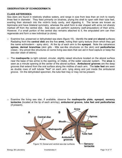
OBSERVATION OF ECHINODERMATA<br />
CLASS ASTEROIDEA<br />
Sea stars are found in relatively shallow waters, and range in size from less than an inch to nearly<br />
three feet in diameter. They feed primarily on bivalves, prying the shell to open with their tube feet,<br />
everting their stomach into the victim's body cavity, and digesting it. The larvae are known as<br />
bipinnaria and have bilateral symmetry, whereas the adult form is star shaped with arms not sharply<br />
marked off from the central disk. Sea stars can perform autotomy (self-amputation) of their arms.<br />
However, if a small portion of the central disc remains attached to it, the amputated arm can then<br />
regenerate and form a new individual (a clone).<br />
o Examine live, preserved or dehydrated sea stars (figure 15). Identify the oral and aboral surfaces.<br />
Radiating from the central disk are the five arms, noting their spiny texture (from which they get<br />
the name echinoderm - spiny skin). At the tip of each arm is the eyespot. Note the calcareous<br />
spines, dermal branchiae (skin gills - little sac-like structures on the skin) and pedicellariae<br />
(claws - tiny pincer-like structures on some living sea stars that can aid in food capture or keep the<br />
sea star clean of debris).<br />
o The madreporite (a light colored, circular, slightly raised structure located on the aboral surface<br />
near the base of two arms) is the opening, or intake, of the water vascular system. The anus is<br />
seen as a minute opening at the center of the aboral surface. Ambulacral grooves are the deep<br />
grooves that extend from the oral surface along the midline of each arm. The tube feet are seen<br />
as double rows of soft tubular "feet" on each arm, lying along and just inside the ambulacral<br />
groove. On the dehydrated specimen, the tube feet may or may not be present.<br />
o Examine the living sea star, if available, observe the madreporite plate, eyespot, sensory<br />
tentacles (located at the tip of each arm/ray), ambulacral groove, tube feet and pedicellariae<br />
(if present).<br />
Figure 15: Dorsal and ventral sea star surfaces<br />
<strong>Biology</strong> <strong>3B</strong> <strong>Laboratory</strong> <strong>Invertebrates</strong> <strong>II</strong> Page 14 of 17