Biology 3B Laboratory Invertebrates II: Annelida, Nematoda ...
Biology 3B Laboratory Invertebrates II: Annelida, Nematoda ...
Biology 3B Laboratory Invertebrates II: Annelida, Nematoda ...

Create successful ePaper yourself
Turn your PDF publications into a flip-book with our unique Google optimized e-Paper software.
<strong>Biology</strong> <strong>3B</strong> <strong>Laboratory</strong><br />
<strong>Invertebrates</strong> <strong>II</strong>: <strong>Annelida</strong>, <strong>Nematoda</strong>, Arthropoda, Onychophora, Echinodermata<br />
Objectives<br />
• To understand the basic differences among the invertebrate animal phyla<br />
• To investigate and learn the obvious external and internal characteristics of annelids,<br />
nematodes, arthropods and echinoderms<br />
• To investigate at the microscopic level the organization and function of selected tissues and<br />
cells within these groups<br />
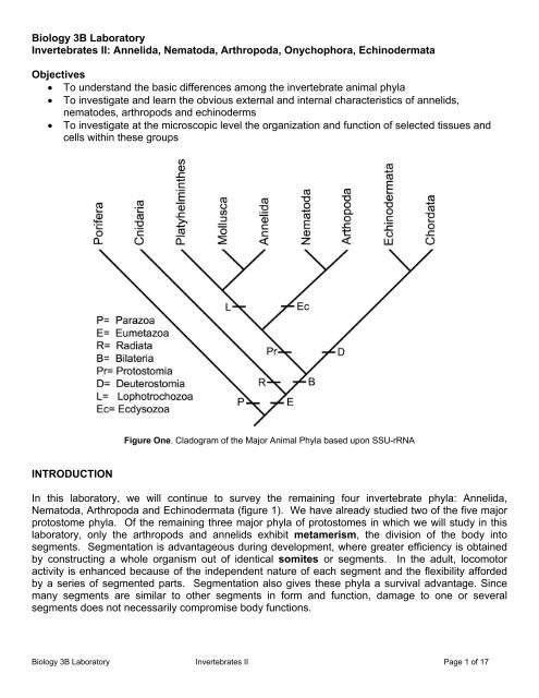
Figure One. Cladogram of the Major Animal Phyla based upon SSU-rRNA<br />
INTRODUCTION<br />
In this laboratory, we will continue to survey the remaining four invertebrate phyla: <strong>Annelida</strong>,<br />
<strong>Nematoda</strong>, Arthropoda and Echinodermata (figure 1). We have already studied two of the five major<br />
protostome phyla. Of the remaining three major phyla of protostomes in which we will study in this<br />
laboratory, only the arthropods and annelids exhibit metamerism, the division of the body into<br />
segments. Segmentation is advantageous during development, where greater efficiency is obtained<br />
by constructing a whole organism out of identical somites or segments. In the adult, locomotor<br />
activity is enhanced because of the independent nature of each segment and the flexibility afforded<br />
by a series of segmented parts. Segmentation also gives these phyla a survival advantage. Since<br />
many segments are similar to other segments in form and function, damage to one or several<br />
segments does not necessarily compromise body functions.<br />
<strong>Biology</strong> <strong>3B</strong> <strong>Laboratory</strong> <strong>Invertebrates</strong> <strong>II</strong> Page 1 of 17
PHYLUM ANNELIDA<br />
Members in the phylum <strong>Annelida</strong> are often referred to as segmented worms because of their<br />
segmentation, a distinguishing characteristic that sets them apart form other animals. The most<br />
recognizable members include the earthworms (terrestrial habitat), leeches (terrestrial and<br />
freshwater), and marine worms. All annelids are triploblastic, bilaterally symmetrical, and<br />
eucoelomate. In addition, annelids exhibit a body wall with both longitudinal and circular muscle<br />
layers (which, along with segmentation mentioned above, allows these animals to be quite mobile).<br />
They have a complete digestive tract. Their nervous system shows some degree of cephalization<br />
with a “brain” and two ventral nerve cords that running the entire length of the body. They have a<br />
closed circulatory system with aortic arches that act as the “heart” to pump blood through muscular<br />
blood vessels. They also have a well developed excretory system which removes waste from the<br />
blood and coelom.<br />
There are three major classes within the phylum <strong>Annelida</strong>, described below.<br />
Class Polychaeta - mostly marine worms, such as Nereis (the clamworm)<br />
Class Hirudinea - the leeches (predominantly freshwater), such as Hirudo<br />
Class Oligochaeta - mostly freshwater and terrestrial worms, such as Lumbricus (the<br />
earthworms)<br />
OBSERVATION OF POLYCHAETA<br />
CLASS POLYCHAETA<br />
Polychaete worms are mostly a marine group of worms characterized by many segments with a pair<br />
of parapodia with numerous setae (figure 2). They have a distinct head with eyes, palps and<br />
tentacles.<br />
o Examine a clamworm (Nerius). These<br />
are the “typical” polychaete worms that<br />
can be found living in the mud and<br />
debris of shallow coastal waters. Using<br />
the dissecting scope, observe the head<br />
region and find the following: eyes,<br />
mouth on the ventral side, jaws, and<br />
tentacles.<br />
o Examine one of the segments. Locate a<br />
parapodium on one side a body<br />
segment. Parapodia function in<br />
locomotion and respiration for<br />
polychaetes. Each parapodium is<br />
comprised of two lobes which bear<br />
numerous setae (the reason for the<br />
class name).<br />
Figure 2: Structure of a clamworm (Nerius)<br />
OBSERVATION OF OLIGOCHAETA<br />
CLASS OLIGOCHAETA<br />
Like polychaete worms, oligochaete worms are also segmented both outside and inside. However,<br />
oligochaetes do not have parapodia, their head is less developed and they have fewer setae. The<br />
<strong>Biology</strong> <strong>3B</strong> <strong>Laboratory</strong> <strong>Invertebrates</strong> <strong>II</strong> Page 2 of 17
most noticeable external feature in this group is the clitellum (figure 3). Most members in this class<br />
are either terrestrial (most) or inhabit freshwater.<br />
o Obtain an earthworm (Lumbricus) and place the animal in a dissecting tray. You may need a<br />
dissecting scope to fully appreciate the external anatomy. The first four segments comprise the<br />
head region. Find the mouth on the first segment (figure 3). The prostomium overhangs the<br />
mouth. They have a complete digestive tract that terminates on the last segment with the anus.<br />
o The most obvious external feature is the clitellum, a swollen area in the anterior third of the<br />
specimen. This region functions in reproduction by secreting a mucous which holds the<br />
participants together during sperm exchange and cocoon formation around the fertilized eggs.<br />
o Orient the worm dorso-ventrally by locating the tiny setae (hairs). Run your fingers along the<br />
animal to feel the rough texture produced by the setae. Four of these structures are found on the<br />
ventral surface of each metamere. They provide traction during locomotion.<br />
o Starting with the segment that holds the mouth, locate segment 14. Observe the openings for the<br />
oviducts (female pore) on the ventral surface. Find the sperm ducts (male pore) on the ventral<br />
surface of segment 15.<br />
Figure 3: Structure of an earthworm.<br />
We will examine the internal structure when we begin the systems.<br />
OBSERVATION OF HIRUDINEA<br />
CLASS HIRUDINEA<br />
The best known member in this class is the freshwater leech. Other members can be found on land<br />
and the marine environments. Members in this class typically have 33-34 segments with a clitellum.<br />
Most do not have setae and no members have parapodia. Members have both anterior and posterior<br />
suckers.<br />
o Examine representative members in this class. The medicinal leech, Hirudo medicinalis, secretes<br />
an anticoagulant on the host at they parasitized them. This leech was commonly used in the<br />
practice of blood-letting. It is still used today to increase circulation to surgical areas, especially<br />
with finger reattachments. Note the smaller oral sucker and larger posterior sucker.<br />
<strong>Biology</strong> <strong>3B</strong> <strong>Laboratory</strong> <strong>Invertebrates</strong> <strong>II</strong> Page 3 of 17
PHYLUM NEMATODA<br />
Triploblastic pseudocoelomates<br />
The development of a body cavity (coelom) is considered a major evolutionary advantage over those<br />
animals which do not possess a body cavity (acoelomate). As you have already learned, body<br />
cavities are advantageous for a number of reasons, such as to provide more room for organ<br />
development, to provide an increased surface area for diffusion of gases and/or nutrients, and to<br />
facilitate locomotion by serving as hydrostatic skeleton.<br />
A body cavity is characteristic of all bilateral animals above the acoelomates. A true coelom is a<br />
cavity in which the inner body wall and the visceral organs are lined with peritoneum. A<br />
pseudocoeIom, found in animals to be examined in the present exercise, is defined as a body cavity<br />
that is lined by mesoderm externally and endoderm internally (figure 4).<br />
The nematodes are one of several phyla<br />
usually discussed together as the<br />
pseudocoelomates because of their shared<br />
possession of this structure. Except for this<br />
one common feature, they are a diverse<br />
group of animals, only distantly related.<br />
Included in this broad group of animals are<br />
the Phyla Rotifera, Nematomorpha,<br />
Gastrotricha, Kinorhyncha, and others. We<br />
will examine members of the phylum<br />
<strong>Nematoda</strong> as a representative<br />
pseudocoelomate.<br />
However, nematodes are grouped with<br />
arthropods as an ectodyzoan due to<br />
molecular evidence supporting ecdysis, the<br />
ability to shed the exoskeleton as the<br />
organism grows.<br />
Nematodes have a worldwide distribution<br />
that include, terrestrial, freshwater, marine<br />
and parasitic forms. They are round worms<br />
with a tough, flexible cuticle that is non;living.<br />
Nematodes are important ecologically for<br />
their recycling and decomposition<br />
capabilities.<br />
OBSERVATION OF NEMATODA<br />
• Examine the male and female intestinal<br />
roundworm, Ascaris lumbricoides, in<br />
Figure 4: Comparison of body cavities. dissecting tray. Gloves should be worn, if<br />
available. If not, handle organism with forceps.<br />
Do not touch with bare skin. Ascaris is sexually dimorphic and sexes are easily differentiated.<br />
Male worms are smaller, typically have a hook-shaped sideways bend near their posterior end,<br />
<strong>Biology</strong> <strong>3B</strong> <strong>Laboratory</strong> <strong>Invertebrates</strong> <strong>II</strong> Page 4 of 17
and may have tiny copulatory spicules protruding slightly from the cloaca. Ascaris is an intestinal<br />
parasite of vertebrates, with A. lumbricoides infecting up to 64% of individuals living in the<br />
southeastern United States. The female lays up to 200,000 eggs/day which passes out of the<br />
host via their feces. The embryo is very resistant, thus be careful handling these worms as you<br />
could potentially infect yourself.<br />
o Examine the slide of a trichina worm, Trichinella spiralis. You will be observing calcified cyst of<br />
the juvenile trichina worm (figure 5) in the muscle of the host. Trichinosis is the disease that is<br />
caused by the trichina worm. Human infestations are typically due to the ingestion of<br />
undercooked meats such as pork. Roughly 2% of the population in the US has a light infection<br />
with trichina worms. Heavy infestations may cause death. Other species that can be infected<br />
include: hogs, rats, dogs, cats and any other omnivorous or carnivorous species.<br />
Figure 5: Encysted juvenile Trichinella spiralis<br />
o Examine the slide of a hookworm, Necator americanus. Note the anterior portion of the worm with<br />
a hook-like appearance in a tissue section. Infestations results when the juvenile hookworm<br />
comes in contact with the skin and burrows into the host. The juvenile then travels via the<br />
bloodstream to the lungs, move up the respiratory tract and then swallowed. In the small<br />
intestines they will mature.<br />
o Examine a slide of pinworms, Enterobius vermicularis. These worms live in the large intestines of<br />
humans and are the most common nematode parasite. These infestations are more<br />
embarrassing than debilitating. The female will travel to the anus at night to deposit eggs around<br />
the anus. Scratching contaminates the hands and bedding. The eggs are then swallowed and<br />
hatch in the duodenum and mature in the large intestines. This is a very common infestation in<br />
children.<br />
<strong>Biology</strong> <strong>3B</strong> <strong>Laboratory</strong> <strong>Invertebrates</strong> <strong>II</strong> Page 5 of 17
o Examine the free-living nematode, the vinegar eel (Tubatrix aceti) on a depression slide, if<br />
available. These worms have a high tolerance to low pH. Describe how the body bends.<br />
PHYLUM ARTHROPODA<br />
The phylum Arthropoda is the largest in the animal kingdom. More than 75% of all living organisms<br />
are arthropods with insects contributing the greatest numbers. Like annelids they are characterized<br />
by metamerism, i.e. the body is segmented. In addition, they have a chitonous exoskeleton. The<br />
segmented body is divisible into functional units called tagmata. In some arthropods three tagmata<br />
are present - a head (involved in feeding and sensory functions), a thorax (involved mostly in<br />
locomotion), and an abdomen (which performs the visceral functions). In many arthropods the head<br />
and thorax are fused, forming a cepahalothorax.<br />
The phylum contains three extant subphyla - Chelicerata, Crustacea, and Uniramia. The subphylum<br />
Chelicerata contains arthropods in which the first appendages are modified into chelicerae (pincerlike<br />
feeding structures). Well-known representatives of this subphylum include the class Arachnida<br />
(scorpions, spiders, ticks, etc.) and the class Merostomata (horseshoe crabs).<br />
Major Arthropoda subdivisions (you are only responsible the taxonomy down to the class level)<br />
• Subphylum Trilobita<br />
• Subphylum Chelicerata<br />
o Class Merostomata - horsecrab<br />
o Class Pycnogonida – sea spiders<br />
o Class Arachnida<br />
• Order Araneae - spiders<br />
• Order Scorpionida - scorpions<br />
• Order Opiliones - harvestmen<br />
• Order Acari – ticks & mites<br />
• Subphylum Crustacea<br />
o Class Branchiopoda<br />
• Order Cladocera – water fleas<br />
o Class Maxillopoda<br />
• Subclass Copepoda - copepods<br />
• Subclass Cirripedia - barnacles<br />
o Class Malacostraca<br />
• Order Isopoda – isopods, pill bugs<br />
• Order Euphausiacea - krill<br />
• Order Decapoda – crabs, shrimp, lobster, crayfish, etc.<br />
• Subphylum Uniramia<br />
o Class Chilopoda - centipedes<br />
o Class Diplopoda - millipeds<br />
o Class Insecta - insects<br />
<strong>Biology</strong> <strong>3B</strong> <strong>Laboratory</strong> <strong>Invertebrates</strong> <strong>II</strong> Page 6 of 17
OBSERVATION OF ARTHROPODA<br />
SUBPHYLUM TRILOBITA<br />
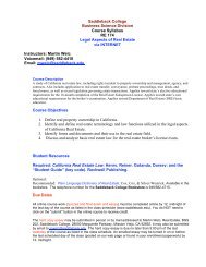
This extinct group has members dating back to the Carboniferous to the Cambrian. The body has<br />
two longitudinal furrows that run down the entire length. There is three distinct body sections: head,<br />
thorax, and abdomen.<br />
o Examine the fossil of a trilobite (Figure 6). Do not get it confuse with chitons.<br />
SUBPHYLUM CHELICERATA<br />
Figure 6. Trilobite<br />
Organisms that you will examine in this group includes: horseshoe crabs, spiders, ticks and scorpions.<br />
These organisms are grouped here because the first pair of appendages is modified into chelicerae<br />
for feeding. They also have a pair of pedipalps for capturing prey and four pairs of legs. There are<br />
two body segments: the cephalothorax and abdomen.<br />
• CLASS MEROSTOMATA<br />
This group contains the aquatic chelicerates such as the horseshoe crab and the extincit<br />
eurypterids.<br />
o Examine the horseshoe crab (Limulus). On the horseshoe shaped carapace comprises<br />
the cephalothorax, the simple eye and pair of compound eyes can be found on the<br />
dorsal surface. Behind the hinge is the abdomen. The telson is the tail.<br />
o Examine the ventral surface of Limulus (figure 7) Find the mouth. The chelicerae are the<br />
first pair of appendage used to manipulate food. The pedipalps are the second pair of<br />
appendages, used to capture prey. The remaining four pairs of appendages are the<br />
walking legs. On the abdomen, find the six leaf-like structures. The first is the genital<br />
opercula and the remaining five are the book gills (used for respiration).<br />
Figure 7: Dorsal and ventral view of Limulus<br />
<strong>Biology</strong> <strong>3B</strong> <strong>Laboratory</strong> <strong>Invertebrates</strong> <strong>II</strong> Page 7 of 17
• CLASS ARACHNIDA<br />
This class consists of members that are rather familiar to most. They include spiders, scorpions,<br />
ticks and mites. All members posses a pair of cherlicera, pedipalps and four pairs of walking legs.<br />
o Examine the Cephalothorax of the garden spider Argiope (figure 8) Note the number of<br />
eyes. Identify the chelicerae. They have been modified into fangs for the injection of<br />
poison. Find the pedipalp. What’s its general function? In males, the pedipalps are<br />
modified as an intromittent organ to deliver sperm to the female. How many walking do<br />
spiders have and what body segment (tagmata) are they located?<br />
o Obtain a dissecting scope and examine the ventral abdominal region of Argiope. Look for<br />
the lung slit at the anterior portion of the abdomen. Towards the posterior end of the<br />
abdomen, you will notice three pairs of spinnerets on a raised surface responsible for silk<br />
production.<br />
Figure 8: Ventral view of Argiope<br />
o Examine a scorpion. The pincers are the pedipalps. Note the stinger with venom sac at<br />
the distal portion of the abdomen.<br />
o Examine the slide of a tick. These are ectoparasites on various vertebrates. Many can<br />
transmit diseases such as Lyme disease and Rocky Mountain spotted fever.<br />
o Examine the slide of a mite. Mites are some of the smallest archnids.<br />
<strong>Biology</strong> <strong>3B</strong> <strong>Laboratory</strong> <strong>Invertebrates</strong> <strong>II</strong> Page 8 of 17
SUBPHYLUM CRUSTACEA<br />
In the subphylum Crustacea, mandibles are the primary feeding appendages. All crustacean<br />
appendages are biramous i.e. they have two processes extending from the base. Gills are used in<br />
respiration. Shrimp, crabs, lobsters, and many microscopic species are included in this subphylum.<br />
• CLASS MAXILLOPODA<br />
You will examine one group of organisms within this class, the barnacles. The body of the<br />
barnacle is sessile as an adult and is housed within a calcareous shell. We will have the<br />
opportunity to see living barnacles at the tidepools. When the tide is out (when we’ll be there);<br />
you will not be able to see the cirri (feeding legs).<br />
o Examine the shell of the acorn barnacle (Balanus). These are attached directly to the<br />
substrate.<br />
o Examine the gooseneck barnacle (Lepas – Figure 9). The main body is attached to the<br />
substrate via a stalk.<br />
Figure 9: Lepas<br />
• CLASS MALACOSTRACA<br />
We examine members in the order Decapoda only in this class. Decapods, as the name implies,<br />
have ten walking legs on the Cephalothorax which is covered by a hard carapace. Many have<br />
the first walking leg modified into a cheliped that is used in capturing prey and defense.<br />
o Examine the dorsal and ventral surface of a preserved crayfish (Cambarus – figure 10).<br />
Locate the following paired structures, then carefully remove them from one side and place<br />
them in the correct sequence on a sheet of paper (figure 11).<br />
Head<br />
‣ Antennules – two, short filamentous structures at the tip of the rostrum for touch,<br />
taste and equilibrium<br />
‣ Antennae – the long filamentous structure lateral to the antennules for touch and<br />
taste<br />
‣ Mandible – bears teeth for crushing food<br />
Thorax<br />
‣ First maxilla – for handling food<br />
‣ Second maxilla – food handling and bailing water from gill chamber<br />
‣ Maxilliped (1 – 3) – touch taste and food handling<br />
• 2 nd & 3 rd maxilliped – have gills for respiration<br />
‣ Cheliped (1 st walking leg) – grasping food, defense and respiration<br />
‣ Walking legs (2 – 4) – walking and respiration<br />
Abdomen<br />
‣ Swimmerets – circulates water<br />
• Males – 1 st is modified to transfer sperm to female seminal receptacle<br />
• Females – assists in carrying eggs and young (2 – 5)<br />
‣ Uropod & Telson – locomotion & protecting eggs (female)<br />
<strong>Biology</strong> <strong>3B</strong> <strong>Laboratory</strong> <strong>Invertebrates</strong> <strong>II</strong> Page 9 of 17
Figure 10 (above): Dorsal and ventral external crayfish structure<br />
Figure 11 (below): Crayfish appendages<br />
<strong>Biology</strong> <strong>3B</strong> <strong>Laboratory</strong> <strong>Invertebrates</strong> <strong>II</strong> Page 10 of 17
o Examine other representative malacostracans (crabs, shrimp, etc.).<br />
We will examine the internal anatomy when we begin the systems.<br />
SUBPHYLUM UNIRAMIA<br />
Organisms in the subphylum Uniramia also have mandibles as the primary feeding appendages,<br />
however their appendages are uniramous (having only one process extending from the base), and a<br />
tracheal system is used for respiration. This large subphylum includes the following classes: lnsecta<br />
(Insects), Diplopoda (millipedes), and Chilopoda (centipedes).<br />
• CLASS CHILOPODA<br />
o Examine preserved centipedes. They do<br />
not have a hundred legs. They do have<br />
one pair of legs per body segment. The<br />
maxilliped is modified into a fang for the<br />
delivery of poison. Centipedes are active<br />
predators living in moist places. If live<br />
ones are available, observe their<br />
locomotion.<br />
• CLASS DIPLOPODA<br />
o Examine preserved millipedes. These do<br />
not have a thousand of legs. They do<br />
Figure 12: Centipede and millipede<br />
have two pairs of legs per body segment.<br />
Like centipedes, millipedes can be found<br />
living in moist habitats. However, they are herbivores or scavengers feeding on decaying<br />
wood or leaves. Some millipedes produce cyanide as a chemical defense mechanism.<br />
Observe the locomotion of live millipedes if available.<br />
• CLASS INSECTA<br />
This is by far the largest group of animals with estimates of over one million named species. The<br />
major characteristics of insects are: three walking legs on the thorax, one pair of antennae, three<br />
body segments (head, thorax and abdomen). Many insects have either one or two pairs of wings.<br />
• Examine a preserved grasshopper and observe its external features (figure 13). The<br />
exoskeleton is divided by sutures into plates called sclerites.<br />
o HEAD: The head consists of fused sclerites forming a cranium and mouth parts. A pair of<br />
antennae arise in front of the compound eyes. Three ocelli (simple eyes) can be seen -<br />
one in the center, between the antennae, and two located above the base of the antennae.<br />
o THORAX: The thorax consists of three segments. The anterior prothorax bears the first<br />
pair of legs. The mesothorax (middle segment) bears a pair of legs and a pair of leathery<br />
wings. The metathorax (third segment) bears a pair of highly modified jumping legs and a<br />
<strong>Biology</strong> <strong>3B</strong> <strong>Laboratory</strong> <strong>Invertebrates</strong> <strong>II</strong> Page 11 of 17
pair of membranous wings which are extension of the respiratory system. The legs are<br />
jointed.<br />
• Examine the wings of a beetle (Orthoptera). The forewing is called the elytra<br />
which functions to protect the membranous hindwing that’s used for flight.<br />
• Examine the wings of a cranefly (Diptera). The forewing is for flight and the<br />
hindwing is reduced and modified for balance.<br />
o ABDOMEN: The abdomen is simple, devoid of appendages, and made up of 10 to 11<br />
segments. Note the terminal structures and use them to determine the sex of the specimen.<br />
Be sure to compare your grasshopper to one of the opposite sex. In the female, the<br />
ovipositor is for laying the eggs inside the earth. At the tip look for a pair of sensory<br />
structures known as cerci.<br />
• Observe the female cricket and notice the long ovipositor for depositing eggs.<br />
o On either side of the first abdominal segment you might see a thin membrane, called the<br />
tympanum - a hearing organ. Spiracles are present on either side of most of the segments.<br />
The spiracles are most prominent in the thorax region. They are the breathing pores of the<br />
elaborate network of the tracheal system.<br />
Figure 13: External features of a female grasshopper.<br />
Insect have mouth parts that are adapted for the type of feeding they specialize in. There are four<br />
basic mouth parts: sucking mouthparts, sponging/lapping mouthparts, siphoning and<br />
chewing/biting mouthparts. You want to be able to differentiate these mouthparts for they type of<br />
feeding (figure 14).<br />
o Examine the slide of the mosquito head (sucking mouthpart)<br />
o Examine the slide of the butterfly head (siphoning mouthpart)<br />
o Examine the slide of the fly head (lapping mouthpart)<br />
o Examine the slide of the honeybee (chewing)<br />
<strong>Biology</strong> <strong>3B</strong> <strong>Laboratory</strong> <strong>Invertebrates</strong> <strong>II</strong> Page 12 of 17
Chewing Sponging/Lapping Siphoning Sucking<br />
Figure 14: Insect mouthparts.<br />
PHLYUM ONYCHOPHORA<br />
Members in this group are often referred to as velvet or walking worms. They are an unusual group<br />
in that they have characteristics of annelids and arthropods. Onychophorans have changed very little<br />
in the past 500 million years. Aysheaia is a fossil onychophoran from the Burgess shale deposit that<br />
dates back to the mid-Cambrian. It looks very much like the modern day onychophorans. Some<br />
have called this the “missing link” between these two phyla.<br />
Onychophorans are a terrestrial species. They are active at night or when there is very high humidity.<br />
o Examine the preserved velvet worm (Peripatus).<br />
PHYLUM ECHINODERMATA<br />
Echinoderms are a group of animals that arose from bilaterally symmetrical ancestors even though<br />
the animals show pentaradial symmetry. Many of them have a bilateral larval stage and hence the<br />
radial feature may be secondarily acquired. As you have already studied, most radially symmetrical<br />
animals are sessile, however echinoderms are free moving. They are triploblastic and<br />
eucoelomate. Echinoderms are marine animals and that include: sea stars, sea urchins, sea<br />
cucumbers, and sea lilies. The body parts are arranged in "fives" around the oral/aboral axis.<br />
The most noticeable characteristics for echinoderms are the calcareous ossicles for the endoskeleton,<br />
the water vascular system with tube feet, pedicellariae, dermal branchiae and pentaradial symmetry.<br />
Major classes of Echinodermata include:<br />
Asteroidea – sea star<br />
Ophiuroidea - brittle stars, basket stars<br />
Echinoidea - sea urchins, sand dollars<br />
Holothuroidea - sea cucumbers<br />
Crinoidea - sea lilies, feather stars<br />
<strong>Biology</strong> <strong>3B</strong> <strong>Laboratory</strong> <strong>Invertebrates</strong> <strong>II</strong> Page 13 of 17
OBSERVATION OF ECHINODERMATA<br />
CLASS ASTEROIDEA<br />
Sea stars are found in relatively shallow waters, and range in size from less than an inch to nearly<br />
three feet in diameter. They feed primarily on bivalves, prying the shell to open with their tube feet,<br />
everting their stomach into the victim's body cavity, and digesting it. The larvae are known as<br />
bipinnaria and have bilateral symmetry, whereas the adult form is star shaped with arms not sharply<br />
marked off from the central disk. Sea stars can perform autotomy (self-amputation) of their arms.<br />
However, if a small portion of the central disc remains attached to it, the amputated arm can then<br />
regenerate and form a new individual (a clone).<br />
o Examine live, preserved or dehydrated sea stars (figure 15). Identify the oral and aboral surfaces.<br />
Radiating from the central disk are the five arms, noting their spiny texture (from which they get<br />
the name echinoderm - spiny skin). At the tip of each arm is the eyespot. Note the calcareous<br />
spines, dermal branchiae (skin gills - little sac-like structures on the skin) and pedicellariae<br />
(claws - tiny pincer-like structures on some living sea stars that can aid in food capture or keep the<br />
sea star clean of debris).<br />
o The madreporite (a light colored, circular, slightly raised structure located on the aboral surface<br />
near the base of two arms) is the opening, or intake, of the water vascular system. The anus is<br />
seen as a minute opening at the center of the aboral surface. Ambulacral grooves are the deep<br />
grooves that extend from the oral surface along the midline of each arm. The tube feet are seen<br />
as double rows of soft tubular "feet" on each arm, lying along and just inside the ambulacral<br />
groove. On the dehydrated specimen, the tube feet may or may not be present.<br />
o Examine the living sea star, if available, observe the madreporite plate, eyespot, sensory<br />
tentacles (located at the tip of each arm/ray), ambulacral groove, tube feet and pedicellariae<br />
(if present).<br />
Figure 15: Dorsal and ventral sea star surfaces<br />
<strong>Biology</strong> <strong>3B</strong> <strong>Laboratory</strong> <strong>Invertebrates</strong> <strong>II</strong> Page 14 of 17
CLASS OPHIUROIDEA<br />
Brittle stars are secretive echinoderms found from tidepools to great depths in the ocean. Although<br />
they are one of the more agile and abundant echinoderm, they are not frequently seen. On our<br />
tidepool trip, you will need to carefully turn over rocks to find brittle stars. If caught, brittle stars will<br />
often detach an arm (autotomize) in hopes that the predator is attracted to the wiggling arm as the<br />
brittle star escapes. Brittle stars differ from sea stars in that brittle star in that their ambulacral<br />
grooves are closed. Their tube feet are reduced and to do not have suckers. As a result, the tube<br />
feet are not used for locomotion. Instead, they “walk” with their arms.<br />
o Examine a preserved brittle (figure 16). Do not handle them roughly. On the oral surface find:<br />
mouth, five triangular jaws, oral shield located between the arms. Find the oral shield that’s<br />
modified into the madreporite plate. Note the spines on each arm.<br />
o Examine the preserved basket star.<br />
Figure 16: Oral view of the central disk of a brittle star.<br />
CLASS ECHINOIDEA<br />
This class includes sea urchins, heart urchins and sand dollars. This group is distinct in that they do<br />
not have arms and are more or less globular. They have tube feet with suckers and movable spines.<br />
Their ambulacral grooves are closed and covered by ossicles.<br />
o Examine the test of sea urchin. On the aboral surface, fine the madreporite plate, anus,<br />
ambulacral groove with tube feet pores, spine tubercle with or without the spine. On the oral<br />
surface, find the mouth and tooth.<br />
<strong>Biology</strong> <strong>3B</strong> <strong>Laboratory</strong> <strong>Invertebrates</strong> <strong>II</strong> Page 15 of 17
Figure 17: External structure of the sea urchin<br />
o Examine the Aristotle’s lantern from the chewing complex of a sea urchin.<br />
o Examine a living sea urchin. Note the moveable spines and the tube feet. Do the tube feet have<br />
suckers?<br />
o Examine the sand dollar and heart urchin or sea biscuit on display.<br />
CLASS HOLOTHUROIDEA<br />
Sea cucumbers are placed into this class and have cucumber shape. There are no arms or spines.<br />
The ambulacral grooves are closed. Sea cucumbers have soft bodies because their ossicles are<br />
microscopic and embedded in the thick muscular wall.<br />
o Examine the sea cucumber (Cucumaria) and find the mouth at one end with the anus at the other.<br />
The mouth is surrounded by modified tube feet called tentacles.<br />
<strong>Biology</strong> <strong>3B</strong> <strong>Laboratory</strong> <strong>Invertebrates</strong> <strong>II</strong> Page 16 of 17
CLASS CRINOIDEA<br />
Most crinoids (seas lilies and feather star) are an ancient group of enchinoderms with few living<br />
members. The oral end is “up” where as the aboral end has the attachment stalk or cirri.<br />
o Examine fossil or preserved specimens as available. Note the 10 arms with pinnules. The arms<br />
radiate from the calyx where the digestive and other organs are located. The calyx and arms are<br />
collectively called the crown.<br />
<strong>Biology</strong> <strong>3B</strong> <strong>Laboratory</strong> <strong>Invertebrates</strong> <strong>II</strong> Page 17 of 17