Successful Dentures Part II - Removable Prosthodontics
Successful Dentures Part II - Removable Prosthodontics
Successful Dentures Part II - Removable Prosthodontics

You also want an ePaper? Increase the reach of your titles
YUMPU automatically turns print PDFs into web optimized ePapers that Google loves.
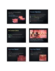
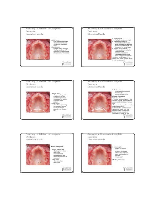
Anatomy in Relation to Complete<br />
<strong>Dentures</strong><br />
Edentulous Maxilla<br />
Anatomy in Relation to Complete<br />
<strong>Dentures</strong><br />
Edentulous Maxilla<br />
e<br />
d<br />
f<br />
i<br />
a<br />
c<br />
k<br />
j<br />
l<br />
g h<br />
d<br />
m<br />
b<br />
a. Labial frenum<br />
•Fold of mucous membrane<br />
•Does not contain muscle<br />
•Labial notch in denture is<br />
narrow<br />
b. Buccal frenum<br />
•Overlies levator anguli oris<br />
•May be moved in an A-P<br />
direction by the actions of the<br />
orbicularis oris and buccinator<br />
e<br />
d<br />
f<br />
i<br />
a<br />
c<br />
k<br />
j<br />
l<br />
g h<br />
d<br />
m<br />
b<br />
c. Labial vestibule<br />
•Reflection contains no muscle<br />
d. Anterior buccal vestibule<br />
•Overlies buccinator muscle<br />
whose fibers are downward and<br />
forward and limit the height and<br />
thickness of the buccal flange<br />
e. Posterior buccal vestibule<br />
•Thickness determined by the<br />
masseter muscle<br />
•Coronoid process of the<br />
mandible encroaches on the<br />
space during lateral excursions<br />
•Labial and buccal flanges of the<br />
denture must contact movable tissues<br />
in order to make a seal<br />
Anatomy in Relation to Complete<br />
<strong>Dentures</strong><br />
Edentulous Maxilla<br />
e<br />
d<br />
f<br />
i<br />
a<br />
l<br />
g<br />
k<br />
j<br />
h<br />
c<br />
d<br />
m<br />
b<br />
f. Hamular notch<br />
•Pterygomaxillary notch<br />
•Does not contain any<br />
muscles or ligaments to<br />
interfere with the addition<br />
of pressure with a<br />
postdam<br />
g. Fovea palatini<br />
•2 small pits representing<br />
mucous gland openings<br />
•Usually located just<br />
posterior to the vibrating<br />
line<br />
Anatomy in Relation to Complete<br />
<strong>Dentures</strong><br />
Edentulous Maxilla<br />
e<br />
d<br />
f<br />
i<br />
a<br />
l<br />
g<br />
k<br />
j<br />
h<br />
c<br />
d<br />
m<br />
b<br />
h. Vibrating line<br />
•Imaginary line across palate<br />
•Connects the<br />
pterygomaxillary notches<br />
Posterior Palatal Seal<br />
•Area not a line<br />
•Functions: border seal, prevent food<br />
impaction beneath, improve retention,<br />
compensate for shrinkage of denture<br />
resin<br />
•Pressure on displaceable mucosa that<br />
covers palatal glands<br />
•Anterior border – junction between<br />
hard and soft palate; ‘blow’ line<br />
•Posterior border (vibrating line) –<br />
junction between movable and<br />
immovable soft palate; ‘ah’ line<br />
Anatomy in Relation to Complete<br />
<strong>Dentures</strong><br />
Edentulous Maxilla<br />
Anatomy in Relation to Complete<br />
<strong>Dentures</strong><br />
Edentulous Maxilla<br />
e<br />
d<br />
f<br />
i<br />
a<br />
c<br />
k<br />
j<br />
l<br />
g h<br />
d<br />
m<br />
b<br />
Denture bearing areas<br />
i. Residual alveolar ridge<br />
•Crest is primary stress<br />
bearing area<br />
•Fibrous CT least<br />
displaceable and best<br />
able to carry the stress of<br />
mastication<br />
j. Palatal rugae<br />
•Secondary stress<br />
bearing area<br />
e<br />
d<br />
f<br />
i<br />
a<br />
c<br />
k<br />
j<br />
l<br />
g h<br />
d<br />
m<br />
b<br />
k. Incisive papilla<br />
•Guards the incisive<br />
canal<br />
•Pressure will interfere<br />
with the blood and nerve<br />
supply causing a burning<br />
sensation<br />
•Provide relief<br />
l. Median palatine raphe