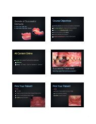
Successful Dentures Part II - Removable Prosthodontics
Successful Dentures Part II - Removable Prosthodontics
Successful Dentures Part II - Removable Prosthodontics

You also want an ePaper? Increase the reach of your titles
YUMPU automatically turns print PDFs into web optimized ePapers that Google loves.
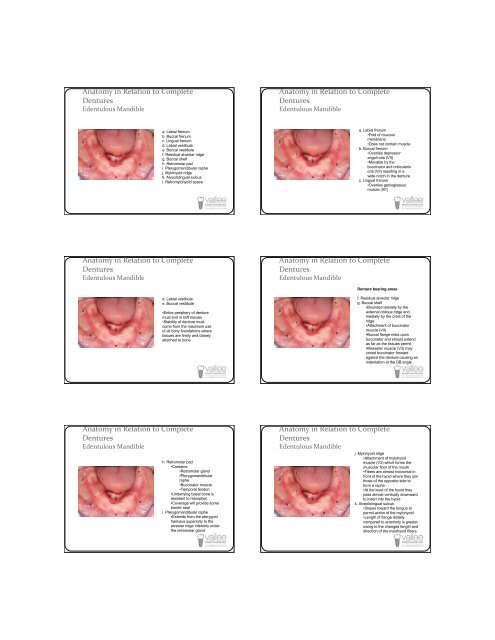
Anatomy in Relation to Complete<br />
<strong>Dentures</strong><br />
Edentulous Mandible<br />
Anatomy in Relation to Complete<br />
<strong>Dentures</strong><br />
Edentulous Mandible<br />
g<br />
b<br />
l<br />
f<br />
j<br />
k<br />
c<br />
a<br />
d<br />
i<br />
h<br />
e<br />
a. Labial frenum<br />
b. Buccal frenum<br />
c. Lingual frenum<br />
d. Labial vestibule<br />
e. Buccal vestibule<br />
f. Residual alveolar ridge<br />
g. Buccal shelf<br />
h. Retromolar pad<br />
i. Pterygomandibular raphe<br />
j. Mylohyoid ridge<br />
k. Alveololingual sulcus<br />
l. Retromylohyoid space<br />
g<br />
b<br />
l<br />
f<br />
j<br />
k<br />
c<br />
a<br />
d<br />
i<br />
h<br />
e<br />
a. Labial frenum<br />
•Fold of mucous<br />
membrane<br />
•Does not contain muscle<br />
b. Buccal frenum<br />
•Overlies depressor<br />
anguli oris (V<strong>II</strong>)<br />
•Movable by the<br />
buccinator and orbicularis<br />
oris (V<strong>II</strong>) resulting in a<br />
wide notch in the denture<br />
c. Lingual frenum<br />
•Overlies genioglossus<br />
muscle (X<strong>II</strong>)<br />
Anatomy in Relation to Complete<br />
<strong>Dentures</strong><br />
Edentulous Mandible<br />
Anatomy in Relation to Complete<br />
<strong>Dentures</strong><br />
Edentulous Mandible<br />
Denture bearing areas<br />
g<br />
b<br />
l<br />
f<br />
j<br />
k<br />
c<br />
a<br />
d<br />
i<br />
h<br />
e<br />
d. Labial vestibule<br />
e. Buccal vestibule<br />
•Entire periphery of denture<br />
must end in soft tissues<br />
•Stability of denture must<br />
come from the maximum use<br />
of all bony foundations where<br />
tissues are firmly and closely<br />
attached to bone<br />
g<br />
b<br />
l<br />
f<br />
j<br />
k<br />
c<br />
a<br />
d<br />
i<br />
h<br />
e<br />
f. Residual alveolar ridge<br />
g. Buccal shelf<br />
•Bounded laterally by the<br />
external oblique ridge and<br />
medially by the crest of the<br />
ridge<br />
•Attachment of buccinator<br />
muscle (V<strong>II</strong>)<br />
•Buccal flange rests upon<br />
buccinator and should extend<br />
as far as the tissues permit<br />
•Masseter muscle (V3) may<br />
crowd buccinator forward<br />
against the denture causing an<br />
indentation at the DB angle<br />
Anatomy in Relation to Complete<br />
<strong>Dentures</strong><br />
Edentulous Mandible<br />
g<br />
b<br />
l<br />
f<br />
j<br />
k<br />
c<br />
a<br />
d<br />
i<br />
h<br />
e<br />
h. Retromolar pad<br />
•Contains:<br />
•Retromolar gland<br />
•Pterygomandibular<br />
raphe<br />
•Buccinator muscle<br />
•Temporal tendon<br />
•Underlying basal bone is<br />
resistant to resorption<br />
•Coverage will provide some<br />
border seal<br />
i. Pterygomandibular raphe<br />
•Extends from the pterygoid<br />
hamulus superiorly to the<br />
alveolar ridge inferiorly under<br />
the retromolar gland<br />
Anatomy in Relation to Complete<br />
<strong>Dentures</strong><br />
Edentulous Mandible<br />
g<br />
b<br />
l<br />
f<br />
j<br />
k<br />
c<br />
a<br />
d<br />
i<br />
h<br />
e<br />
j. Mylohyoid ridge<br />
•Attachment of mylohyoid<br />
muscle (V3) which forms the<br />
muscular floor of the mouth<br />
•Fibers are almost horizontal in<br />
front of the hyoid where they join<br />
those of the opposite side to<br />
form a raphe<br />
•At the level of the hyoid they<br />
pass almost vertically downward<br />
to insert into the hyoid<br />
k. Alveololingual sulcus<br />
•Slopes toward the tongue to<br />
permit action of the mylohyoid<br />
•Length of flange distally<br />
compared to anteriorly is greater<br />
owing to the changed length and<br />
direction of the mylohyoid fibers