

PDF Download - Glidewell Dental Labs
PDF Download - Glidewell Dental Labs
PDF Download - Glidewell Dental Labs

You also want an ePaper? Increase the reach of your titles
YUMPU automatically turns print PDFs into web optimized ePapers that Google loves.
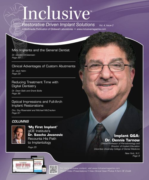
Inclusive<br />
Restorative Driven Implant Solutions Vol. 4, Issue 2<br />
A Multimedia Publication of <strong>Glidewell</strong> Laboratories • www.inclusivemagazine.com<br />
Mini Implants and the General Dentist<br />
Dr. Gordon Christensen<br />
Page 20<br />
Clinical Advantages of Custom Abutments<br />
Dr. Jack Hahn<br />
Page 29<br />
Reducing Treatment Time with<br />
Digital Dentistry<br />
Dr. Dean Saiki and Grant Bullis<br />
Page 36<br />
Optical Impressions and Full-Arch<br />
Implant Restorations<br />
Drs. Guy Rosenstiel and Michael McCracken<br />
Page 67<br />
COLUMNS<br />
‘My First Implant’<br />
gIDE Institute’s<br />
Dr. Sascha Jovanovic<br />
Recounts His Path<br />
to Implantology<br />
Page 25<br />
Implant Q&A:<br />
Dr. Dennis Tarnow<br />
Clinical Professor of Periodontology and<br />
Director of Implant Education<br />
Columbia University College of <strong>Dental</strong> Medicine<br />
New York, N.Y.<br />
Page 9<br />
For more exclusive content, visit www.inclusivemagazine.com<br />
Watch Video Presentations • View Clinical Case Photos • Earn CE Credit
On the Web<br />
Here’s a sneak peek at additional<br />
Inclusive magazine content available online<br />
ONLINE Video Presentations<br />
■ Dr. Dennis Tarnow reflects on the changing world of implant dentistry,<br />
exploring dental education, implant spacing, the international<br />
implant marketplace and more.<br />
■ Dr. Gordon Christensen offers his thoughts on the growing role of<br />
general practitioners in placing implants, the benefits of mini implants<br />
and the ongoing debate over restorative materials.<br />
■ Dr. Timothy Kosinski illustrates the clinical flexibility of the Inclusive<br />
® Tooth Replacement System by summarizing the restoration of<br />
an edentulous anterior maxilla with an implant-retained bridge.<br />
■ Dr. Perry Jones discusses how digital scanning, CBCT, guided surgery<br />
and treatment planning software are improving the predictability of<br />
restorative outcomes.<br />
■ Dzevad Ceranic, CDT, details the laboratory production of a<br />
Screw-Retained Hybrid Denture, demonstrating the efficiency of an<br />
all-in-one approach to implant therapy.<br />
■ Two-Day Custom Abutments and Crowns: Learn about taking advantage<br />
of the latest digital impression systems and CAD/CAM technology<br />
with digitally produced custom abutments and crowns from<br />
<strong>Glidewell</strong> Laboratories.<br />
Inclusive Magazine Digital Edition<br />
Inclusive magazine is now optimized for all<br />
popular desktop, tablet and smartphone<br />
platforms. To try out the new digital edition from<br />
your computer or favorite mobile device, visit<br />
www.inclusivemagazine.com.<br />
Look for these icons on the pages that follow<br />
for additional content available online<br />
■ R&D Corner: Catch a glimpse of digital implant restorations from taking<br />
and submitting the digital impression through CAD/CAM design<br />
and fabrication of the abutment and crown in “Digital in a Day.”<br />
gide lecture-on-demand preview<br />
■ Dr. Egon Euwe presents key considerations involved in effectively<br />
managing soft tissue when treatment planning and completing an<br />
implant restoration in this excerpt from the gIDE video lecture “Soft<br />
Tissue Management Around Implants.”<br />
ONLINE CE credit<br />
■ Get free CE credit for the material in this issue with each test you<br />
complete and pass. To get started, visit our website and look for the<br />
articles marked with “CE.”<br />
– www.inclusivemagazine.com –
Contents<br />
9<br />
20<br />
29<br />
36<br />
Implant Q&A: An Interview with Dr. Dennis Tarnow<br />
Dr. Dennis Tarnow — In this exclusive interview, Dr. Dennis<br />
Tarnow, clinical professor of periodontology and director of implant<br />
education at Columbia University College of <strong>Dental</strong> Medicine<br />
in New York, shares his experience as a clinician, researcher and<br />
educator. Discover what this expert thinks on topics ranging from<br />
the relationship between endodontics and implantology, to training<br />
the general dentist to place implants, to immediate loading, biologic<br />
width, guided surgery and digital scanning.<br />
Mini Implants: Insight from Dr. Gordon Christensen<br />
Dr. Gordon Christensen — Dr. Gordon Christensen, director of<br />
Practical Clinical Courses and CEO of CLINICIANS REPORT, has<br />
shared his knowledge and experience with <strong>Glidewell</strong> Laboratories<br />
on numerous occasions. This article recounts his recent insights on<br />
the current and future state of implants, detailing his thoughts on<br />
mini implants as a permanent solution, the advantages of mini implants<br />
for both patients and clinicians, and the factors that can lead<br />
to their failure or success.<br />
The Clinical Advantages of Custom Abutments<br />
Dr. Jack Hahn — Custom abutments are an excellent option for<br />
implant treatment, with advantages including patient-specific soft<br />
tissue management during the healing phase and final restorations<br />
that adhere precisely to the patient’s gingival architecture. Even so,<br />
many doctors are hesitant to make the switch from stock abutments.<br />
Dr. Jack Hahn discusses his conversion to custom abutments,<br />
highlighting the efficiency, precision, cost-effectiveness, and optimal<br />
functional and esthetic outcomes these customized, digitally produced<br />
components provide over their generic counterparts.<br />
Reducing Treatment Time with Digital Dentistry<br />
Dr. Dean Saiki and Grant Bullis — For edentulous patients with<br />
ill-fitting lower dentures, persistent soreness can make wearing<br />
their denture difficult, even with periodic relines. Screw-retained<br />
fixed implant restorations can greatly improve comfort and chewing<br />
function for these patients. Dr. Dean Saiki and <strong>Glidewell</strong> Laboratories<br />
Director of Implant R&D and Digital Manufacturing Grant<br />
Bullis use a case example to explain how advanced treatment protocols<br />
that leverage digital impressions, treatment planning, guided<br />
surgery and dental CAD/CAM technology can transform implant<br />
therapy, shorten treatment times and improve prosthetic outcomes.<br />
– Contents – 1
Contents<br />
49<br />
55<br />
Bridging the Gap: Restoring Partially Edentulous<br />
Spaces and Maximizing Treatment Options with the<br />
Inclusive ® Tooth Replacement System<br />
Dr. Timothy Kosinski — Continual improvements in surgical and<br />
restorative technology, materials and methodology are allowing<br />
clinicians to approach implant cases with more certainty and<br />
predictability than ever before. Dr. Timothy Kosinski explores<br />
how these advances in implantology are maximizing the range of<br />
treatment options available to patients, presenting an edentulous<br />
maxillary anterior case that illustrates how a comprehensive<br />
approach to implant dentistry that uses patient-specific components<br />
and CAD/CAM technology can help to guide any case toward<br />
predictable success.<br />
Clinical Case Report: iTero ® Digital Scanning<br />
Technology and Tooth-Supported Surgical Guides<br />
Dr. Perry Jones and Zach Dalmau — Digital scanning technology<br />
used in conjunction with CBCT scanning can offer a high level of<br />
confidence to practitioners both in terms of implant placement and<br />
implant restoration. With a clinical case report involving the replacement<br />
of two missing maxillary left bicuspids, Dr. Perry Jones<br />
and Zach Dalmau show how digital technology can be used to capture<br />
data and design and fabricate tooth-supported surgical guides,<br />
temporary abutments, cement-retained provisional crowns and final<br />
zirconia restorations with the highest level of accuracy.<br />
ALSO IN THIS ISSUE<br />
8 Trends in Implant Dentistry<br />
Model-Less Restorations<br />
25 My First Implant<br />
gIDE Institute’s<br />
Dr. Sascha Jovanovic<br />
46 Product Spotlight<br />
Two-Day Custom Abutments and<br />
Crowns from Intraoral Scans<br />
73 Lab Sense<br />
All Together Now: Inclusive ® Tooth<br />
Replacement System Removables<br />
79 Clinical Tip<br />
Managing Implants in<br />
Patients with Bruxism<br />
83 Small Diameter Implants<br />
Benefits of CBCT-Assisted<br />
Guided Surgery<br />
67<br />
Optical Impressions and<br />
Full-Arch Implant Restorations: A Case Study<br />
Drs. Guy Rosenstiel and Michael McCracken — When restoring<br />
implant cases, clinicians strive for accurate impressions and wellfitting<br />
restorations, yet highly accurate implant impressions can<br />
be difficult to achieve using conventional methods. In a case<br />
study involving a full-arch maxillary implant restoration, Drs. Guy<br />
Rosenstiel and Michael McCracken detail a cutting-edge impression<br />
technique that utilizes scanning abutments and an optical scanner,<br />
demonstrating how this all-digital process can increase impression<br />
accuracy and improve the fit of the final restoration, while saving<br />
the clinician time and money.<br />
2<br />
– www.inclusivemagazine.com –
Letter from the Editor<br />
This issue of Inclusive magazine showcases some of the leaders in the<br />
field of implant dentistry. Implant dentistry is in a state of constant<br />
evolution, with breakthroughs in digital and CAD/CAM technologies<br />
at the forefront. Coupled with practice decisions relying on evidencebased<br />
dentistry, strong scientific research and a focus on improved dental<br />
materials, these technologies position implantology at the leading edge<br />
of dentistry.<br />
An exclusive interview with Dr. Dennis Tarnow of Columbia University<br />
College of <strong>Dental</strong> Medicine touches on his journey in implant dentistry,<br />
offering a historical perspective, current trends and what he believes<br />
the future holds. Continuing education is the basis for personal growth<br />
for clinicians and desired outcomes for patients. As one of the most wellrespected<br />
practitioners and educators in our field, Dr. Tarnow speaks to this<br />
importance and other implant-related topics that are well worth the read.<br />
As clinicians, we’re constantly looking for ways to improve quality of life<br />
for our patients. Providing solutions to patients with moderate to severe<br />
bone loss who are not candidates for bone grafting procedures and face<br />
socioeconomic challenges has always been difficult. The validation of<br />
the use of small-diameter implants for the retention of tissue-supported<br />
removable prostheses allows practitioners to address these patients’<br />
concerns. Drawing from his years of experience as a prosthodontist and<br />
educator, Dr. Gordon Christensen discusses his experience with smalldiameter<br />
implants and current trends in patient treatment.<br />
Several articles highlight the use of intraoral scans to initiate a completely<br />
digital workflow, from the diagnostic phase through delivery of the<br />
temporary and final restorations. Complemented with the merging of<br />
intraoral scan data and DICOM data from CBCT scans, precision in<br />
treatment planning and implant placement has reached a new level.<br />
Take note of the digital workflow incorporated at the laboratory level to<br />
fabricate case-specific custom temporary components and final abutments,<br />
allowing for efficiency in tissue contouring.<br />
As you’ll discover in this issue, the combination of esthetics, science,<br />
materials and technology makes for an interesting and exciting future for<br />
implant dentistry.<br />
With kind regards,<br />
Dr. Siamak Abai<br />
Editor-in-Chief, Clinical Editor<br />
inclusivemagazine@glidewelldental.com<br />
– Letter from the Editor – 3
Contributors<br />
■ SIAMAK ABAI, DDS, MMedSc<br />
Dr. Siamak Abai earned his DDS degree from<br />
Columbia University in 2004, followed by<br />
two years of residency in general dentistry.<br />
After two years of general private practice<br />
in Huntington Beach, Calif., he returned to<br />
academia and received an MMedSc degree<br />
and a certificate in prosthodontics from<br />
Harvard University. Dr. Abai brings nearly 10 years of clinical,<br />
research and lecturing experience to his role as director of<br />
clinical research and development for the Implant division<br />
of <strong>Glidewell</strong> Laboratories. He is also editor-in-chief and<br />
clinical editor of Inclusive magazine. Before joining <strong>Glidewell</strong><br />
in January 2012, Dr. Abai practiced at the Wöhrle <strong>Dental</strong><br />
Implant Clinic in Newport Beach, Calif. Contact him at<br />
inclusivemagazine@glidewelldental.com.<br />
■ DZEVAD CERANIC, CDT<br />
Dzevad began his career at <strong>Glidewell</strong> Laboratories<br />
in 1999 as a waxer and metal finisher,<br />
working his way up to ceramist. In 2007, he was<br />
promoted to general manager of the Full-Cast<br />
department, successfully conducting a complete<br />
turnaround of that business in 18 months.<br />
Dzevad completed the eight-month “Implants<br />
A to Z” course at UCLA School of Dentistry in 2009, while<br />
assuming a new role as general manager of the <strong>Glidewell</strong> Laboratories<br />
Implant department. Dzevad has spearheaded an annual<br />
department growth of more than 38 percent for four consecutive<br />
years, leading to his 2013 promotion to vice president of lab<br />
operations. Dzevad has a certificate in dental technology from<br />
Pasadena City College and recently completed a management<br />
development program at the USC Marshall School of Business.<br />
Contact him at inclusivemagazine@glidewelldental.com.<br />
■ GRANT BULLIS, MBA<br />
Grant Bullis, director of implant R&D and digital<br />
manufacturing at <strong>Glidewell</strong> Laboratories,<br />
began his dental industry career at Steri-Oss<br />
(now Nobel Biocare) in 1997. Since joining<br />
the lab in 2007, Grant has been integral in<br />
obtaining FDA 510(k) clearances for the company’s<br />
Inclusive ® Mini Implants, Tapered Implant<br />
System and Custom Implant Abutments. In 2010, he was<br />
promoted to director and now oversees all aspects of CAD/CAM,<br />
implant product development and manufacturing for more<br />
than 1,000 implant and prosthetic components at the lab. He<br />
has an MBA from the Keller Graduate School of Management.<br />
Contact him at inclusivemagazine@glidewelldental.com.<br />
■ Zach Dalmau<br />
Zach Dalmau began his dental career in 2006<br />
at the nSequence Center for Advanced Dentistry<br />
in Reno, Nev. As the director of guided implant<br />
surgery and 3-D diagnostic imaging at<br />
nSequence, he played a key role in building the<br />
CT guided implant surgery and 3-D diagnostic<br />
imaging departments there from the ground<br />
up. In September 2009, he moved to Baltimore, Md., to work<br />
at Materialise <strong>Dental</strong> Inc. There, he managed the design and<br />
production of all SimPlant ® SurgiGuides ® for the North American<br />
market. Zach joined <strong>Glidewell</strong> Laboratories in October 2011<br />
and plays a key role in the company’s R&D efforts for digital<br />
treatment planning and implant manufacturing. Contact him<br />
at inclusivemagazine@glidewelldental.com.<br />
■ David Casper, MBA<br />
David Casper is president of San Diegobased<br />
IOS Technologies Inc., a wholly owned<br />
subsidiary of <strong>Glidewell</strong> Laboratories and<br />
manufacturer of the IOS FastDesign System.<br />
After beginning his dental industry career<br />
at CeraMed in 1990, David went on to hold<br />
various positions in sales and global marketing<br />
for Nobel Biocare, GE and Sybron Implant Solutions. He joined<br />
<strong>Glidewell</strong> in 2011 as vice president of implant sales & business<br />
development, contributing significantly to the lab’s growth<br />
through his business development efforts. David assumed his<br />
current role as president of IOS Technologies in June 2012.<br />
He has an MBA from LaSalle University. Contact him at<br />
inclusivemagazine@glidewelldental.com.<br />
■ GORDON J. CHRISTENSEN, DDS, MSD, Ph.D.<br />
Dr. Gordon Christensen is a practicing<br />
prosthodontist in Provo, Utah. He is director<br />
of Practical Clinical Courses and CEO of<br />
CLINICIANS REPORT ® . He is also a Diplomate<br />
of the American Board of Prosthodontics;<br />
Fellow and Diplomate of the ICOI; Fellow of<br />
the AO, ACD, ICD, ACP and Royal College of<br />
Surgeons of England; Honorary Fellow of the AGD; and an<br />
Associate Fellow of the AAID. Dr. Christensen and his wife<br />
Rella are co-founders of the nonprofit Gordon J. Christensen<br />
CLINICIANS REPORT (formerly CRA Newsletter). Contact him<br />
at 801-226-6569 or info@pccdental.com.<br />
4<br />
– www.inclusivemagazine.com –
■ JACK A. HAHN, DDS<br />
Dr. Jack Hahn earned his DDS from The Ohio<br />
State University College of Dentistry, and<br />
completed postgraduate coursework at Boston<br />
University, New York University, the University<br />
of Michigan and the University of Kentucky.<br />
A pioneer in the field of implant dentistry,<br />
Dr. Hahn developed the NobelReplace dental<br />
implant system for Nobel Biocare and has been actively involved<br />
in placing and restoring implants for 40 years. In addition<br />
to lecturing to dentists around the world, he maintains a<br />
private practice in Cincinnati, Ohio, focused on placing and<br />
restoring implants. He received the Aaron Gershkoff Lifetime<br />
Achievement Award in implant dentistry in 2004. Contact him<br />
at replace7@mac.com.<br />
■ TIMOTHY F. KOSINSKI, DDS, MAGD<br />
Dr. Timothy Kosinski graduated from the University<br />
of Detroit Mercy School of Dentistry<br />
and received a Master of Science degree in biochemistry<br />
from Wayne State University School<br />
of Medicine. An adjunct assistant professor<br />
at UDM School of Dentistry, he serves on the<br />
editorial review board of numerous dental<br />
journals and is a Diplomate of the ABOI/ID, ICOI and AO.<br />
Dr. Kosinski is a Fellow of the AAID and received his Mastership<br />
in the AGD, from which he received the 2009 Lifelong Learning<br />
and Service Recognition award. Contact him at 248 -646 -8651,<br />
drkosin@aol.com or www.smilecreator.net.<br />
■ PERRY E. JONES, DDS, MAGD<br />
Dr. Perry Jones received his DDS from Virginia<br />
Commonwealth University School of Dentistry,<br />
where he has held adjunct faculty positions<br />
since 1976. He maintains a private practice in<br />
Richmond, Va. One of the first GP Invisalign ®<br />
providers, Dr. Jones has been a member of<br />
Align’s Speaker Team since 2002, presenting<br />
more than 250 Invisalign presentations. He has been involved<br />
with CADENT optical scanning technology since its release to<br />
the GP market and is currently beta testing its newest software.<br />
Dr. Jones belongs to numerous dental associations and is a<br />
fellow of the AGD. Contact him at perry@drperryjones.com.<br />
■ SASCHA A. JOVANOVIC, DDS, MS<br />
Dr. Sascha Jovanovic received his training in<br />
periodontics, implant dentistry and prosthodontics<br />
at UCLA School of Dentistry, Loma<br />
Linda University and University of Aachen in<br />
Germany, respectively, and holds a Master of<br />
Science degree in oral biology from UCLA. He<br />
maintains a private practice in Los Angeles,<br />
specializing in dental implants and periodontics, and is academic<br />
chairman of the Global Institute for <strong>Dental</strong> Education<br />
(gIDE) and course director of implant dentistry for UCLA Continuing<br />
<strong>Dental</strong> Education. Dr. Jovanovic lectures worldwide<br />
and has published numerous materials on implant dentistry.<br />
Contact him at 310-820-9641 or sascha@jovanoviconline.com.<br />
■ MICHAEL McCRACKEN, DDS, Ph.D.<br />
Dr. Michael McCracken is co-director of the<br />
Comprehensive Implant Residency Program<br />
(CIRP), a yearlong comprehensive implant<br />
education institute in Birmingham, Ala. After<br />
completing dental school at University of<br />
North Carolina at Chapel Hill and a prosthodontic<br />
residency at University of Alabama<br />
at Birmingham, he received a Ph.D. in biomedical engineering.<br />
Dr. McCracken is a part-time professor at UAB, where<br />
he has served as associate dean for education, director of<br />
graduate prosthodontics and director of the implant training<br />
program. He maintains an active research program within<br />
the university and a private practice focused on implant dentistry.<br />
He lectures internationally on dental implants and<br />
complex oral rehabilitation. Contact him at 256-797-1964 or<br />
inclusivemagazine@glidewelldental.com.<br />
– Contributors – 5
Contributors<br />
■ Guy Rosenstiel, DMD, MAGD<br />
Dr. Guy Rosenstiel received his dental degree<br />
from the University of Alabama School of<br />
Dentistry and completed a general practice<br />
residency at the Birmingham VA Medical<br />
Center. He teaches continuing dental education<br />
involving implant surgery and implant prosthodontics<br />
to private practitioners and AEGD<br />
residents through CIRP in Bessemer, Ala. His interests in IV<br />
sedation, surgery and all aspects of general dental practice<br />
enable him to continually enjoy his career as a dentist, both as<br />
a faculty member and as a private practitioner. Contact him at<br />
205-979-8655 or drguy@agd.org.<br />
■ Dean H. Saiki, DDS<br />
Dr. Dean Saiki graduated from the USC<br />
School of Dentistry in 1988. He maintains a<br />
private practice in North County San Diego,<br />
Calif., specializing in cosmetic, laser, implant<br />
and digital dentistry. He has been a member<br />
of the ADA, CDA and San Diego County<br />
<strong>Dental</strong> Society since 1989, as well as other<br />
advanced study clubs including the Trojan <strong>Dental</strong> Study Club.<br />
Dr. Saiki is trained and certified in dental soft tissue lasers<br />
and CAD/CAM technology. Contact him at 760-732-3456 or<br />
dentist@deansaiki.com.<br />
■ Dennis P. Tarnow, DDS<br />
Dr. Dennis Tarnow is currently clinical<br />
professor of periodontology and director of<br />
implant education at Columbia University<br />
College of <strong>Dental</strong> Medicine. Formerly, he was<br />
a professor and chairman of the Department<br />
of Periodontology and Implant Dentistry<br />
at New York University College of Dentistry<br />
(NYUCD). Dr. Tarnow has certificates in periodontics and<br />
prosthodontics from NYUCD and is a Diplomate of the American<br />
Board of Periodontology. He also has a private practice in<br />
New York City. Dr. Tarnow has published more than<br />
100 articles on perio-prosthodontics and implant dentistry,<br />
co-authored several textbooks and lectured extensively in the<br />
U.S. and internationally. Contact him at 212-752-7937 or<br />
www.nycsdonline.com.<br />
Publisher<br />
Jim <strong>Glidewell</strong>, CDT<br />
Editor-in-Chief, clinical editor<br />
Siamak Abai, DDS, MMedSc<br />
Inclusive Marketing & Education Manager<br />
Jennifer Archer<br />
Managing Editors<br />
Grant Bullis; David Casper;<br />
Dzevad Ceranic, CDT; Greg Minzenmayer<br />
Creative Director<br />
Rachel Pacillas<br />
Copywriters/copy editors<br />
David Frickman, Jennifer Holstein,<br />
Chris Newcomb, Keith Peters, Tina Quan,<br />
Megan Strong, Eldon Thompson<br />
digital marketing manager<br />
Kevin Keithley<br />
Graphic Designers/Web Designers<br />
Emily Arata, Jamie Austin, Deb Evans,<br />
Juan Gallardo, Kevin Greene, Joel Guerra,<br />
Tony Hsiao, Audrey Kame, Allison Newell,<br />
Phil Nguyen, Ty Tran, Makara You<br />
Photographers/Videographers<br />
Sharon Dowd, Mariela Lopez;<br />
James Kwasniewski, Andrew Lee,<br />
Marc Repaire, Stanford Southall, Sterling Wright,<br />
Maurice Wyble, Peter Yun<br />
Illustrator<br />
Phil Nguyen<br />
coordinatorS/AD Representatives<br />
Teri Arthur, Suzeanne Harms, Vivian Tsang<br />
If you have questions, comments or suggestions, e-mail us at<br />
inclusivemagazine@glidewelldental.com. Your comments may<br />
be featured in an upcoming issue or on our website.<br />
© 2013 <strong>Glidewell</strong> Laboratories<br />
Neither Inclusive magazine nor any employees involved in its publication<br />
(“publisher”) make any warranty, expressed or implied, or assumes any<br />
liability or responsibility for the accuracy, completeness, or usefulness of<br />
any information, apparatus, product, or process disclosed, or represents<br />
that its use would not infringe proprietary rights. Reference herein to<br />
any specific commercial products, process, or services by trade name,<br />
trademark, manufacturer or otherwise does not necessarily constitute<br />
or imply its endorsement, recommendation, or favoring by the publisher.<br />
The views and opinions of authors expressed herein do not necessarily<br />
state or reflect those of the publisher and shall not be used for advertising<br />
or product endorsement purposes. CAUTION: When viewing the<br />
techniques, procedures, theories and materials that are presented, you<br />
must make your own decisions about specific treatment for patients and<br />
exercise personal professional judgment regarding the need for further<br />
clinical testing or education and your own clinical expertise before trying<br />
to implement new procedures.<br />
Inclusive is a registered trademark of Inclusive <strong>Dental</strong> Solutions.<br />
6<br />
– www.inclusivemagazine.com –
Online Exclusive<br />
R&D<br />
CORNER<br />
“Digital in a Day”<br />
See how your implant case can be turned around in a single day!<br />
View the video “Digital in a Day” as we follow an implant<br />
restoration in real time from scanning in the doctor’s<br />
office to receipt in the laboratory, design, manufacture and<br />
shipping back to the doctor. Every step of the process will<br />
have a time stamp in the upper right-hand corner of the<br />
screen showing the elapsed time since the start. This video<br />
presents a convincing argument for the adoption of digital<br />
dentistry into your practice.<br />
You can view “Digital in a Day” and other Inclusive<br />
magazine videos on your computer or favorite mobile<br />
device. To watch these videos anytime, anywhere, visit<br />
www.inclusivemagazine.com.<br />
www.inclusivemagazine.com
Trends in<br />
Implant Dentistry<br />
Model-Less Restorations<br />
70 %<br />
Year-Over-Year Growth Rate<br />
for Model-Less Crowns<br />
March 2012–March 2013<br />
Growing ranks of clinicians are using chairside digital impression<br />
systems to save money and time on their restorations. They benefit<br />
from working with a fully integrated digital dental lab that can<br />
produce restorations entirely from a digital impression — without a<br />
physical model. Ease of use and accuracy make digital impressions<br />
a superior option for a wide range of restorations. In addition to<br />
crowns, bridges and veneers, custom implant abutments can be<br />
produced directly from a digital impression. Model-less restorations<br />
are priced lower, exhibit a better fit and cut the in-lab working time<br />
in half. They deliver more benefits for less money, and the dental<br />
community is taking notice.<br />
4000<br />
Total Model-Less Restorations — June 2010–March 2013<br />
3500<br />
3000<br />
2500<br />
2000<br />
1500<br />
1000<br />
500<br />
0<br />
879 934 942 958<br />
817 787<br />
729<br />
85 115 131 212 309<br />
1171<br />
995<br />
1644<br />
1520<br />
1278 1276 1355<br />
3241<br />
2897 2937<br />
2713<br />
2665<br />
2492<br />
2369<br />
2272 2314<br />
2178 2145<br />
1863 1923 1964 1971<br />
June<br />
July<br />
August<br />
September<br />
October<br />
November<br />
December<br />
January<br />
February<br />
March<br />
April<br />
May<br />
June<br />
July<br />
August<br />
September<br />
October<br />
November<br />
December<br />
January<br />
February<br />
March<br />
April<br />
May<br />
June<br />
July<br />
August<br />
September<br />
October<br />
November<br />
December<br />
January<br />
February<br />
March<br />
2010 2011<br />
2012 2013<br />
Source: <strong>Glidewell</strong> Laboratories internal data<br />
Watch here for emerging trends<br />
Check back here for more observations in the next issue.<br />
8<br />
– www.inclusivemagazine.com –
Implant&<br />
Q A:<br />
Go online for<br />
in-depth content<br />
An Interview with Dr. Dennis Tarnow<br />
Interview of Dennis P. Tarnow, DDS<br />
by Managing Editor David Casper<br />
Photo courtesy of Columbia University<br />
Dr. Dennis Tarnow, expert in the field of implantology and leader<br />
in dental implant continuing education, is currently clinical<br />
professor of periodontology and director of implant education<br />
at Columbia University College of <strong>Dental</strong> Medicine in New York.<br />
Here, the seasoned practitioner shares his experience on<br />
topics ranging from the relationship between endodontics and<br />
implantology, to training the general dentist to place implants,<br />
to immediate loading, biologic width around implants, guided<br />
surgery and digital scanning.<br />
David Casper: Dr. Tarnow, you’re known<br />
around the world as the leader in dental<br />
implant continuing education. Everywhere<br />
we go, whether it’s undergraduate<br />
or postgraduate, it’s clear you’ve touched<br />
the lives of so many dentists around the<br />
world. Tell us about this journey. How<br />
did it start, and what ignited this passion<br />
for continuing education?<br />
Dr. Dennis Tarnow: Well, I graduated<br />
in 1972 and went for a general practice<br />
residency at Brookdale University<br />
Hospital in Brooklyn. There I realized I<br />
was being trained beautifully as an intern<br />
and as a general practitioner, but<br />
I wanted to do a lot more. So I wound<br />
up pursuing a double specialty in<br />
both periodontology and prosthodontics.<br />
And I loved education, which<br />
stimulated my entire way of thinking.<br />
– Implant Q&A: An Interview with Dr. Dennis Tarnow – 9
I knew I was very fortunate to be exposed<br />
to some of the world’s greatest<br />
teachers who also had great dedication,<br />
and I learned to see their passion,<br />
and what their teaching did for me<br />
and for my fellow students. So I think<br />
what gave me my own passion for<br />
education was seeing how many people<br />
you can impact as a teacher.<br />
I have a very active group practice<br />
in New York City, and when I teach,<br />
whether I lecture to 10 people or<br />
2,000, I think of how many people I<br />
may indirectly touch — to me, that is<br />
the beauty of being an educator. If they<br />
can learn even one thing in an hour<br />
lecture and take it home, they will do<br />
it for the rest of their lives. Hopefully,<br />
they will not only propagate better<br />
dentistry, but they will also feel better<br />
about themselves. So, education was<br />
really inspired in me by my teachers<br />
— people like Dr. Sigmund Stahl<br />
in periodontology, and Drs. Harold<br />
Litvak, Frank Celenza Sr. and Sidney<br />
Silverman in prosthodontics — these<br />
people were great mentors to me.<br />
Today, I think of doing their quality<br />
of work and continuing that thought<br />
process.<br />
DC: When did you first start the emphasis<br />
in dental implants as part of your<br />
curriculum in the universities?<br />
DT: I completed my internship — we<br />
called it an internship then, what is<br />
now a general practice residency or<br />
GPR — at Brookdale Hospital under<br />
Dr. Norman Cranin. Unfortunately,<br />
Norman passed away last year. But<br />
Norman was one of the pioneers in<br />
implant dentistry, particularly in subperiosteal<br />
and blade implants. He was<br />
the first to develop what was called an<br />
“anchor implant,” which was a modification<br />
of the blade. During that year,<br />
we worked with him and did some<br />
subperiosteals and some blades. So<br />
my first implant was with Norman<br />
Cranin and it was quite an interesting<br />
thing — we were placing blade<br />
implants into edentulous ridges. That<br />
was in 1972 or ’73. It wasn’t until 1982<br />
that I was exposed to them again in<br />
Toronto at a continuing education<br />
course given by Drs. Per-Ingvar Brånemark<br />
and George Zarb.<br />
DC: You were at that original Toronto<br />
conference on tissue-integrated prostheses?<br />
DT: I was at that original conference.<br />
I was being trained as a periodontist<br />
and a prosthodontist, but what was<br />
always interesting to me was that, at<br />
the time, no periodontist was allowed<br />
to take the course. So I actually took<br />
it legally as a prosthodontist! It was<br />
quite a learning experience. I knew<br />
that this was a paradigm shift. And<br />
when I saw what George Zarb was<br />
showing, which was a duplicate of<br />
what the Brånemark group had done<br />
in Sweden, I realized that this was<br />
something totally different.<br />
As a periodontist and prosthodontist I<br />
was saving root tips. I was doing hemisections<br />
and root resections, and these<br />
teeth were holding fine for a few years<br />
— five years, seven years, even 10<br />
years. But in attending this conference<br />
I realized there was another world out<br />
there. To me, this was the next step<br />
in perio and prosthetics. We now had<br />
something that looked amazing and<br />
was different from what I saw with the<br />
blades. These were being what was<br />
termed “osseointegrated” — and we<br />
never looked back. We learned what it<br />
was that Brånemark did that allowed<br />
integration, and of course since then<br />
we have modified it from there, along<br />
with the rest of the world. Everybody<br />
has improved on what was there, from<br />
that fantastic work and great thinking<br />
of Brånemark.<br />
DC: A paradigm shift, for sure.<br />
DT: It was a paradigm shift. Recently, I<br />
was lecturing on when to save a tooth,<br />
and when I went back and looked<br />
at the post-ops from some of my old<br />
cases from 20 to 30 years ago, I realized<br />
that there are not many of the rootresected<br />
teeth around anymore. But<br />
the implants that I did in 1982, 1984,<br />
1985 — most of them are still in and<br />
functioning well. And so, especially<br />
with the original machined implant,<br />
once it took, there were very minimal<br />
periodontal problems. With many of<br />
the teeth that we were holding on to,<br />
in addition to periodontal disease, the<br />
people also had problems with decay.<br />
So how nice it is to give somebody<br />
a new root that doesn’t decay — it’s<br />
called an implant. Whatever implant<br />
you choose, there’s still no decay. So, in<br />
most people, the crown on an implant<br />
definitely lasts longer than the crown<br />
on a tooth, especially if the patient is<br />
prone to decay or periodontal disease.<br />
10<br />
– www.inclusivemagazine.com –
DC: We often hear that endodontics is<br />
pre-implant therapy. Do you share that<br />
view?<br />
DT: [Laughing] Not quite, no. As a<br />
matter of fact, the person I lectured<br />
with today is a great endodontist —<br />
Dr. John West. We supposedly had a<br />
head-to-head debate on endo versus<br />
implants, but it’s not that way. Endo<br />
is just one aspect of treating a tooth.<br />
Great endodontists today have a 95 to<br />
97 percent success rate. So, that’s not<br />
the question. It’s not about the apex<br />
— that’s about a 3 to 5 percent failure<br />
rate — it’s about what’s left of the<br />
natural tooth that becomes the real<br />
treatment planning problem. You can<br />
seal an apex, but what does the rest<br />
of the tooth look like? Is the patient<br />
prone to decay? Is the patient prone to<br />
periodontal issues? Is the patient still<br />
susceptible even though the apex was<br />
sealed beautifully? Is the tooth strong<br />
enough to withstand the occlusion?<br />
What’s the fracture rate of posts in<br />
general and the fracture rate of teeth?<br />
So, I see it as what they call the “etiological<br />
pile.” The pile builds up on<br />
a given patient. If you have a patient<br />
sitting in your chair, and the patient<br />
is prone to periodontal disease, prone<br />
to tooth decay, and they’re in your<br />
chair because they have lost teeth and<br />
are having problems in their mouth,<br />
it’s not just whether the apex can<br />
be sealed. Decay rate in five years<br />
might cause a problem, certainly in<br />
10 years. Post fractures, post loosenings,<br />
debonding of teeth — all of these<br />
things become an additional pile. And<br />
if the patient has a few of these things<br />
on their list, not just the apex of a<br />
tooth being the problem, the pile suddenly<br />
can become overwhelming and<br />
you’re leaning toward an implant. So<br />
it’s not endo versus implants. Some of<br />
my best friends are endodontists!<br />
DC: [Laughing] And your best referring<br />
doctors, too?<br />
DT: And my best referring doctors,<br />
yes. We work very closely together.<br />
But if they can’t save a tooth, we go to<br />
an implant. But, clearly, for a lot of the<br />
teeth that I used to treat endodontically<br />
— hemisections and things like<br />
that — I don’t need it anymore, other<br />
than to hold a temporary while I<br />
transition these patients out of those<br />
teeth and put in implants. Long-term,<br />
an implant is a far superior restoration<br />
in our hands, and I’ve been doing<br />
both. Being a periodontist and a<br />
prosthodontist, I saved all those teeth<br />
for so many years. I built my practice<br />
on that originally. And if you ever<br />
want a great lecture on furcations, I’ll<br />
be glad to give it to you. But the reality<br />
is that nobody wants to listen to that.<br />
If I did a lecture today on furcations,<br />
nobody would show up. Or if I did a<br />
lecture today on implant esthetics versus<br />
teeth — forget it.<br />
In most people,<br />
the crown on an<br />
implant definitely<br />
lasts longer than<br />
the crown on a<br />
tooth, especially<br />
if the patient is<br />
prone to decay<br />
or periodontal<br />
disease.<br />
DC: How would you assess the state of<br />
dental education on implants at the<br />
university level today?<br />
DT: I think it’s gotten a lot better.<br />
When we first started with implants,<br />
we realized this needed to be taught<br />
more universally because the students<br />
were not getting the right kind of<br />
information. Some of them were just<br />
getting lectures. There were six or<br />
eight lectures they used to get back in<br />
the 1980s, but nothing that was handson.<br />
That was only for the grad students<br />
— the prosthodontists, periodontists<br />
and oral surgeons. So the general<br />
dentists who were coming out of school<br />
were totally untrained. And I really<br />
mean untrained. They didn’t know<br />
how to do it. So if a patient came to<br />
one of these former students missing a<br />
tooth and needing it replaced, and the<br />
patient was their first since graduating<br />
from dental school and opening their<br />
own office, what was the new dentist<br />
going to tell them?<br />
DC: A bridge?<br />
DT: If you’ve never even done one implant<br />
restoration, then you’re going<br />
to do a 3-unit bridge. Because that is<br />
what you were trained to do in school.<br />
But this has finally changed. Now it’s<br />
a requirement to restore some missing<br />
teeth with implants while in dental<br />
school. Even if you’re not doing the<br />
surgery, you should at least be able to<br />
do the restoration. So it’s still minimal<br />
compared to the number of patients,<br />
and it’s more costly in general, although<br />
not much more costly than a<br />
three-unit bridge. <strong>Dental</strong> insurance is<br />
just starting to cover implants, as you<br />
know. But before that, people would<br />
say, “Well, it’s the same price for a<br />
3-unit bridge or a single implant.” But<br />
the insurance didn’t cover the implant<br />
part. They’d think, “Well, my insurance<br />
will give me some money for the<br />
3-unit bridge, so I’m going to have<br />
to go that route.” So, they were still<br />
cutting down perfectly good virgin<br />
teeth to put 3-unit bridges on.<br />
DC: What are your thoughts on training<br />
general dentists at the undergraduate<br />
level to place implants?<br />
DT: This is a politically charged question<br />
in some respects because it takes<br />
the place of what an oral surgeon and<br />
a periodontist want to do. Today, even<br />
prosthodontists are being cross-trained<br />
for certain easier cases. It is ethical<br />
now in their code of responsibility to<br />
get trained in simpler cases of placing<br />
implants. Right now, many schools<br />
– Implant Q&A: An Interview with Dr. Dennis Tarnow – 11
are struggling just to get the implant<br />
cases restored by the undergraduates,<br />
believe it or not. But you probably see<br />
it routinely in a laboratory the size<br />
of <strong>Glidewell</strong>, that everybody is doing<br />
prosthetics on implants. But that’s not<br />
so easy yet. The young practitioners<br />
are still gun-shy and still feel a little<br />
overwhelmed by it. This is also true for<br />
many of the older practitioners, who<br />
were never exposed to implants during<br />
their training. So it’s more like the<br />
practitioners who are five to 10 years<br />
out of school who are into the new<br />
technology. They have learned about it<br />
and know they have a whole career to<br />
expose themselves to it, so they’re the<br />
ones doing most of the work now. But<br />
to go back to your original question, as<br />
far as training people in school, I think<br />
we’re just doing a very limited amount<br />
when someone has to have the training.<br />
We don’t want anybody to take a<br />
two-day course and then think they’re<br />
an implantologist.<br />
DC: Right.<br />
DT: That’s where we see the mistakes.<br />
Sixty percent of my new cases in my<br />
private office today are redoing other<br />
people’s work. This is an absolutely<br />
frightening scenario. And it’s gotten<br />
worse since a few of the companies<br />
started offering a weekend course.<br />
One company in particular was<br />
just into sales, and so they said,<br />
essentially, “Come to us to take the<br />
course for two days, and we’ll sell<br />
you all the equipment, we’ll give you<br />
some implants, you give us a whole<br />
bunch of money — and then you’re<br />
an implantologist.” And the reality is<br />
these people didn’t know what they<br />
were doing. They had no surgical<br />
knowledge; they had no background<br />
in biology and wound healing. They<br />
were just mechanics. And many of<br />
them were bad mechanics. What we<br />
saw was just a horror show. It keeps<br />
me busy, but believe me, it’s almost<br />
like gut money. It’s hard because the<br />
patients are unhappy. They’re angry.<br />
And that’s not good for dentistry, and<br />
it’s certainly not good for the patient,<br />
which is really the main story.<br />
But I think that the proper training<br />
is now available. I started a program<br />
at Columbia to train general dentists<br />
who want to learn how to do implant<br />
procedures properly.<br />
DC: Is it a four-year or a two-year program?<br />
DT: We do both. We do it with the<br />
undergrads if they’re in an honors<br />
program for placing implants. For<br />
restorative, they always should restore<br />
and do clip-ons and attachments,<br />
things of that sort. But for the surgical<br />
part, we are just doing a pilot program.<br />
Those with the desire to learn more<br />
and who want to do the surgery go<br />
through courses, and during their<br />
senior year they take an elective in<br />
implants and implant surgery. By the<br />
end of the year we involve them in the<br />
placement of implants for some simple<br />
cases. But they have to take more than<br />
just their regular courses.<br />
DC: Are they partnered with a mentor<br />
— maybe one of the post-graduate residents?<br />
DT: They partner with the faculty.<br />
Columbia is a small enough school that<br />
we have that personal touch. We get<br />
to know the students, we work with<br />
them, and they come up and watch the<br />
periodontists and the surgeons and<br />
eventually do the procedures themselves.<br />
But they need the didactics, too.<br />
So they are allowed to join the elective<br />
courses and get more education.<br />
I started a program at Columbia in<br />
2010 for the general dentist to learn<br />
how to place implants. It’s either one<br />
weekend a month for six months,<br />
which is a 12-day program; or two<br />
one-week programs, the first of which<br />
is six days, and then they come back<br />
about eight weeks later for another six<br />
days. That’s 12 days of didactics. So<br />
it’s definitely not a two-day course. It<br />
teaches the biology of implants, wound<br />
healing around implants, anatomy and<br />
the entry-level basics to get you feeling<br />
comfortable with incision designs.<br />
These are the things people need to be<br />
comfortable, to know what they should<br />
and shouldn’t do. So, as important as<br />
knowing which cases to take on, they<br />
need to learn when to say, “OK, this is<br />
for the periodontist, and this is for the<br />
oral surgeon — or someone trained<br />
with appropriate skills.” So they know<br />
how not to get into trouble.<br />
There is no reason why a general dentist<br />
can’t learn how to place implants.<br />
If they can prep a tooth, and they are<br />
good at that, then there’s no reason<br />
why they can’t angulate an implant.<br />
But they must know the biology; they<br />
must know the diagnosis and treatment<br />
planning of the case. And therein<br />
lies the difference. I could teach a<br />
high school student how to place an<br />
implant. If they have any kind of manual<br />
dexterity, give me a week or so<br />
and I can teach them how to place an<br />
implant. But they wouldn’t know what<br />
they’re doing. They would be like a little<br />
monkey: monkey see, monkey do.<br />
If you say, “Now go do this,” the monkey<br />
goes and does it. But he won’t<br />
know what he’s doing. He just knows<br />
that he is supposed to do it. You can<br />
train someone to carve anatomy into<br />
an amalgam, and to carve an occlusal<br />
surface; they can be an artist and<br />
carve it, but they have no idea what<br />
amalgam is, or what composite is, or<br />
what zirconium is, and which material<br />
should be used and why. That’s the<br />
doctor. Otherwise, they’re just doing it<br />
because they were told to do it, and<br />
they become like a carpenter. That’s<br />
the level we want to try to avoid by<br />
offering proper education. But do I see<br />
people doing it in the future in undergrad?<br />
Yes. Do I think it’s tomorrow? No.<br />
It’s just because of the lack of patients<br />
available. But they will just do the simple<br />
cases, and they will know when to<br />
refer. This is very important. The periodontist<br />
and the oral surgeon should<br />
not feel threatened. If you become the<br />
teacher, you will be busy — busier<br />
than you can imagine.<br />
As you know, we’ve only penetrated<br />
about 8 percent of the market in the<br />
U.S. today — 10 percent at best. And it’s<br />
been that way for a while. The expansion<br />
is not there, but it could be. Other<br />
12<br />
– www.inclusivemagazine.com –
markets have been highly penetrated,<br />
such as Korea and Japan. Statistically<br />
speaking, per 10,000 people, they have<br />
more people who need and are getting<br />
implants. So why are we behind? Part<br />
of it is the general dentist population,<br />
who patients go to and trust —<br />
DC: The quarterback.<br />
DT: More like the gatekeeper. The guy<br />
you trust, who does not feel comfortable<br />
doing an implant, so he does a<br />
bridge. And the patient doesn’t question<br />
it, unless the patient is well educated<br />
and realizes there is another<br />
possible route. Today, the Internet is<br />
helping. I’ve said this to my own academy<br />
in periodontology: “Don’t hesitate<br />
to train the general dentist now.”<br />
They’ve been fighting general dentists,<br />
and I’m on record now saying this, as<br />
a teacher: “If people want to learn<br />
something, they’re going to learn it.”<br />
When I at first was not allowed to take<br />
the course that Brånemark gave, I had<br />
to take it as a prosthodontist, not as<br />
a periodontist. But we periodontists<br />
had a will. This was something that<br />
we wanted, and we found a way to<br />
learn. The general dentist who wants<br />
to learn is going to do the same thing.<br />
So why don’t we be the ones to teach<br />
them in a way that is accepted, so they<br />
can decide whether they want to do<br />
implants? They’ll still come to us for<br />
referrals in the more difficult cases,<br />
and even in some regular cases — and<br />
for their mother and their father. What<br />
I’m saying is, if we had penetrated the<br />
market by 50, 60, 70 percent, everybody<br />
would be looking at each other’s<br />
cases, and stealing each other’s cases.<br />
But here’s the key: Because we’ve<br />
only penetrated the market by about 8<br />
percent, there’s such room for expansion,<br />
for the whole pie to grow. And<br />
that’s when everybody will be happy,<br />
including the general dentists who<br />
may want to do something different,<br />
as well as the periodontists, the oral<br />
surgeons and the prosthodontists. The<br />
market will expand for everybody.<br />
The key is to get the word out to the<br />
general dentist and to the patient that<br />
Because we’ve only penetrated the market<br />
by about 8 percent, there’s such room for<br />
expansion … And that’s when everybody<br />
will be happy, including the general<br />
dentists who may want to do something<br />
different, as well as the periodontists, the<br />
oral surgeons and the prosthodontists.<br />
implants are safe and reliable when<br />
done properly. The periodontists,<br />
prosthodontists and oral surgeons can<br />
all do their part, and everybody should<br />
be happy because the pie will grow.<br />
DC: We’ve seen that the metric you mentioned<br />
— the number of implants placed<br />
per 10,000 inhabitants — is pretty common<br />
within the industry. One thing<br />
we’ve noticed is that those countries<br />
with the highest level of penetration are<br />
also the countries where the cost is the<br />
lowest — not only the treatment cost to<br />
the patient, but also the cost of the components,<br />
the materials, the implants. Do<br />
you think there is a direct correlation,<br />
or is this a coincidence?<br />
DT: Well, I think it may be part<br />
coincidence, due to some of these<br />
countries tending to be more<br />
nationalistic from a business standpoint.<br />
Whereas the big companies had market<br />
share in many parts of the world —<br />
often total market share — some places<br />
like Korea, to give an example, began<br />
to develop their own industry, and<br />
they began making their own implants.<br />
So there were Korean companies<br />
making their own implants, and they<br />
were moderately priced — not even<br />
inexpensively priced. Some people in<br />
these countries said, “OK, the quality is<br />
there, I’m Korean, and I prefer to buy a<br />
Korean implant.” So the big companies<br />
lost some of their market share there.<br />
The same thing is happening in Brazil.<br />
Part of that is because it’s inexpensive,<br />
but part of that is a feeling of national<br />
pride in being able to place a Korean or<br />
a Brazilian implant, for example. If you<br />
look at the implant marketplace around<br />
the world, let’s say in Germany — which<br />
is a big implant market, as you know<br />
— one of the German-made implants<br />
is always one of the biggest sellers,<br />
certainly one of the top two or three.<br />
In Switzerland, it’s Swiss implants. In<br />
Sweden, the Swedish implant is number<br />
one. So there’s a bit of a nationalistic<br />
flavor to the implant world.<br />
DC: It’s the World Cup of dental implants.<br />
DT: That’s about right. In the U.S. for<br />
a while, it was that way, too. We were<br />
certainly strong contenders. Americanmade<br />
implants just felt comfortable.<br />
That’s what I think was going on. Of<br />
course the economy today is so difficult,<br />
certainly in Europe. There are<br />
ups and downs in Asia. And in America<br />
now, we are just slowly starting to<br />
come out of the recession. But we’re<br />
getting there. I certainly see that in<br />
my practice. Three years ago, when<br />
we were all about to go over the cliff,<br />
almost literally, people just stopped<br />
spending. You couldn’t tell patients<br />
they needed a full-mouth rehabilitation.<br />
The words just wouldn’t come<br />
out of your mouth. The question was<br />
more, “What do I need now to hold<br />
me off?” They weren’t ready to spend.<br />
– Implant Q&A: An Interview with Dr. Dennis Tarnow – 13
The same thing is happening in Europe<br />
right now. Some of my graduate students<br />
who are in top practices in<br />
Spain, for example, are in such tough<br />
situations because people are just not<br />
spending. They don’t know what’s happening<br />
to the euro; they don’t know<br />
what’s happening to the economy.<br />
Patients are not coming in except for<br />
the most critical needs — and I’m talking<br />
about really established practices,<br />
not clinicians who are just starting out.<br />
So, clearly, if the economy is affected,<br />
people are going to say: “Hey, can I<br />
get the same quality for less money?<br />
And if it happens to be a company in<br />
my own nation, even better.”<br />
DC: That’s an interesting take on things.<br />
If we look at research for a second, and<br />
the library’s worth of articles you’ve<br />
written over the last several years, we see<br />
that many of the papers you’ve authored<br />
have changed the way doctors treat patients<br />
when it comes to dental implants.<br />
DT: Hopefully, for the better [laughs].<br />
DC: Well, we all think so. One of the<br />
articles that comes to mind for me is<br />
that first paper in 1997 that refers to<br />
immediate loading. 1 It was the first paper<br />
that had good long-term data showing<br />
pretty good results with immediate<br />
loading and immediate function given<br />
the right parameters. What’s your take<br />
on immediate loading now, immediate<br />
provisionalization? Has it changed?<br />
DT: Dr. Paul Schnitman was the first<br />
to do something like that for implants.<br />
He did seven cases and published an<br />
article — in 1990. 2 He published that<br />
article with seven cases with three<br />
implants: one in the midline and<br />
two small ones in the back. And he<br />
buried the other five in the front. So<br />
if the three failed, the patient would<br />
still have five implants to finish that<br />
case — a regular hybrid case. So 21<br />
implants were loaded. And when I read<br />
that, I nearly fell off my chair. Why?<br />
Because this was 1990, and we were<br />
all doing Brånemark-style implants<br />
and we submerged everything. Here<br />
he took three implants and an old<br />
denture — the previous denture —<br />
and just secured it like a tripod, with<br />
one implant in the front and two in<br />
the back. And we assumed that all<br />
of those implants would fail because<br />
it went against every principle that<br />
Brånemark had taught us: no loading,<br />
submergence — all of the things that<br />
Brånemark taught us were being<br />
violated for those three implants.<br />
Immediate loading. Non-submerged.<br />
Everything was wrong. And yet the<br />
three implants took. Everybody said,<br />
“Overall, three out of 21 have failed,<br />
so it wasn’t a high success rate.” But<br />
I was amazed that 18 survived! I’m a<br />
glass-half-full kind of guy. For me the<br />
amazing thing was that 18 survived.<br />
They were not supposed to survive.<br />
In fact, Dr. Leonard Linkow was teaching<br />
with me at New York University<br />
when I inherited the job as director<br />
of implantology. Lenny is one of the<br />
fathers, or pioneers, of implants. He<br />
was involved with blades and screws<br />
— many people don’t know he had<br />
patents on screws. Anyway, I was so<br />
amazed that 18 of the implants hadn’t<br />
failed, that they osseointegrated — not<br />
fibrous encapsulation. So I said to Lenny,<br />
“Look at what Schnitman is doing.”<br />
And he said, “Yeah, well, that works.”<br />
“Really?” I asked. And he went on to<br />
say: “I’ve been doing it for years. I may<br />
have more failures than anybody else,<br />
but I also have more successes than<br />
anybody else.” When I asked him why<br />
it worked, he said: “It went around<br />
the turn of the arch. As long as you<br />
go around the turn of the arch, you’ll<br />
have a high success rate. It doesn’t<br />
matter. Just don’t move or take off the<br />
temporary and everything will be fine.<br />
Wait three or four months, and then,<br />
even if you have to drill the temporary<br />
off, the implants are going to be tight.”<br />
And I said, “Tight, like osseointegrated<br />
tight?” He said: “Yeah, osseointegrated<br />
tight. No problem.”<br />
So right after this, I started the process<br />
of looking at a very standardized<br />
way of going about this. The biggest<br />
problem that Schnitman said he had<br />
on these three was that they were<br />
short implants. The extra ones he put<br />
posteriorly behind the mental foramen<br />
were short, and in softer bone. So I<br />
said: “What if we loaded four or<br />
five? Let’s pick mandibles that have<br />
plenty of bone.” So as a process at<br />
the school, we picked mandibles that<br />
we could put lots of implants in, 8 or<br />
10 — more than we would normally<br />
need — but we would load half of<br />
them and submerge the other half. So,<br />
medical-legally, the patient would be<br />
good either way. So we loaded four to<br />
six implants on these 10 consecutive<br />
cases over five years. And what was<br />
amazing to us is we had success after<br />
success after success. By going around<br />
the turn of the arch we had enough<br />
support. It was more than just a tripod<br />
effect. By having something similar<br />
to the legs on a chair, we had a good<br />
A-P spread, as we call it, or a good<br />
anterior-posterior spread. The farther<br />
forward the anterior implants are, and<br />
the farther back the implants are in the<br />
posterior, the more stable they are, just<br />
like a chair. So we realized that as long<br />
as you can decrease the torque during<br />
loading, lateral forces get diminished.<br />
And today we know that if there is less<br />
than 150 microns of lateral motion<br />
during the healing phase, we don’t<br />
If you go around<br />
the turn of an<br />
arch with a good<br />
A-P spread,<br />
during the<br />
healing phase<br />
the implants will<br />
take. I am very<br />
comfortable<br />
saying that.<br />
14<br />
– www.inclusivemagazine.com –
get fibrous encapsulation. By going<br />
around the turn of an arch, just like<br />
splinting loose teeth around the turn<br />
of an arch, the mobility stops because<br />
the fulcrum of rotation is not through<br />
all of the teeth, or not through all of<br />
the implants. The fulcrum of rotation<br />
is in the tongue area in the center. So<br />
if there’s no easy fulcrum to rotate<br />
around, they don’t move, they don’t<br />
rotate and, therefore, they’re stable.<br />
And today it still stands.<br />
I’ll tell you exactly what Lenny told<br />
me. It’s a different understanding of<br />
what we need to do. If you go around<br />
the turn of an arch with a good A-P<br />
spread, during the healing phase the<br />
implants will take. I am very comfortable<br />
saying that.<br />
Schnitman once did a review on this,<br />
presenting his article from 1990 and<br />
my article from 1997, but between<br />
that there’s almost nothing written<br />
on immediate loading in the 1990s.<br />
It’s amazing. There was one article by<br />
some people from the University of<br />
Pennsylvania. Of course, Henry Salama<br />
did one, but that was only one<br />
case report — just an oddball case.<br />
Now there are hundreds of articles.<br />
But it wasn’t until ’97 that it all broke<br />
loose. So I feel good about that. It’s<br />
a long answer, but a meaningful one<br />
because people get into trouble loading<br />
single teeth or straight-line splints.<br />
When they go around the turn of an<br />
arch, they’re going to be cooking. You<br />
go around the turn of an arch and<br />
splint them together (not originally),<br />
and let everything integrate for two<br />
or three months; then you can do<br />
whatever you want. They integrate<br />
at the same percentage rate as everything<br />
else. Everybody knows that<br />
now, of course. But you don’t want<br />
to do a single tooth and then put it<br />
into occlusion. That’s stupid. Let the<br />
other teeth take on the load during<br />
the healing phase. You just want to<br />
avoid lateral forces during the healing<br />
phase. So, as long as you have good<br />
stability, put a temporary on, keep it<br />
out of occlusion, and then let the other<br />
teeth take on the load during the<br />
healing phase, you can do whatever<br />
you want after that.<br />
DC: In 2000, you wrote an article that<br />
has become the widely adopted standard<br />
that remains unchallenged. The<br />
article talks about the spacing between<br />
implants and preservation of the relative<br />
inter-implant bone height. Why did<br />
it take so long for that to be evaluated?<br />
What caused you to look at that?<br />
DT: That’s a great question. What<br />
caused me to look at it was a patient<br />
named Anne-Marie. Anne-Marie was a<br />
patient in my office, and we did two<br />
implants right next to each other. She<br />
had a high smile line, and I didn’t<br />
know any better. Previously, I’d had<br />
patients with low smile lines and I’d<br />
put two in, and if the papilla was a<br />
little short, it never bothered anybody.<br />
As long as you filled it up and the patient<br />
didn’t hiss during their speaking,<br />
they accepted it as long as the lip line<br />
was low. But Anne-Marie had a high<br />
smile line, and then you could see<br />
normal teeth on the right side, but on<br />
the left side the papilla was too short.<br />
And no matter what I did to stimulate<br />
the papilla like I would between two<br />
teeth with my 5 mm rule, I couldn’t<br />
get it to grow 5 mm. It wasn’t happening.<br />
So the problem was right in front<br />
of us but we didn’t see what it was.<br />
So, I did the study with Sang-Choon<br />
Cho and Steven Wallace, and we<br />
started to look at the distance between<br />
implants. 3 The reason why that paper<br />
was very significant was that, up<br />
to that time, we were all looking<br />
at the threads down the side of the<br />
implant. Think of the shoulder of<br />
the implant. Dr. Jan Lindhe, Dr. Bo<br />
Bergman, Dr. Daniel Buser and all the<br />
– Implant Q&A: An Interview with Dr. Dennis Tarnow – 15
2 mm<br />
1.5 mm<br />
1.5 mm<br />
others were looking at the implant<br />
from the shoulder down, and we all<br />
wanted to know how much bone<br />
loss took place after you connected<br />
the abutment and took it on and off<br />
a few times. Dr. Joachim Hermann,<br />
Dr. Ingemar Abrahamsson and others<br />
did great studies on this. And they<br />
were all looking at different implants,<br />
but they were looking at the vertical<br />
bone loss from where the implantabutment<br />
interface was. This usually<br />
went down to the first thread of these<br />
older implants. Now there’s nothing<br />
magical about the first thread; it’s just<br />
that there’s a 1.5 mm distance. That’s<br />
the biologic width reformation. On the<br />
Brånemark implant, that happened<br />
to be where the first thread was, so<br />
everybody thought it was magical, but<br />
it was pure coincidence.<br />
What we weren’t paying attention to<br />
was something I noticed with my patient<br />
Anne-Marie. When I looked at the<br />
case, I realized I had lost the bone between<br />
the two implants because they<br />
were too close. That’s what it looked<br />
like to me. So I went back, because I<br />
couldn’t get the papilla to grow back.<br />
In fact, on that case I eventually had to<br />
submerge a perfectly healthy implant.<br />
I just published this case recently in<br />
Clinical Advances in Periodontics with<br />
Dr. Paul Fletcher, one of my partners.<br />
And I realized, when I went back to<br />
look at the research, what we were<br />
missing: Everybody was looking at<br />
how much bone loss we had vertically,<br />
but there is a horizontal component to<br />
that bone loss. By having a horizontal<br />
component to bone loss, the question<br />
became how deep was it? That article<br />
with Cho and Wallace was the first to<br />
measure that. It was 1.4 mm on that<br />
typical Brånemark-style implant —<br />
1.4 mm horizontally. If you lose bone<br />
not just vertically down the side but<br />
also horizontally, the biologic width<br />
is three-dimensional. The inflammation<br />
and irritation from the abutment<br />
connection moves the bone away from<br />
it vertically and also horizontally. So<br />
what happens is, if two implants are<br />
3 mm apart, each of them are separate<br />
horizontal components and the<br />
bone peak is not affected. But if two<br />
implants are closer than 3 mm, they’re<br />
going to overlap, and so the 1.5 mm<br />
from one implant and the 1.5 mm<br />
from the other implant overlap and the<br />
bone crest will go down; therefore, the<br />
support for the papilla will be missing.<br />
I thought that was the answer, that I’d<br />
solved the world’s problem. So I told<br />
everybody, “Keep the implants 3 mm<br />
apart, and the bone will be higher and<br />
the papilla will come back.” So I figured<br />
that was the problem. And it still<br />
is a problem; 13 years later it’s still one<br />
of the things you need to pay attention<br />
to. But I wrote that and everybody<br />
went and did that, yet in many cases<br />
the papilla still didn’t come back. So<br />
it wasn’t just that horizontal distance.<br />
Even though the peak of bone wasn’t<br />
being lost, the papilla still didn’t come<br />
back fully. What else was I missing,<br />
along with the rest of the world?<br />
The article written in 2003 4 was really<br />
the pivotal one that finally helped<br />
explain it. I went to five different offices<br />
— Drs. David Garber and Henry<br />
Salama in Atlanta, our office in New<br />
York City, Drs. Stuart Froum, Ann<br />
Magna and Paul Fletcher — and what<br />
we found was the same. We all came<br />
up with the same curve. What was<br />
the height of the tissue between any<br />
two implants? What was the peak of<br />
tissue? Not to the contact point, because<br />
those are two artificial crowns<br />
and it could be any shape. But I asked,<br />
“How much tissue forms over the crest<br />
of bone between any two implants?<br />
What’s the average height?” And we<br />
found the average height was 3.4 mm,<br />
not 5 mm and 6 mm as it is between<br />
two teeth. And that’s when it hit me.<br />
We almost never got to 5 mm between<br />
any two implants, no matter what<br />
16<br />
– www.inclusivemagazine.com –
type they were. And what was wrong?<br />
There was a missing piece. Even if the<br />
implants were 3 mm apart, we still had<br />
a problem. The problem was that we<br />
were missing the supracrestal attachment<br />
that a natural tooth has. That’s<br />
why a single-tooth implant looks so<br />
good on the lecture circuit. Everybody<br />
can’t wait to show you a single-tooth<br />
implant because if there’s a healthy<br />
tooth on either side, the biologic<br />
width on a healthy tooth, the connective<br />
tissue fibers and epithelium are<br />
supracrestal. So they actually wind up<br />
helping to support the papilla. That’s<br />
2 mm of biologic width supracrestal,<br />
not subcrestal. But if I have an implant<br />
there instead of a tooth, the biologic<br />
width is subcrestal down to the first<br />
thread. So the biologic width is below<br />
the crest of bone, not above the crest.<br />
That’s why we’re 2 mm short. And<br />
that’s what we need.<br />
So, where are we today? The real challenge<br />
today that we’re all working on<br />
is getting attachments onto an abutment<br />
and keeping them there, and<br />
not continuing to break the seal. And<br />
keeping the biologic width, which we<br />
can get to form on an abutment — we<br />
have enough data to show that certainly<br />
happens. The issue is when we<br />
take an abutment on and off multiple<br />
times as opposed to “one abutment,<br />
one time” — I think Dr. Henry Salama<br />
quoted that. If we keep breaking that<br />
seal, the biologic width goes down. If<br />
we get it right with the initial seating<br />
of the abutment, we want to keep it<br />
there if we can. So, based on Abrahamsson’s<br />
work, maybe one change is<br />
OK, but more than that, and it starts to<br />
drop down. So we’re at a point where<br />
what we must do in order to put two<br />
implants next to each other in the esthetic<br />
zone, with a high smile line and<br />
to keep the papilla, is get supracrestal<br />
biologic width. This is a very big challenge,<br />
and that’s what everybody is<br />
trying to work on now. Between two<br />
centrals we get away with it because<br />
if the papilla is a little short, it’s right<br />
down the middle. So you don’t see any<br />
comparison, you just close it down<br />
with a little fatter contact point and it’s<br />
OK. As long as you have a little scallop<br />
and no black spot, you’re OK, even if<br />
it’s a little short. But central lateral, in<br />
a high smile, with a lateral cuspid? It’s<br />
a nightmare waiting to happen. A high<br />
smile with the natural teeth on the<br />
other side, with good papilla, always<br />
looks lopsided to the eye. So that’s<br />
what we have to be careful about.<br />
People used to<br />
say, ‘Look at my<br />
implant. There’s<br />
no biologic width<br />
on my implant.’<br />
That’s ridiculous.<br />
There’s always a<br />
biologic width on<br />
an implant.<br />
DC: Does implant design play into any<br />
of this?<br />
DT: It sure does. That original article in<br />
2000 that you asked about was without<br />
a platform switch. So now, what we’re<br />
looking at — myself with Dr. Xavier<br />
Vela Nebot and others at the Barcelona<br />
Osseointegration Research Group — is<br />
platform switching. We looked at this<br />
and saw that when you put a platform<br />
switch on an implant, the horizontal<br />
component is no longer 1.4 mm or 1.5<br />
mm like the old Brånemark implant<br />
was. What we realize now is that the<br />
horizontal component is only about<br />
0.3 mm to 0.5 mm. This is because the<br />
platform switch is there, and there’s<br />
an abutment in the middle, and there’s<br />
a shoulder to the implant instead of<br />
being straight up, and the abutment<br />
connection between the bevel, or<br />
shoulder, allows room so the bone<br />
doesn’t have to move as far down to<br />
protect itself from the irritant. The<br />
bone can stay up there higher because<br />
the extra coverage of soft tissue acts<br />
as a buffer between the possible<br />
irritation of the abutment connection<br />
to the bone. Long story short, we lose<br />
less bone down the side when we<br />
have a platform switch; we only see<br />
about 0.3 mm to 0.6 mm, depending<br />
on the system, but under 0.5 mm or in<br />
that range, whereas we used to see 1.5<br />
mm to 2 mm. Now we’re seeing about<br />
0.5 mm down from the shoulder.<br />
That’s because there’s still a biologic<br />
width there.<br />
People used to say, “Look at my implant.<br />
There’s no biologic width on my<br />
implant.” That’s ridiculous. There’s always<br />
a biologic width on an implant.<br />
Once an implant is exposed to the oral<br />
cavity, there’s always a biologic width<br />
— there has to be one. The bone covers<br />
itself with connective tissue in the<br />
periosteum. The periosteum covers itself<br />
with epithelium. So there’s always<br />
a biologic width. We will never change<br />
that. The question is, “Where is it?” Not<br />
whether it exists. On an X-ray it might<br />
look great. The bone may be covering<br />
the shoulder of an implant, if you<br />
place it deep enough. There’s always a<br />
biologic width, but is it on the shoulder,<br />
or even on the abutment? If you<br />
never took off the abutment, it could<br />
be on the abutment. So the bone could<br />
look great, but once you take the abutment<br />
off a few times, the bone will go<br />
down a little — but not as far with a<br />
platform switch. So I think you’re going<br />
to see that platform switching is<br />
here to stay, and I think it will keep<br />
growing, and that almost every company<br />
will eventually have one to offer.<br />
DC: Let’s talk about guided surgery. Is<br />
that something you embrace? How do<br />
you decide when to do a case using a<br />
surgical guide?<br />
DT: That’s an important question. I<br />
think that it is going to get better, but<br />
that it got pushed too far, too quickly.<br />
One of the companies pushed way<br />
– Implant Q&A: An Interview with Dr. Dennis Tarnow – 17
too far, way too quickly: “Teeth in a<br />
day.” “Teeth in a minute.” “Teeth in an<br />
hour.” As soon as I saw that I thought:<br />
“You’ve got to be kidding me. You’re<br />
going to do the final bridge in one<br />
day? That’s a joke.” You’re not going<br />
to be happy with that, not routinely.<br />
You might have a case or two that<br />
work, but not on a regular basis. Not<br />
for everybody, not even for advanced<br />
clinicians. You’re going to suffer problems.<br />
And sure enough, I was right.<br />
Then they backed off to only temporaries<br />
in a day, not the final bridge.<br />
But why? They had to. They were<br />
getting killed, even by some of their<br />
own great researchers who were doing<br />
their prototype work. Why? They<br />
were only getting a 90 percent success<br />
rate in some of these cases. Drs.<br />
Osamu Komiyama and Peter Moy<br />
— great clinicians — were only getting<br />
an 89 to 90 percent survival rate<br />
of these implants, even though they<br />
would normally get 95 to 97 percent.<br />
Part of the problem is the full-arch<br />
case. The full-arch case is the most<br />
difficult, yet that’s where you need<br />
guided surgery the most. And this is<br />
the problem. For the single tooth, I<br />
have all my guides right there — the<br />
adjacent teeth, the opposing dentition<br />
— and I can see where I need to go.<br />
For an advanced clinician, it’s not as<br />
critical. No matter how good you are,<br />
though, when you have all the markers<br />
gone, and you have an edentulous<br />
ridge, you have to ask yourself where<br />
you place it buccal-lingually. Where do<br />
you place it mesiodistally so it doesn’t<br />
come out in an embrasure space?<br />
That’s where I need more information.<br />
So that’s where you’re going to<br />
see guided surgery being used, in the<br />
more difficult cases.<br />
What’s the problem, then? Well, first<br />
you do the proper setup. You know<br />
where the teeth are going to be —<br />
proper bite, proper position. So you<br />
duplicate the denture and make a<br />
radiographic guide with markers on<br />
it. You send the patient to the radiologist,<br />
or you do it in your office.<br />
Whoever does it better be seating the<br />
mock-up denture with the markers on<br />
it in the proper place because if it’s<br />
0.1 mm or 0.2 mm off in the wrong<br />
position mesiodistally, it’s not hooking<br />
onto anything. It just depends on<br />
how the patient is biting. So they get<br />
a CAT scan by a radiologist. But the<br />
radiologist isn’t a dentist, so they put it<br />
in and ask, “Does it feel OK?” And the<br />
patient says: “Well, I guess so. I think<br />
it’s fine. How does it look?” The radiologist<br />
says, “Just hold still.” And they<br />
take a picture. But even if it’s 2 mm<br />
forward, or 2 mm slipped back, the<br />
position of the markers to the ridge is<br />
completely wrong — and you’ve spent<br />
all this time on it. Then you go to your<br />
computer, you’re marking the position<br />
where you want the teeth based on<br />
where that radiographic template was<br />
during the X-ray, and all your markers<br />
are off. So then the surgical template<br />
that you’re going to make now includes<br />
holes for drilling in a place<br />
that you told them was correct, but it<br />
isn’t correct. It’s off because the radiographic<br />
template was put in the wrong<br />
spot. If the radiographic template is<br />
fine, then it’s very accurate, and you’ve<br />
got plenty of bone and plenty of<br />
attached gingiva. But if that’s off, it’s<br />
a nightmare, and that’s when it really<br />
becomes a problem.<br />
So temporaries in an hour, fine. But<br />
not final bridges in an hour. There are<br />
too many variables, too many things<br />
that you might want to change and<br />
modify. Temporaries in an hour are<br />
fine. That’s immediate loading with a<br />
fancy version of the immediate loading<br />
we discussed before. I have no<br />
problem with that, and I recommend<br />
it. But be careful.<br />
When we do these fully edentulous<br />
cases where we’re worried about this<br />
— and other people do this, too — before<br />
we put the radiographic template<br />
in, we’ll put mini implants in: a couple<br />
of implants in the ridge, at least two,<br />
one on each side. Then we reline the<br />
denture that’s going to be used, or the<br />
duplicate denture, so it’s in the right<br />
spot — with just a doughy mix over<br />
the top with something to lock into it.<br />
So now when it goes in, it can only go<br />
in one spot.<br />
DC: It’s indexed.<br />
DT: It’s indexed. We add fixed index<br />
material, so the radiographic template<br />
and then the surgical template get<br />
hooked onto it.<br />
DC: As long as it’s not too stable.<br />
DT: Right [laughing]. So now, we’ll<br />
sometimes save even one tooth in<br />
the treatment plan if the person’s not<br />
edentulous. Sometimes we’ll just save<br />
one or two teeth, just to stabilize the<br />
guide, even though the teeth are going<br />
to come out after the implants are in.<br />
And we do that to help stabilize the<br />
radiographic template, and that stabilizes<br />
the surgical template.<br />
DC: So it leads to a more accurate result.<br />
DT: Absolutely. All the time.<br />
DC: Do you see technology changing the<br />
role of dental labs in restoring implant<br />
cases?<br />
DT: Obviously there are different ways<br />
to come into the digital world. For example,<br />
almost all of your crowns now<br />
at <strong>Glidewell</strong> are digital. You’re not the<br />
old technician sitting there with wax,<br />
knowing how to use the wax, and<br />
which type of wax —<br />
DC: We don’t have any of those.<br />
DT: Well, if you think about that, you’re<br />
certainly one of the largest labs in the<br />
country, if not the world — you’re all<br />
digitized. I think this is great. What it<br />
has done is that even if someone gives<br />
the lab an old type of impression, not<br />
one that’s scanned, the general dentist<br />
or even a prosthodontist can enter the<br />
digital world right away just by sending<br />
you a model. Everything is digitized after<br />
that. So, I think it’s just going to be<br />
faster and more direct. It can be done<br />
via e-mail and then your electronic waxup,<br />
so to speak, is transferred back. I<br />
think there’s no question that this is the<br />
way it will all go in the future.<br />
18<br />
– www.inclusivemagazine.com –
The young dental professionals of<br />
today are so computer literate that<br />
they’re not going to be intimidated<br />
by this technology. Some of the oldtimers,<br />
both laboratory technicians<br />
and dentists, are intimidated. They<br />
want to see a little wax. They want to<br />
take off the corner, round if off a little<br />
bit. But the computer does it in about<br />
10 seconds. Rotate it. Like it. OK —<br />
Boom! — and the computer makes it.<br />
There’s no question that it’s here to<br />
stay, and it’s only going to get better<br />
and will soon be used by everybody.<br />
It’s already being used by all of your<br />
people today. The only question<br />
is when the scanning is going to<br />
take place. Does it take place in the<br />
laboratory off of the model? Or does it<br />
take place in the mouth? Everything is<br />
virtual. Pretty soon you’ll never make<br />
a model. All of the plaster I saw being<br />
poured up will probably become<br />
obsolete — I think soon.<br />
DC: We hope so.<br />
DT: But there’s no question that it<br />
makes sense, especially when the margins<br />
are readable.<br />
DC: Is there any impact on the dentist?<br />
They now see their margins eight inches<br />
around on a screen.<br />
DT: It’s a little intimidating when you<br />
really look at it. That’s why in our<br />
prosthetic program we always have<br />
the students trim their own dies. It’s<br />
the most humbling part of the day.<br />
You may think to yourself, “Did I really<br />
miss that margin? Did I really prep<br />
that tooth that way?” As part of a grad<br />
student program, this is how they become<br />
refined. And in the same way,<br />
the doctors will see their own errors. I<br />
think it will make for better dentistry.<br />
Because if they can do the scanning<br />
without using the old goop and do all<br />
the scanning in the mouth, then they<br />
can look at it right away and do a<br />
digital pick-up, and in five seconds it’s<br />
on the computer, if not sooner. They<br />
can see it on the computer and say: “I<br />
missed that margin, that’s not good. I<br />
didn’t really prep that lingual as well<br />
as I thought. Let me go back right now<br />
and fix it.” So they can take care of it<br />
before the patient has walked out the<br />
door. It’s almost like having loupes<br />
with magnification at the chair. It’s<br />
like doing stuff with a microscope —<br />
DC: — a microscope on steroids.<br />
DT: Right. It blows everything up so<br />
you can see your mistakes or the areas<br />
where you need some fine-tuning. I<br />
think it will lead to better dentistry.<br />
What used to happen is the patient<br />
would leave after you did the goop,<br />
and the doctor never even saw the<br />
poured model during that visit. Then<br />
after they got the model, they’d say,<br />
“Oh, I can’t see the margin.” There was<br />
a bubble there, or something like that.<br />
And they couldn’t do it. The lab would<br />
come back and say they couldn’t read<br />
the margin, or they were afraid to say<br />
it. Then a technician would play with<br />
it, and it just wouldn’t fit well, or it<br />
wasn’t accurate. So, I think it’s here<br />
to stay. I think the biggest problem<br />
is still subgingival, the placement<br />
of margins, and so on. But with<br />
implants having standardized<br />
sizes for the most part so you<br />
know their size and shape, you<br />
can put impression copings on<br />
and not worry. So for implants, as<br />
opposed to teeth, it’s a no-brainer.<br />
Just scan.<br />
DC: No margins.<br />
DT: No margins. So I think digital<br />
scanning is here to stay. IM<br />
References<br />
1. Tarnow DP, Emtiaz S, Classi A. Immediate loading<br />
of threaded implants at stage 1 surgery in<br />
edentulous arches: ten consecutive case reports<br />
with 1- to 5-year data. Int J Oral Maxillofax<br />
Implants. 1997 May-Jun;12(3)319-24.<br />
2. Schnitman PA, Wöhrle PS,<br />
Rubenstein JE. Immediate fixed<br />
interim prostheses supported by<br />
two-stage threaded implants:<br />
methodology and results. J Oral<br />
Implantol. 1990;16(2):96-105.<br />
3. Tarnow DP, Cho SC, Wallace SS.<br />
The effect of inter-implant<br />
distance on the height of<br />
inter-implant bone crest.<br />
J Periodontol. 2000<br />
Apr;71(4)546-9.<br />
4. Tarnow D, Elian N, Fletcher P, Froum S, Magner A,<br />
Cho SC, Salama M, Salama H, Garber DA. Vertical<br />
distance from the crest of bone to the height of the<br />
interproximal papilla between adjacent implants. J<br />
Periodontol. 2003 Dec;74(12):1785-8.<br />
– Implant Q&A: An Interview with Dr. Dennis Tarnow – 19
Mini Implants: Insight from Dr. Gordon Christensen<br />
20<br />
– www.inclusivemagazine.com –
Go online for<br />
in-depth content<br />
Mini Implants: Insight from<br />
Dr. Gordon Christensen<br />
with<br />
Gordon J. Christensen, DDS, MSD, Ph.D.<br />
Dr. Gordon Christensen, a pioneer in the field of<br />
implantology and a leading proponent of smalldiameter,<br />
or mini, implants, has shared his knowledge<br />
and experience during his visits to the laboratory. The<br />
following details his recent insights on the current<br />
and future state of mini implants.<br />
Mini implants as a permanent solution<br />
Initially, I was using minis as transitional implants when I<br />
had placed conventional implants and just wanted something<br />
to hold the denture or the fixed bridge in place while<br />
the conventional-diameter implants integrated. I found,<br />
after three or four months of waiting for the conventionaldiameter<br />
implants to integrate, that I seldom could take the<br />
mini implants out easily. In fact, I had a couple that I practically<br />
had to cut out. That was the turning point.<br />
When the early transitional implants were introduced, they<br />
were pure titanium. They were so weak that you could bend<br />
them with your finger. They were not adequate. However,<br />
when Dr. Victor Sendax (one of the first proponents of<br />
small-diameter implants) got together with the IMTEC<br />
Corporation and made them from titanium alloy, they were<br />
significantly stronger. The combination of strength and<br />
– Mini Implants: Insight from Dr. Gordon Christensen – 21
Mini Implants: Insight from Dr. Gordon Christensen<br />
ease of placement, and the fact that they could be loaded<br />
immediately, made me change my mind about using smalldiameter<br />
implants.<br />
Current state of implant placement<br />
My observation is that we are only treating a small fragment<br />
of the American population who could benefit from dental<br />
implants — it’s abominable. At most, maybe 2 percent of<br />
Americans have had an implant. A lot of countries I visit<br />
have 10 percent, 15 percent, 18 percent. We are basically at<br />
nearly zero.<br />
We have aimed at the boutique level, but what about the<br />
other 98 percent? What about the rest of the people who<br />
make around $55,000 a year, the average American salary<br />
for a family? We need to get involved with people who have<br />
a typical income.<br />
Implant dentistry is on fire! It’s going to continue to grow<br />
and grow. It’s going completely beyond what I expected.<br />
But we are only hitting the market with the large-diameter<br />
implants. We’re not involved with the other part of the<br />
population because of high costs. We are going with the big<br />
implants and they only fit in patients with 6 mm of bone<br />
facial-lingual. Who has that? Not people at age 50 or 60 or<br />
70. They usually don’t have that much bone. So the obvious<br />
change is going from just serving the boutique level to<br />
treating typical people.<br />
Why so few dentists are placing implants<br />
I’ve been doing surgical placement of implants for about<br />
25 years. As a prosthodontist, it was a very difficult thing<br />
to get into because the surgeons wanted to dominate the<br />
area. For many years, they did. Now, we are starting to see<br />
general practitioners become more involved. But still, there<br />
is frustration on their part. Are they going to hit a tooth? Are<br />
Implant dentistry is on fire! ...<br />
But we are only hitting<br />
the market with the<br />
large-diameter implants.<br />
We’re not involved with the<br />
other part of the population<br />
because of high costs.<br />
Courtesy of Christopher P. Travis, DDS<br />
they going to hit the sinus? Will they perforate the inferior<br />
alveolar canal? And all the other negatives that are pretty<br />
obvious to any one of us.<br />
Yet, a statistic that I’m going to give you right now is very<br />
clearly oriented toward the desirability of general practitioners<br />
placing implants. We know that about 69 percent of<br />
general dentists will do a third molar impaction. According<br />
to our legal experts, that is potentially far more legally<br />
threatening and problematic than placing an implant — and<br />
I’ll qualify this — in a healthy person with good bone.<br />
We did a survey recently of 140 procedures in dentistry to<br />
identify the difficulty of each on a scale of 1 to 10, 1 being<br />
simple and 10 being extremely difficult. An implant, in a<br />
healthy person with good bone, got a rating of 5. And so<br />
did a Class II composite.<br />
Who does most of the routine surgery in dentistry? The<br />
general dentist. Most general dentists will take out a third<br />
molar, but less than 10 percent will do an implant in a<br />
healthy person with good bone. There is an educational<br />
demand there.<br />
Advantages of mini implants for patients<br />
The primary benefit of mini implants is for a person who<br />
is too debilitated to undergo the surgery necessary for conventional<br />
implant placement; the person who does not have<br />
the money for a complex case, which very often might be<br />
better; or the person who will not accept, or cannot have for<br />
health reasons, a major bone graft.<br />
Advantages of mini implants for clinicians<br />
Simplicity. In the November 2007 CRA ® Newsletter (now<br />
CLINICIANS REPORT ® ), when asked about difficulty of implant<br />
placement, respondents reported placement without<br />
22<br />
– www.inclusivemagazine.com –
Courtesy of Christopher P. Travis, DDS<br />
Minimal invasiveness is<br />
one of the major benefits<br />
of small-diameter implants.<br />
Another significant advantage<br />
is that they can be<br />
immediately loaded in bone<br />
that is adequate.<br />
a flap as “simple” and placement with a flap as “slightly more<br />
difficult.” That’s about what we saw as the major advantage<br />
to the clinician. I delivered a program at the World Congress<br />
of Minimally Invasive Dentistry (WCMID) on about 20 different<br />
minimally invasive techniques. Minimal invasiveness<br />
is one of the major benefits of small-diameter implants.<br />
Another significant advantage is that they can be immediately<br />
loaded in bone that is adequate. With Type I bone, there’s<br />
no question; I’ve loaded hundreds of them immediately.<br />
Conditions that cause mini implants to fail<br />
Improper radiography and lack of thorough treatment planning<br />
are often the cause of mini implant failure. I strongly<br />
suggest a facial-lingual radiograph for any treatment plan<br />
— either a tomograph or a CBCT scan. The quality and<br />
quantity of bone, as well as the ideal location of the implant,<br />
can be evaluated pre-surgically.<br />
Too much soft tissue thickness on the ridge is another<br />
issue. If the thickness of the soft tissue is more than 2 mm,<br />
the clinician should take a V-wedge out and allow the soft<br />
tissue to heal before even considering making any kind of<br />
an impression. About 2 mm of soft tissue should be the<br />
maximum on the crest.<br />
Too few implants placed can also be a major problem. For<br />
Type I bone, four mini implants in the anterior region of<br />
an edentulous mandible is more than enough. I usually say<br />
two small-diameter implants would equal one conventionaldiameter<br />
implant.<br />
Besides diameter, the length of the implant must be<br />
considered; 10 mm is very borderline. If you’re going<br />
through 2 mm of soft tissue, you really don’t have enough<br />
bone-to-implant contact. The average length used by the<br />
profession is 13 mm.<br />
Regarding lining up the implants — and this is totally empirical<br />
— I do not like anything greater than 15 degrees<br />
from parallel. Usually, the housings will compensate for that<br />
quite nicely. If the divergence is too great, the O-rings will<br />
wear more quickly.<br />
And poorly adjusted occlusion is a total killer. If clinicians attempt<br />
to put minis into a bruxer, they’re kidding themselves.<br />
Finding success with mini implants<br />
Making minis succeed means adequate numbers of implants<br />
and proper treatment planning. It means parallelism. It also<br />
means not having too much soft tissue coronal to the bone.<br />
It means adjusting occlusion well after the prosthesis is<br />
delivered. And, in the event that the bone is of questionable<br />
quality, it means waiting, with a soft denture reline, for<br />
several months before loading. However, most of the smalldiameter<br />
implants I have placed were loaded immediately.<br />
But there is a cautionary note, and that is: recognizing what<br />
makes them fail. If clinicians respect the several points I’ve<br />
mentioned, minis will serve patients well for years.<br />
Small-diameter implants are a revolution in implant therapy.<br />
This is where the action is! Small-diameter implants will<br />
satisfy two-thirds of the population who are not well<br />
served by larger diameter implants. When we wake up and<br />
recognize that and learn how to use those small-diameter<br />
implants, we will have a whole new marketplace in our<br />
practices. IM<br />
Dr. Christensen is founder and director of Practical Clinical Courses (PCC). PCC<br />
offers step-by-step continuing education videos for dentists that show how to place<br />
and restore both conventional and mini implants. For a list of titles, such as “Placing<br />
Mini Implants — Simple, Safe, and Effective” (V2360), and additional information on<br />
these valuable resources, call 800-223-6569 or visit www.pccdental.com.<br />
– Mini Implants: Insight from Dr. Gordon Christensen – 23
my first<br />
implant<br />
with Sascha A. Jovanovic, DDS, MS<br />
Most clinicians who place implants<br />
can easily recall the first<br />
implant case they ever treated. For<br />
Dr. Sascha Jovanovic, the memory is<br />
clear. Dr. Jovanovic has contributed<br />
to the advancement of implantology<br />
for more than two decades. From<br />
founding the Global Institute for<br />
<strong>Dental</strong> Education (gIDE) to serving<br />
as academic faculty, chairman<br />
and president for various universities,<br />
dental education boards and<br />
organizations, Dr. Jovanovic has<br />
significantly impacted the implant<br />
field, dedicating his clinical work<br />
to dental implant therapy and soft<br />
and hard tissue reconstruction.<br />
Here he recalls where it all began.<br />
After graduating from the University<br />
of Amsterdam in 1986, where I fell in<br />
love with the idea of titanium implants,<br />
my father, a cardiologist, encouraged<br />
me to go to the U.S. Upon his urging, I<br />
packed my things and headed to Loma<br />
Linda University in sunny Southern<br />
California. While there, a patient came<br />
to me with an edentulous lower jaw.<br />
Uncertain of the appropriate treatment<br />
plan, I sought counsel from my<br />
co-resident, Dr. Jaime Lozada, who is<br />
now the director of the implantology<br />
program at Loma Linda. He instructed<br />
me to speak with Dr. Robert James —<br />
the professor I studied under at Loma<br />
Linda — so I could plan the case with<br />
him. Dr. James had a reputation for<br />
his clinical and research work, and<br />
developed the first worldwide, postgraduate<br />
implant dentistry program,<br />
which offered training to general dentists<br />
and specialists.<br />
Dr. Sascha Jovanovic in 1987 (left) during his postgraduate implant training at Loma Linda University in Loma<br />
Linda, Calif., with Dr. Robert James (middle) and Dr. Jaime Lozada (right).<br />
After graduating from the University of<br />
Amsterdam ... where I fell in love with<br />
titanium implants ... I packed my things<br />
and headed to Loma Linda University.<br />
While there, a patient came to me<br />
with an edentulous lower jaw.<br />
– My First Implant: Dr. Sascha Jovanovic – 25
When I reached out to Dr. James, he<br />
invited me to his house to review<br />
the case plan. He took me to his<br />
garage, where he had a machining<br />
lathe, and proceeded to take out a titanium<br />
rod. He looked at the panoramic<br />
radiograph, which was the diagnostic<br />
standard at the time, to estimate the<br />
length of the implants and dimensions<br />
for the edentulous jaw, and started to<br />
make the implants right on the spot<br />
out of the titanium rod. They were customized,<br />
screw-type implants. Most of<br />
my experience stemmed from what I<br />
learned from Dr. Per-Ingvar Brånemark<br />
and the periodontal faculty at the<br />
University of Amsterdam, so I didn’t<br />
know how to react to this.<br />
The next day, I went back to Jaime<br />
Lozada, and I asked, “What else can<br />
we do if I want to have another option?”<br />
He again told me to meet with<br />
Dr. James because he knew of a commercially<br />
available implant from the<br />
Denar Corporation in Anaheim, Calif.<br />
The company later became Steri-Oss,<br />
and then Nobel Biocare. Denar had<br />
consulted with Dr. James on the design<br />
of the implant, and Jaime thought<br />
that particular implant could be a<br />
good fit. I made an appointment to<br />
see Dr. James, and when I mentioned<br />
the Denar implant during our meeting,<br />
he agreed that it might be a suitable<br />
alternative and suggested that I call<br />
the company.<br />
At the young and audacious age of 23,<br />
I called Denar and told them I would<br />
like to place some of their commercially<br />
available implants, asking them<br />
if they would be interested in helping<br />
me. To my surprise, they agreed<br />
and came to Loma Linda, where they<br />
donated a kit to the school’s implant<br />
department. I got started on my first<br />
case and successfully placed six<br />
screw-type tapered implants in the anterior<br />
lower jaw. I still remember the<br />
patient’s name, and as far as I know, it<br />
was a successful case for many years.<br />
So, that was my first experience placing<br />
implants and the start of an exciting<br />
journey in the field of implant dentistry.<br />
The rest, as they say, is history. IM<br />
Dr. Sascha Jovanovic at the gIDE Institute.<br />
I got started on my first case<br />
and successfully placed six screw-type<br />
tapered implants in the anterior lower<br />
jaw. I still remember the patient’s name,<br />
and as far as I know, it was a successful<br />
case for many years.<br />
26<br />
– www.inclusivemagazine.com –
Go online for<br />
in-depth content<br />
The Clinical Advantages of Custom Abutments:<br />
Why Customization is Rendering<br />
Stock Abutments Obsolete<br />
by<br />
Jack A. Hahn, DDS<br />
As implant therapy continues to expand its role and<br />
popularity as a treatment option for replacing missing<br />
teeth, doctors are dedicating considerable time<br />
and resources toward simplifying clinical methodology and<br />
improving clinical outcomes. Along the way, preferences are<br />
inevitably developed for certain implant systems and restorative<br />
components. Familiarity with the parts we use plays a<br />
key role in approaching cases with confidence. As technological<br />
innovations offer tools for enhancing the restorative<br />
process and outcomes of implant therapy, it is important to<br />
leverage these advancements to better serve patients. At the<br />
same time, efficiencies brought about by these clinical and<br />
technological breakthroughs can help increase the number<br />
of patients we are able to treat.<br />
Custom abutments are just such an advancement. Their advantages,<br />
including patient-specific soft tissue management<br />
during the healing phase and final restorations that adhere<br />
precisely to the patient’s gingival architecture, make them<br />
an excellent option for implant treatment. But many doctors<br />
are hesitant to transition to custom components, having<br />
spent years — even decades — utilizing stock abutments<br />
– The Clinical Advantages of Custom Abutments – 29
the clinical advantages of custom Abutments<br />
for implant cases. This is understandable given the ease<br />
of maintaining an inventory of stock abutments and the<br />
assumption that custom abutments are prohibitively expensive.<br />
Plus, clinicians have successfully restored implant<br />
cases for years with stock abutments. So why switch?<br />
After spending 38 years restoring implant cases with<br />
stock abutments, I was reluctant to switch. I had grown<br />
accustomed to having my own lab with two experienced<br />
technicians. They would take stock abutments and modify<br />
them according to the soft tissue model. Though this protocol<br />
produced successful outcomes, my practice was spending<br />
valuable resources modifying prefabricated components. In<br />
addition, the peri-implant tissue, which had healed around<br />
the generic shape of stock healing components, did not<br />
conform well to the contours of the final abutment, making<br />
delivery of the final restoration more difficult and the<br />
experience less comfortable for the patient. I frequently had<br />
to make chairside adjustments to the abutments to achieve<br />
the proper fit and ideal emergence profile I demand of the<br />
cases I restore, requiring additional chair time.<br />
Death of the Stock Abutment?<br />
Since last May, I have been restoring my implant cases<br />
with custom abutments and all-ceramic crowns and bridges<br />
designed and fabricated by the dental laboratory utilizing<br />
CAD/CAM software. The results have been eye-opening<br />
to say the least. The fit and contours of the final custom<br />
abutments have been close to perfect, adhering nicely to the<br />
soft tissue architecture sculpted by the custom components<br />
used during the healing phase and requiring very little<br />
to no chairside adjustments (Fig. 1). Additionally, the<br />
final abutment margins of these restorations demonstrate<br />
exceptional support of the soft tissue immediately upon<br />
placement (Fig. 2). This result is created by custom abutments<br />
during the healing phase, which train the soft tissue to<br />
achieve optimal gingival contours and help establish ideal<br />
emergence profiles (Figs. 3, 4).<br />
Figure 1: Final custom abutment immediately following delivery. The abutment seated<br />
perfectly and required no chairside adjustments, adhering nicely to the gingival<br />
architecture and margins established by the custom healing abutment.<br />
Figure 2: Final custom abutments, milled from titanium using CAD/CAM technology,<br />
exhibiting tremendous support of the soft tissue around the margins of<br />
the abutments.<br />
Many doctors are hesitant<br />
to transition to custom<br />
components, having spent<br />
years — even decades —<br />
utilizing stock abutments<br />
for implant cases.<br />
3<br />
4<br />
Figures 3, 4: By the time the final custom abutments (top) and crowns (bottom)<br />
are delivered, the custom healing abutments have established optimal emergence<br />
profiles for the restorations, illustrated by the natural fit of the final restorations<br />
for tooth #29 & #30.<br />
30<br />
– www.inclusivemagazine.com –
Custom components begin guiding a case toward a predictable<br />
outcome from the very beginning, after an implant is<br />
placed. During the healing phase, the patient-specific contours<br />
of a custom abutment sculpt the peri-implant tissue to<br />
accommodate the final restoration, setting up a smooth and<br />
atraumatic transition when the time comes to deliver the<br />
final custom abutment and crown (Figs. 5–7).<br />
Final custom abutments are available in several materials,<br />
including zirconia, titanium and zirconia with a titanium<br />
base. Though there is considerable and ongoing debate<br />
over the advantages of zirconia versus titanium, zirconia<br />
abutments offer esthetic advantages for patients with a thin<br />
tissue biotype and for cases in the anterior. Zirconia abutments<br />
allow for restorations where the gray color of metal<br />
does not show at the gingival margins or through the soft<br />
tissue, which is a key consideration for cases in the esthetic<br />
zone (Fig. 6). Additionally, crowns and bridges can<br />
be fabricated with more translucency when a tooth-colored<br />
foundation is used. Regardless of which material is used,<br />
the abutment height, margins, contours and emergence profile<br />
are designed and milled with precision using CAD/CAM<br />
technology to meet the needs of individual patients.<br />
5<br />
During the healing phase,<br />
the patient-specific contours<br />
of a custom abutment sculpt<br />
the peri-implant tissue to<br />
accommodate the final<br />
restoration, setting up<br />
a smooth and atraumatic<br />
transition when the time<br />
comes to deliver the final<br />
custom abutment and crown.<br />
6<br />
7<br />
Figures 5–7: Even when patients present with severe gingival trauma (top) — in<br />
this case, damage from a bicycle accident — custom healing abutments effectively<br />
sculpt the gingiva to adhere to the parameters of the final restoration and facilitate<br />
optimal healing. By the time the final restorations are delivered, the contours and<br />
margins of the peri-implant tissue have been trained to accommodate the final custom<br />
abutments (middle) and crowns (bottom). By eliminating concerns about metal<br />
showing through the gingiva, custom abutments fabricated from zirconia (middle)<br />
are an especially attractive option for cases in the esthetic zone.<br />
– The Clinical Advantages of Custom Abutments – 31
the clinical advantages of custom Abutments<br />
The use of delivery jigs when placing custom abutments<br />
further streamlines the restorative process. A delivery jig<br />
serves as a positioning index, eliminating guesswork and<br />
helping to ensure full seating and accurate positioning of the<br />
abutment during placement (Fig. 8). When the appropriate<br />
tooth number is indicated on the labial or buccal wall of<br />
both the jig and the abutment, the task of orienting the<br />
abutment properly is simplified. This is especially helpful in<br />
cases involving multiple implants adjacent to one another,<br />
where the lack of anatomical landmarks can complicate the<br />
placement of the abutments. The proper orientation of the<br />
jig and abutment indicated on the model is easily conveyed<br />
to the patient’s mouth upon delivery, saving valuable<br />
chair time that might otherwise be spent repositioning the<br />
abutment to ensure proper alignment (Figs. 9, 10).<br />
Now that I have converted to using custom abutments, the<br />
final restorations I place typically exhibit a precise fit upon<br />
initial seating. When the tissue has been contoured with<br />
custom healing abutments, chairside adjustments to the<br />
final restorative components are minimized, saving valuable<br />
chair time. And this shouldn’t be a surprise. By guiding cases<br />
toward the final outcome during the healing phase, custom<br />
abutments make delivery of the final restoration a smooth<br />
and predictable endeavor (Figs. 11, 12). I have received many<br />
compliments on the beautiful restorations I have placed<br />
with these custom abutments, and the laboratory certainly<br />
deserves its fair share of the credit.<br />
While the precision, predictability, soft tissue management<br />
and restorative-driven approach offered by patient-specific<br />
components produce optimal functional and esthetic outcomes,<br />
the clinical efficiencies yielded by custom abutments<br />
make them cost-competitive with stock abutments. Though<br />
the apparent convenience and lower price of stock abutments<br />
might suggest that they are the most cost-effective<br />
option, much of the savings offered by generic components<br />
are lost to the expenses associated with the modifications<br />
and chairside adjustments required to establish an acceptable<br />
fit. Because custom abutments promote tissue healing<br />
and sculpt the gingival contours to set up the final restoration,<br />
practices save on the clinical resources and lab fees<br />
involved in modifying stock abutments. Moreover, the prices<br />
for custom abutments continue to improve as CAD/CAM<br />
technologies make additional gains in restorative precision<br />
and efficiency.<br />
Figure 8: The delivery jig helps verify complete and accurate seating of the abutment.<br />
9<br />
10<br />
Figures 9, 10: By carrying over the correct position of the implant from the model<br />
(top) to the patient’s mouth (bottom), the delivery jig simplifies the delivery of the<br />
custom abutment.<br />
32<br />
– www.inclusivemagazine.com –
With stock abutments, soft tissue is left to heal around a<br />
generic cylindrical shape, making it difficult to achieve the<br />
ideal emergence profiles fostered by customization. Custom<br />
abutments offer a patient-specific design that sculpts the<br />
gingiva to form the ideal contours, helping to transition cases<br />
toward the final restoration. Components designed and<br />
fabricated with CAD/CAM technology accommodate the<br />
gingival contours of patients with a degree of precision that<br />
is simply not possible when stock abutments are modified<br />
on a conventional soft tissue model.<br />
My conversion to custom abutments is only a microcosm<br />
of what is taking place throughout the industry. Ultimately,<br />
the clinical advantages offered by custom abutments are<br />
rendering the stock abutment obsolete. As more and<br />
more doctors give patient-specific components a try and<br />
experience the efficiency, precision and cost-effectiveness<br />
of custom abutments, the industry’s conversion to digital<br />
customization can only become more pronounced. Practices<br />
run more smoothly and efficiently when abutments arrive at<br />
the clinic ready to place without any adjustments. Though<br />
stock abutments have played an essential role in the rise<br />
of implant dentistry over the years, custom abutments will<br />
inevitably become the restorative components of choice as<br />
their clinical benefits become more widely known. IM<br />
11<br />
12<br />
Figures 11, 12: The final custom abutments (top) and all-ceramic, implant-retained<br />
bridge (bottom) in this case illustrate how effectively custom healing abutments<br />
establish ideal fits, margins and contacts.<br />
– The Clinical Advantages of Custom Abutments – 33
educing treatment time with digital dentistry<br />
36<br />
– www.inclusivemagazine.com –
Go online for<br />
in-depth content<br />
Reducing Treatment Time<br />
with Digital Dentistry<br />
by<br />
Dean H. Saiki, DDS and<br />
Grant Bullis, Director of Implant R&D and Digital Manufacturing<br />
Edentulous patients with loose and ill-fitting mandibular dentures are<br />
a common sight in the dental office. Often, the maxillary denture fits<br />
well, but the deficient fit of the lower denture limits these patients’<br />
ability to chew certain foods. Even with periodic relines, persistent<br />
soreness makes it difficult for these patients to wear their lower<br />
denture on a regular basis.<br />
provisional fixed denture supported by<br />
four implants. The final fixed restoration<br />
would be delivered after the implants<br />
were fully integrated. The provisional<br />
restoration was designed based on the<br />
patient’s existing, approved esthetics,<br />
occlusion and vertical dimension of occlusion<br />
(VDO).<br />
Case Description<br />
A 72-year-old female patient complained<br />
of a loose lower denture that<br />
was painful to wear and chew with.<br />
A routine examination revealed a pronounced<br />
lack of bone volume in the<br />
lower ridge in conjunction with a relatively<br />
high floor of the mouth, making<br />
relines ineffective in resolving the patient’s<br />
chief complaint.<br />
After a comprehensive examination, including<br />
cone beam computed tomography<br />
(CBCT) imaging, and an in-depth<br />
consultation with the patient, her family<br />
and her physician, the decision was<br />
made to proceed with a screw-retained,<br />
The restorative protocol for this case<br />
used state-of-the-art techniques to<br />
improve the accuracy of implant<br />
placement, optimize the function and<br />
esthetics of the provisional, and reduce<br />
the time required for treatment.<br />
The diagnosis, treatment planning,<br />
surgery, provisional denture and final<br />
restorative steps of her treatment plan<br />
– Reducing Treatment Time with Digital Dentistry – 37
educing treatment time with digital dentistry<br />
all leveraged digital dentistry technologies.<br />
The convergence of these<br />
technologies is enabling simpler, more<br />
convenient and more affordable restorative<br />
protocols.<br />
Diagnosis<br />
The patient had no medical contraindications<br />
for implant surgery. She<br />
was on blood thinners, which were<br />
discontinued for a period of four days<br />
before and two days after the surgery<br />
to mitigate the amount of bleeding<br />
during and after implant placement.<br />
Her periodontal tissues were generally<br />
healthy and free from irritation<br />
because of a recent reline and adjustment<br />
of the lower denture to relieve<br />
sore spots. Her occlusion with the relined<br />
dentures was good, and she had<br />
been functioning in these dentures<br />
for years without any TMJ issues. Her<br />
lower denture’s lack of retention was<br />
her primary motivation for considering<br />
implant therapy.<br />
Figure 1: Though scan appliances must often be<br />
fabricated for CBCT scanning, the patient’s existing<br />
denture was functionally and esthetically sufficient to<br />
serve as the radiographic guide.<br />
Figure 2: Gutta-percha markers were placed in the<br />
patient’s lower denture to serve as radiographic reference<br />
points for the CBCT scans.<br />
Treatment Objectives<br />
The objective of the treatment plan<br />
was to improve patient comfort and<br />
chewing function by replacing the<br />
patient’s existing mandibular denture<br />
with a screw-retained fixed implant<br />
bridge. With sufficient primary stability,<br />
the patient could receive a fixed<br />
provisional at the time of surgery, providing<br />
her with a stable lower denture.<br />
The provisional denture would be<br />
designed with dental CAD software,<br />
using the setup from the existing denture.<br />
The final restoration would also<br />
be produced digitally, beginning with<br />
the CAD/CAM provisional design and<br />
incorporating any adjustments made<br />
to the provisional post-surgery.<br />
Figure 3: A CBCT scan was taken of the scan appliance (in this case, the patient’s existing denture) in the<br />
patient’s mouth.<br />
4<br />
Treatment Planning<br />
Because the occlusion, esthetics and<br />
VDO of the patient’s existing denture<br />
were correct, it was modified to serve<br />
as the CBCT scan appliance (Fig. 1).<br />
Prior to the imaging appointment, six<br />
5<br />
Figures 4, 5: Following dual-scan protocol, in addition<br />
to scanning the patient with the scan appliance<br />
in place, the patient’s existing maxillary denture and<br />
lower denture were scanned outside of the patient’s<br />
mouth.<br />
Figure 6: The scan of the bite registration was used<br />
to digitally articulate the lower and upper dentures.<br />
38<br />
– www.inclusivemagazine.com –
The patient’s<br />
existing denture ...<br />
was modified to<br />
serve as the CBCT<br />
scan appliance.<br />
fiducial markers were placed in the lingual<br />
flanges of the patient’s lower denture.<br />
The indentations were made with<br />
a #6 round bur to one-half depth, with<br />
spacing approximated equally around<br />
the flanges. The indentations were then<br />
filled with warm gutta-percha and the<br />
excess was polished off using a rubber<br />
wheel (Fig. 2). The imaging center then<br />
scanned the patient with the marked<br />
denture in place (Fig. 3).<br />
Figure 7: Treatment planning software was used to merge the anatomical data obtained from scanning the<br />
patient with the radiographic guide in place with the STL files of the scan appliance.<br />
To ensure maximum accuracy of the<br />
surgical guide, an accurate scan of<br />
the denture was then taken following<br />
dual-scan protocol, including the intaglio<br />
surface and the fiducial markers.<br />
A NewTom CBCT scanner (Cefla North<br />
America; Charlotte, N.C.) was used to<br />
scan the maxillary denture (Fig. 4),<br />
lower denture (Fig. 5) and bite. From<br />
these DICOM data sets, stereolithography<br />
(STL) files that described the<br />
surface geometry of the scanned objects<br />
were extracted. The bite scan<br />
was used to articulate the scans of the<br />
lower denture scan appliance and the<br />
maxillary denture (Fig. 6). Then, the<br />
CBCT DICOM anatomical data was<br />
carefully registered to the STL files of<br />
the scan appliance (Fig. 7).<br />
Once the data sets were accurately<br />
merged in the treatment planning<br />
software, the implants were virtually<br />
selected and placed at the optimal<br />
positions and angulations for the<br />
available bone volume and prosthesis<br />
support (Fig. 8). The two posterior<br />
implants were angled to fit within the<br />
greatest volume of bone and increase<br />
the anterior-posterior spread of the<br />
Figure 8: The 3-D imaging offered by CBCT scanning was invaluable in evaluating the patient’s underlying anatomy<br />
and determining the ideal placement of the implants.<br />
– Reducing Treatment Time with Digital Dentistry – 39
educing treatment time with digital dentistry<br />
restoration support. Multi-unit abutments<br />
were used to correct the angle<br />
of the two posterior implants and to<br />
provide a common restorative platform<br />
across all implant sites (Fig. 9).<br />
The provisional prosthesis and surgical<br />
guide were designed using the<br />
occlusal and intaglio surface data from<br />
the scan appliance (Fig. 10).<br />
After segmenting the DICOM data<br />
for density, models of the patient’s<br />
mandibular arch (Fig. 11), provisional<br />
denture (Fig. 12) and surgical guide<br />
were 3-D printed, and then articulated<br />
so the entire surgical and prosthetic<br />
stack could be examined (Fig. 13).<br />
Figure 9: After digitally evaluating the quality and<br />
quantity of mandibular bone, implants and multi-unit<br />
abutments were virtually placed with the appropriate<br />
angling and depth for the patient’s bone morphology.<br />
Figure 10: CAD technology was used to design the<br />
provisional denture and surgical guide. Treatment<br />
planning software helped ensure an accurate fit and<br />
conformity to the patient’s anatomy and the virtually<br />
placed implants.<br />
The provisional was designed with<br />
relief for the multi-unit temporary<br />
cylinders (Fig. 14). The surgical guide<br />
incorporated occlusal anatomy to function<br />
with a bite index for anatomically<br />
correct fixation of the guide. A surgical<br />
index was constructed for precise<br />
placement of the surgical guide at the<br />
time of surgery (Fig. 15).<br />
11 12<br />
The provisional<br />
prosthesis and<br />
surgical guide were<br />
designed using the<br />
occlusal and intaglio<br />
surface data from<br />
the scan appliance.<br />
13<br />
Figures 11–13: Models of the patient’s restorative<br />
arch (upper left) and provisional prosthesis (above)<br />
were articulated along with the patient’s bite registration<br />
and a maxillary cast (left) for analysis.<br />
Figure 14: Multi-unit temporary cylinders (shown<br />
here on the mandibular soft tissue model) allowed the<br />
patient’s provisional to function as a fixed prosthesis.<br />
Figure 15: A surgical index was fabricated on the articulated<br />
model between the guide and maxillary cast.<br />
40<br />
– www.inclusivemagazine.com –
Implant Placement<br />
The surgical index proved reliable in<br />
accurately positioning the guide for<br />
surgery (Fig. 16). After administering<br />
mandibular anesthesia, the surgical<br />
guide was placed with the aid of the<br />
index, and fixation pins were installed<br />
to hold the guide firmly in place to<br />
begin the surgery.<br />
After using a gingival tissue punch,<br />
the guide was removed to facilitate<br />
removal of tissue at the implant sites.<br />
The index was again used to position<br />
the guide following tissue removal,<br />
and the fixation pins realigned to their<br />
holes without incident. The surgical<br />
guide was used to prepare the osteotomies<br />
and guide the placement of four<br />
4.7 mm implants. Primary stability of<br />
all four implants was acceptable, and<br />
multi-unit abutments were mounted<br />
on top of the implants (Fig. 17).<br />
The temporary prosthesis was held<br />
in place with a luting index, and cold<br />
cure acrylic was used to fix the prosthesis<br />
to the multi-unit temporary<br />
cylinders (Fig. 18). After curing, the<br />
prosthesis was removed and finished<br />
extraorally before being reattached to<br />
the multi-unit abutments (Fig. 19).<br />
Minimal occlusal adjustments were<br />
made immediately after delivery of<br />
the provisional prosthesis. Because<br />
the patient was still under anesthesia,<br />
obtaining an accurate, repeatable<br />
centric occlusion proved challenging.<br />
The patient’s bite was adjusted during<br />
a routine postoperative check a<br />
few days later, and by the third visit,<br />
her bite was comfortable and stable<br />
(Figs. 20a–20c).<br />
The provisional was designed to<br />
remain out of contact with the periimplant<br />
tissue to allow for cleansing. It<br />
also did not cantilever past the distalmost<br />
implant. This abbreviated design<br />
reduced the forces transmitted to the<br />
implants, even though the patient’s<br />
provisional prosthesis was opposed<br />
Figure 16: Before installing the fixation pins, the<br />
surgical index was used to help ensure accurate<br />
positioning of the surgical guide.<br />
Figure 18: A luting index was installed below the provisional<br />
denture, and the prosthesis was processed<br />
to the temporary cylinders with cold cure acrylic.<br />
20a<br />
20c<br />
Figure 17: After verifying primary stability of the<br />
implants, the multi-unit abutments were torqued into<br />
place.<br />
Figure 19: After being finished extraorally, the denture<br />
was reattached to the multi-unit abutments,<br />
serving the patient well as a fixed overdenture until<br />
delivery of the final restoration.<br />
20b<br />
Figures 20a–20c: Retracted lateral and anterior<br />
views of the patient’s mouth with adjusted provisional<br />
denture in place exhibit the proper occlusion and<br />
centric relation that were achieved by the third visit.<br />
– Reducing Treatment Time with Digital Dentistry – 41
educing treatment time with digital dentistry<br />
by a full upper denture. She was<br />
instructed to maintain a soft diet for the<br />
first three months. The implants were<br />
allowed to integrate for six months.<br />
Throughout the entire healing phase,<br />
the patient reported a very stable bite<br />
and was free of TMJ symptoms.<br />
Final Restoration<br />
The final restoration protocol made use<br />
of intraoral scanning, dental CAD/CAM<br />
and 3-D printing to deliver the final<br />
prosthesis in just three appointments.<br />
Digital impressions were taken with<br />
an intraoral scanner at the first appointment.<br />
The second appointment<br />
was for try-in of the combined denture<br />
setup and milled titanium bar. The<br />
definitive prosthesis was delivered at<br />
the final appointment.<br />
Figure 21: A digital impression of the patient’s temporary overdenture in place was taken with an intraoral<br />
scanner.<br />
❯ First Appointment<br />
The esthetics, occlusion and VDO of<br />
the patient’s provisional prosthesis<br />
were correct, so it was used to guide<br />
the design of the final restoration. First,<br />
a scan was taken of the provisional in<br />
the mouth, taking care to capture adjacent<br />
anatomical landmarks (Fig. 21).<br />
Next, the opposing dentition was<br />
scanned. Because a denture served as<br />
the opposing dentition, it was scanned<br />
extraorally (Fig. 22). The patient was<br />
then asked to close, and the bite was<br />
scanned in maximum intercuspation<br />
(Fig. 23).<br />
Next, the provisional was removed,<br />
scanning fixtures were attached to the<br />
multi-unit abutments and the restorative<br />
arch was scanned (Fig. 24). Care<br />
was taken to capture the same adjacent<br />
anatomical landmarks as the first<br />
scan of the provisional. After intraoral<br />
scanning of the edentulous arch was<br />
complete, the provisional denture was<br />
reattached to the multi-unit abutments.<br />
Figure 22: The maxillary denture scan was performed<br />
extraorally.<br />
Figure 23: Scanning the patient’s bite.<br />
At the laboratory, technicians used<br />
the scan data to design the final<br />
prosthesis. The denture and the milled<br />
titanium bar were designed together<br />
Figure 24: After removing the temporary overdenture and attaching scanning fixtures, the edentulous arch was<br />
scanned to capture the position of the multi-unit abutments.<br />
42<br />
– www.inclusivemagazine.com –
The final restoration<br />
protocol made<br />
use of intraoral<br />
scanning, dental<br />
CAD/CAM and 3-D<br />
printing to deliver<br />
the final prosthesis<br />
in just three<br />
appointments.<br />
Figure 25: Utilizing the scans of the provisional overdenture, opposing denture and edentulous arch with scanning<br />
fixtures, the laboratory used CAD to design the titanium framework and final prosthesis.<br />
in dental CAD using the scans of the<br />
provisional prosthesis and opposing<br />
denture as design guides (Fig. 25). The<br />
intersecting volume of the milled bar<br />
was then subtracted from the bottom<br />
of the denture to complete the design.<br />
After design, a 3-D model of the final<br />
denture was printed (Fig. 26), and the<br />
titanium bar was milled (Fig. 27). The<br />
denture and bar were mated together,<br />
and a denture mold was made from the<br />
assembly (Figs. 28, 29). After removing<br />
the printed denture model from the<br />
mold, denture teeth were inserted<br />
into the mold, and then affixed to the<br />
milled bar with wax (Fig. 30).<br />
Figure 26: CAD/CAM software and a 3-D printer<br />
were used to produce a prototype of the final prosthesis.<br />
Figure 27: The denture framework was milled from<br />
titanium for a precise fit of less than 20 microns.<br />
❯ Second Appointment<br />
An appointment was made for try-in<br />
of the denture setup. After removing<br />
the provisional prosthesis, the denture<br />
setup was placed with one screw<br />
tightened on the milled bar, and radiographs<br />
were taken to verify passive fit<br />
of the substructure (Figs. 31a–31c). The<br />
denture setup was checked for proper<br />
fit, and a small adjustment was made.<br />
Then, the provisional was reinstalled<br />
and the verified denture setup was<br />
sent back to the lab.<br />
28 29<br />
Figures 28, 29: A mold was fabricated from the combined titanium bar and final denture model.<br />
– Reducing Treatment Time with Digital Dentistry – 43
educing treatment time with digital dentistry<br />
❯ Third Appointment<br />
The lab processed the denture to the<br />
titanium bar with acrylic to finish the<br />
final prosthesis (Fig. 32). The provisional<br />
was removed and the final fixed<br />
implant denture was delivered. The<br />
patient was extremely happy with the<br />
fit, comfort and function of the final<br />
prosthesis (Fig. 33).<br />
Discussion<br />
Achieving adequate primary stability is<br />
a must before proceeding with an implant-retained<br />
provisional or final prosthesis.<br />
Had sufficient primary stability<br />
not been achieved for all of the implants<br />
upon surgical placement, the patient’s<br />
existing denture could have been<br />
used as a provisional after relieving it<br />
to accommodate healing abutments or<br />
cover screws and sutures. Patient comfort<br />
was the primary consideration in<br />
the decision to construct a new provisional<br />
denture instead of modifying the<br />
existing denture, which lacked retention<br />
and would have posed problems if<br />
placed on a new surgical site.<br />
Conclusion<br />
Guided surgery and dental CAD/CAM<br />
are complementary technologies that<br />
can make the surgical and restorative<br />
phases of implant therapy more<br />
efficient and predictable. Guided surgery<br />
offers extremely accurate implant<br />
placement relative to the treatment<br />
plan. Because we can predict the implant<br />
position postsurgically, prosthesis<br />
design can be done pre-surgically.<br />
Advanced treatment protocols that<br />
leverage digital impressions, treatment<br />
planning, guided surgery and dental<br />
CAD/CAM technology are transforming<br />
implant therapy, shortening treatment<br />
times and improving prosthetic<br />
outcomes (Fig. 34). As the technologies<br />
behind implant digital dentistry continue<br />
to evolve and converge, we can<br />
expect further reductions in the time<br />
required to restore implant cases and<br />
increasingly consistent results. IM<br />
Achieving adequate<br />
primary stability<br />
is a must before<br />
proceeding with an<br />
implant-retained<br />
provisional or<br />
final prosthesis.<br />
31a<br />
31c<br />
Figure 32: The framework and setup were processed<br />
into acrylic at the lab to produce the final denture.<br />
Figure 30: Denture teeth were affixed to the denture<br />
setup with wax.<br />
31b<br />
Figures 31a-31c: Radiographs confirmed a passive<br />
fit of the titanium framework.<br />
Figure 33: The final fixed restoration met the esthetic<br />
and functional needs of the patient while eliminating<br />
the retention problems the patient experienced prior<br />
to implant treatment.<br />
44<br />
– www.inclusivemagazine.com –
Past Protocol<br />
1 st APPOINTMENT<br />
Preliminary impressions<br />
2 nd APPOINTMENT<br />
Jaw relation records and shade selection<br />
3 rd APPOINTMENT<br />
Trial denture setup try-in<br />
4 th APPOINTMENT<br />
Verification jig try-in/final impression<br />
5 th APPOINTMENT<br />
Denture setup/milled framework try-in<br />
6 th APPOINTMENT<br />
Deliver final prosthesis<br />
Current Protocol<br />
Future Protocol<br />
1 st APPOINTMENT<br />
Digital impression<br />
2 nd APPOINTMENT<br />
Milled framework try-in<br />
1 st APPOINTMENT<br />
Digital impression<br />
3 rd APPOINTMENT<br />
Deliver final prosthesis<br />
2 nd APPOINTMENT<br />
Deliver final prosthesis<br />
Figure 34: Digital technologies are streamlining restorative protocols and enhancing the accuracy of implant dentistry, allowing for fewer appointments and more predictable<br />
prosthetic outcomes.<br />
– Reducing Treatment Time with Digital Dentistry – 45
System-specific intraoral workflow<br />
guides are available online.<br />
Intraoral Scanning Guides…<br />
46<br />
– www.inclusivemagazine.com –
Go online for<br />
in-depth content<br />
PRODUCT<br />
SPOT<br />
light<br />
Two-Day Custom Abutments and Crowns from Intraoral Scans<br />
Clinicians are leveraging their investments in chairside<br />
digital impression systems to reduce the time and cost<br />
of implant therapy. <strong>Dental</strong> technicians produce custom<br />
abutments and crowns directly from digital impressions<br />
taken with these systems, eliminating the need for physical<br />
models. Doctors and patients alike can benefit from this<br />
simple, convenient and affordable restorative process.<br />
For system-specific information on how to send digital<br />
implant impressions to <strong>Glidewell</strong> Laboratories, intraoral<br />
workflow guides are available by visiting the “Digital<br />
Impression Systems” page under Dentist, then Services, at<br />
www.glidewelldental.com. IM<br />
From a chairside digital impression, <strong>Glidewell</strong> Laboratories<br />
will digitally design and fabricate a custom abutment or<br />
crown in two days or less. Doctors save on the cost of impression<br />
materials, shipping to the lab and model work,<br />
and they benefit from lower lab prices on restorations. The<br />
process for taking the digital implant impression and sending<br />
it to the lab is straightforward.<br />
The chairside procedure is as follows:<br />
1. Remove the healing abutment and attach an Inclusive<br />
® Scanning Abutment (available through <strong>Glidewell</strong><br />
Direct) to the implant. For Bellatek ® Encode ®<br />
Impression System (Biomet 3i ; Warsaw, Ind.) digital<br />
impressions, it is not necessary to remove the healing<br />
abutment.<br />
2. Take buccal, lingual and occlusal scans of the implant<br />
site.<br />
3. Scan the opposing dentition.<br />
4. Remove the scanning abutment and scan the bite.<br />
5. Electronically submit the digital implant impression<br />
to the lab.<br />
Chairside digital impression scans communicate patient anatomy to the lab with<br />
improved accuracy and efficiency.<br />
CAD/CAM crowns and abutments ship in two days or<br />
less when derived from digital implant impressions.<br />
– Product Spotlight: Two-Day Abutments and Crowns from Intraoral Scans – 47
Go online for<br />
in-depth content<br />
BRIDGING THE GAP<br />
Restoring Partially Edentulous Spaces and Maximizing Treatment<br />
Options with the Inclusive ® Tooth Replacement System<br />
by Timothy F. Kosinski, DDS, MAGD<br />
<strong>Dental</strong> implants have become<br />
an important method of restoring<br />
missing teeth with<br />
function and esthetics. Many edentulous<br />
and partially eden tulous patients<br />
are requesting — some even demanding<br />
— implant therapy. Clinicians are<br />
leveraging the continual improvements<br />
in surgical and restorative technology,<br />
materials and methodology,<br />
and approaching implant cases with<br />
more certainty and predictability than<br />
ever. At the same time, these trends are<br />
broadening clinical flexibility while<br />
allowing us to treat an increasing variety<br />
of den tal conditions with implant therapy.<br />
Whether a case involves a partially<br />
eden tulous space or a fully edentulous<br />
arch, advances in implantology are<br />
maximizing the range of treatment options<br />
available to patients.<br />
The Inclusive ® Tooth Replacement System<br />
(<strong>Glidewell</strong> Laboratories; New port<br />
Beach, Calif.) offers the many benefits<br />
of clinical flexibility while bringing together<br />
all of the elements required to<br />
provide implant treatment in a single<br />
package, with a streamlined clinical<br />
workflow that can be followed for single<br />
or multi-unit restorations. Regardless of<br />
the indication, this comprehensive approach<br />
to implant dentistry is focused<br />
on the final result throughout the restorative<br />
process, using patient-specific<br />
components and CAD/CAM technology<br />
to help guide any case toward predictable<br />
success.<br />
The placement of dental implants involves<br />
a comprehensive understand ing<br />
of both surgical and prosthetic applications.<br />
Today’s implant dentistry is<br />
restorative driven, and there must be<br />
a clear visualization of the completed<br />
case prior to any surgical intervention.<br />
With the Inclusive Tooth Replacement<br />
System, the final restoration is conveyed<br />
through every phase, from initial<br />
placement of the implant, through<br />
healing or temporization, all the way<br />
to delivery of the final crown; or, as<br />
seen in the case presented here, the<br />
final bridge.<br />
The case that follows involves the<br />
restoration of an edentulous anterior<br />
maxilla. As with all cases, limitations<br />
need to be recognized prior to surgical<br />
– Restoring Partially Edentulous Spaces and Maximizing Treatment Options with the Inclusive Tooth Replacement System – 49
Restoring Partially Edentulous Spaces and Maximizing Treatment Options<br />
placement of any dental implants.<br />
Anatomic considerations must be addressed,<br />
including the position of<br />
nerves and undercuts. The thickness<br />
and angulation of bone, as well as<br />
the integrity of the buccal and lingual<br />
plates, must be studied and clearly<br />
understood. The esthetic zone of the<br />
anterior maxilla is a critical consideration<br />
in achieving an acceptable<br />
restoration for the patient. Simply<br />
placing an implant where there is<br />
adequate bone is no longer acceptable.<br />
Smile design and emergence<br />
profile have developed into art forms<br />
unto themselves. With the Inclusive<br />
Tooth Replacement System, soft tissue<br />
is trained with patient-specific components<br />
from the outset, making establishment<br />
of the emergence profile and<br />
the transition to the final restoration a<br />
smooth and predictable endeavor.<br />
The patient-specific contours of the<br />
Inclusive Tooth Replacement System<br />
custom healing abutments begin training<br />
the soft tissue immediately following<br />
placement of the implants. In<br />
the case presented here, a composite<br />
transitional bridge was fabricated<br />
to further encourage proper tissue<br />
healing while meeting the esthetic<br />
needs of the patient. Following full<br />
osseointegration and healing of the<br />
soft tissue, final impressions were taken<br />
to convey the gingival architecture<br />
and precise position of the implants<br />
to the laboratory for fabrication of the<br />
final restoration.<br />
CASE REPORT<br />
The patient in this case is a 42-year-old<br />
white female who presented with a<br />
mobile conventional bridge spanning<br />
teeth #7–10. She has a fairly high<br />
smile line that made our final prosthetic<br />
esthetics a challenge. Although<br />
With the Inclusive Tooth Replacement System, soft tissue<br />
is trained with patient-specific components from the outset,<br />
making establishment of the emergence profile and the transition<br />
to the final restoration a smooth and predictable endeavor.<br />
her existing bridge was done well,<br />
the patient felt that the prosthetic<br />
teeth were too yellow and too short.<br />
There were no significant health findings<br />
except for her slightly elevated<br />
blood pressure and a prosthetic joint<br />
that required premedication. A bovine<br />
graft had been placed previously by<br />
another clinician following extraction<br />
of non-restorable tooth #8 and #9. The<br />
patient was anxious to obtain a more<br />
stable prosthesis.<br />
As with any edentulous maxillary<br />
anterior space, conventional treatment<br />
options to consider included a removable<br />
partial denture, fabrication of<br />
a longer-span anterior conventional<br />
bridge, or placement of two dental<br />
implants following extraction of the<br />
remaining non-restorable, mobile teeth.<br />
For this particular case, we chose a<br />
4-unit, implant-retained bridge seated<br />
on the maxillary right and left lateral<br />
incisors, combining the use of a<br />
traditional bridge with the clinical<br />
flexibility afforded by recent advances<br />
in implantology.<br />
50<br />
– www.inclusivemagazine.com –
1 2<br />
6 7<br />
Figures 1, 2: Preoperative digital periapical radiographs illustrating weakened<br />
right and left maxillary lateral incisors maintaining a 4-unit conventional fixed bridge.<br />
These teeth were mobile and required extraction. The maxillary central incisors<br />
had been extracted and grafted with Bio-Oss ® (Geistlich Pharma North America;<br />
Princeton, N.J.) several years earlier by another clinician.<br />
8<br />
3<br />
Figures 6–8: Physics ® Forceps (Golden <strong>Dental</strong> Solutions Inc.; Detroit, Mich.) were<br />
used to atraumatically remove the remaining left and right maxillary lateral incisors.<br />
Maintenance of the buccal plates was critical to the ability to immediately place two<br />
Inclusive ® Tapered Implants (<strong>Glidewell</strong> Laboratories).<br />
4<br />
Figures 3, 4: The patient’s conventional porcelain-fused-to-metal bridge was yellow<br />
in color and no longer stable.<br />
Figure 9: A 2.3 mm diameter pilot drill was positioned palatal to the socket site<br />
and approximately 3 mm palatal to the facial plane, maintaining the integrity of the<br />
buccal plate. Implant depth was determined with the pilot drill.<br />
Figure 5: The pontics were sectioned from the conventional maxillary anterior<br />
bridge.<br />
Figure 10: A 2.8 mm diameter drill was used to widen the osteotomy site.<br />
– Restoring Partially Edentulous Spaces and Maximizing Treatment Options with the Inclusive Tooth Replacement System – 51
Restoring Partially Edentulous Spaces and Maximizing Treatment Options<br />
Figure 11: The final 3.4 mm diameter drill made the final osteotomy site. The site<br />
was slightly undersized, making immediate stability of the first 3.7 mm Inclusive<br />
Tapered Implant possible.<br />
Figure 15: After placement, both implants were torqued to a little more than<br />
35 Ncm, which provided excellent initial implant stability, even in the immediate<br />
extraction sites.<br />
16<br />
17<br />
Figure 12: The 3.7 mm x 11.5 mm Inclusive Tapered Implant was initially hand<br />
tightened into place using the tactile carrier.<br />
Figures 16, 17: Digital periapical radiographs illustrating the implants in proper<br />
position.<br />
18<br />
Figure 13: Inclusive Tapered Implant in place in the surgical site for tooth #7 after<br />
completion of hand tightening.<br />
19 20<br />
Figure 14: A torque wrench was used to tighten the implant to proper depth.<br />
Figures 18–20: Custom impression copings in place over the implants. The<br />
Inclusive Tooth Replacement System provides for fabrication of custom impression<br />
copings that match the patient-specific soft tissue contours of the system’s custom<br />
temporary components. The impression copings are fabricated prior to any surgical<br />
intervention using the hard tissue preoperative casts and proper radiographs.<br />
Because the procedure was atraumatic, with little or no bleeding, the impression<br />
copings approximated proper tissue height, providing a good starting point for ideal<br />
prosthetic reconstruction.<br />
52<br />
– www.inclusivemagazine.com –
Figure 21: A polyvinyl siloxane impression was made using the prefabricated<br />
custom impression copings.<br />
Figure 25: The final Inclusive ® Zirconia Custom Abutments (<strong>Glidewell</strong> Laboratories)<br />
included with the system were fabricated by the dental laboratory and torqued into<br />
place at 25 Ncm.<br />
Figure 22: The prefabricated BioTemps ® provisional bridge (<strong>Glidewell</strong> Laboratories)<br />
spanning teeth #7–10 was seated over the custom temporary abutments attached<br />
to the implants. Although not perfect at the margins, it allowed for nice tissue healing<br />
prior to final impressions for the final implant-retained bridge.<br />
23<br />
24<br />
Figure 26: Digital periapical radiograph showing one of the fully seated final zirconia<br />
abutments.<br />
Figures 23, 24: After approximately four months of integration, the transitional<br />
appliance and custom temporary abutments were removed, revealing very healthy<br />
pink tissue with trained tissue contours. There was no need for extra anesthesia or<br />
surgical cutting of the tissue.<br />
– Restoring Partially Edentulous Spaces and Maximizing Treatment Options with the Inclusive Tooth Replacement System – 53
Restoring Partially Edentulous Spaces and Maximizing Treatment Options<br />
Figure 27: The final implant-retained bridge was cemented into place.<br />
Figure 30: The final implant-retained bridge accommodates the patient’s high<br />
smile line. The patient was allowed to wear the transitional appliance up until the<br />
final bridge was placed and was minimally inconvenienced during the procedure.<br />
Conclusion<br />
Figure 28: Occlusal view of final implant-retained bridge.<br />
Success with dental implants is based on the need to<br />
achieve primary stabilization and secondary integration of<br />
the titanium fixtures, while maintaining hard and soft tissue<br />
contours, to create long-term function and esthetics. Achieving<br />
these standards is made easier by the customization and<br />
comprehensive approach of the Inclusive Tooth Replacement<br />
System, which allows for surgical predictability, terrific<br />
initial implant stability and reliable osseointegration. Simple<br />
preoperative prosthetic techniques made fabrication of the<br />
final implant-retained bridge in this case as easy as, or easier<br />
than, conventional crowns.<br />
Patients come to us looking for a better smile and improved<br />
function. As practitioners seeking to meet the needs of our<br />
patients, implant therapy has opened up a world of options.<br />
With modern dental technology, predictable restorative<br />
phases and custom components that transition cases<br />
toward patient acceptance of the final restoration, we can<br />
approach every case with confidence in achieving a successful<br />
and esthetic result. IM<br />
Figure 29: Digital periapical radiograph of fully integrated implant and the<br />
final restoration. The clinical flexibility, restorative phases and patient-specific<br />
components of the Inclusive Tooth Replacement System were key in making this<br />
treatment option a predictable success.<br />
54<br />
– www.inclusivemagazine.com –
Clinical Case Report<br />
Go online for<br />
in-depth content<br />
iTero ® Digital Scanning Technology and<br />
Tooth-Supported Surgical Guides<br />
by<br />
Perry E. Jones, DDS, MAGD<br />
and Zach Dalmau, R&D Project Manager<br />
echnologies such as digital intraoral scanning, CAD software and digital treatment planning are enhancing the<br />
precision of implant therapy while producing predictable restorative outcomes. Patient anatomy can be understood<br />
with remarkable detail through the digital collection of scanning data, and cases can be virtually planned<br />
using computer software.<br />
The following case exhibits the ability of these technologies to create a highly accurate tooth-supported surgical<br />
guide in a 100 percent digital environment, without the use of conventional modeling. Furthermore, the immediate<br />
placement of provisionals demonstrates the accuracy of computer-assisted implant placement because it requires<br />
the screw-retained abutments and temporary crowns to fit in all dimensions of space, including depth, in order to<br />
be delivered properly. Relining of the provisional crowns was not necessary to compensate for fit inaccuracy, further<br />
emphasizing the precision of these convergent technologies.<br />
Ultimately, this case demonstrates how digital technology can be used to capture data and design and fabricate<br />
tooth-supported surgical guides, temporary abutments, cement-retained provisional crowns and final zirconia<br />
restorations with the highest level of accuracy.<br />
Case Description<br />
A 78-year-old male patient presented with two missing<br />
maxillary right bicuspids (Fig. 1). The patient stated that he<br />
had success with past implants on the opposite side of his<br />
upper jaw and wished to have implants placed to restore<br />
the areas of missing dentition. The patient was concerned<br />
about provisionalization for his missing bicuspids. A comprehensive<br />
assessment was completed to evaluate his<br />
condition and provide treatment options.<br />
The patient’s medical history was noncontributory. He<br />
appeared to be a good candidate for implant placement<br />
and implant restoration, especially given his success on<br />
Figure 1: Occlusal view of the patient’s two missing bicuspids (#4 and #5).<br />
– iTero Digital Scanning Technology and Tooth-Supported Surgical Guides – 55
CLINICAL CASE REPORT<br />
the maxillary left side. The three implant fixtures that were<br />
placed some 10 years prior were restored with a screwretained,<br />
cast gold framework prosthesis with an acrylic<br />
matrix and prosthetic teeth. The fixed prosthesis spanned<br />
from the second molar area to the midline.<br />
Given recent advancements in the accuracy of implant placement<br />
using merged-file, tooth-supported surgical guides, it<br />
was decided to provide the patient with an optimized provisional<br />
solution. The option of immediate placement of<br />
custom, provisional screw-retained abutments and temporary<br />
full-coverage crowns was presented to the patient. After<br />
a discussion of the alternatives, benefits and possible complications,<br />
the patient was pleased with this treatment plan.<br />
Figure 2: CBCT scan, axial view.<br />
DIAGNOSTIC FINDINGS<br />
Medical history: Normal healthy patient with no contraindications<br />
to dental treatment or implant placement.<br />
Periodontal: The patient’s periodontal health was within<br />
normal limits, with probing depths of 3–4 mm in posterior<br />
areas with generalized bone loss and recession. Radiographic<br />
data confirmed generalized bone loss. Gingival health was<br />
satisfactory; several areas bled when probed.<br />
Restorative: Examination revealed multiple lost teeth and<br />
restorations. The maxillary left side had been restored with<br />
a screw-retained cast bar with an acrylic matrix and acrylic<br />
teeth. There was poor marginal adaptation of several of<br />
the restorations, but the patient expressed his need for a<br />
phased treatment plan to help defray costs.<br />
Figure 3: iTero scan, occlusal view.<br />
TMJ/Muscles of mastication: Evaluation revealed an apparently<br />
healthy TMJ with no limitation of range of motion, and<br />
no joint pain or joint noise. The patient exhibited dental<br />
wear patterns consistent with bruxism or clenching.<br />
Occlusion: Intercuspation was observed to be acceptable,<br />
given the patient’s past wear history. Maximum intercuspation<br />
appeared to coincide with a position of centric<br />
relation as best as could be determined manually. A full<br />
range of mandibular motion was observed, and the patient<br />
reported no areas of discomfort in positions of maximum<br />
intercuspation, protrusive, eccentric movements or maximum<br />
opening.<br />
Figure 4: Merge of DICOM and STL files.<br />
Treatment Objectives<br />
The overall treatment objective was to use digital technology<br />
to scan, plan, place and immediately provisionalize the areas<br />
of the two missing maxillary left bicuspids, tooth #4 and<br />
#5. Upon satisfactory implant integration, final restorations<br />
Figure 5: Virtual wax-up.<br />
56<br />
– www.inclusivemagazine.com –
would be designed and milled from the digital scanning<br />
data without the use of stone models.<br />
Treatment<br />
The patient agreed to the placement of two implants in the<br />
areas of tooth #4 and #5 with the aid of a tooth-supported<br />
surgical guide, and the immediate insertion of two provisional<br />
screw-retained abutments and cement-retained crowns. The<br />
treatment was divided into four phases: planning, placement,<br />
provisionalization and final restoration.<br />
Phase I: Planning<br />
Cone beam computed tomography (CBCT) imaging was<br />
performed to produce digital data in the DICOM file format<br />
(Fig. 2). In order to produce an accurate data field of the<br />
surface morphology of the coronal portions of the maxillary<br />
arch, a digital scan was performed with the iTero ® digital<br />
scanning system (Align Technology; San Jose, Calif.) (Fig. 3).<br />
The iTero scan produced STL files, the standard CAD/CAM<br />
file format. These STL files are easily exported directly from<br />
the iTero scanning machine to the user’s computer. Using<br />
file merge software, the DICOM files and STL files were<br />
merged to create an accurate 3-D rendering for the computer-aided<br />
treatment planning and design phase (Fig. 4).<br />
Figure 6: CAD software used to determine optimal crown and implant positions.<br />
Figure 7: Software design of tooth-supported surgical guide.<br />
Digital scanning produces a virtual model with sufficient<br />
accuracy to ensure that the tooth-supported surgical guide<br />
designed with CAD software fits properly on the tooth surfaces<br />
of the mouth. It must fit with a high level of accuracy<br />
or the guided surgery will be inaccurate.<br />
Merging the STL files with the DICOM files creates an<br />
accurate, virtual, volumetric rendering that can be used for<br />
“crown down” planning of implant placement and implant<br />
restoration. Using CAD software, a virtual wax-up was<br />
created to locate the ideal positions of the crown restorations<br />
(Fig. 5). The software was used to select the implant types<br />
and sizes that would fit properly into the available alveolar<br />
bone without violating structures such as the sinus. The<br />
CAD software was then used to position the implants<br />
optimally in the bone and properly coordinate their<br />
locations with the retention screw access holes planned for<br />
the implant crown restorations (Fig. 6).<br />
After all placement parameters were checked, the digital<br />
plan was saved. A guide was designed using the CAD software<br />
and sent to a 3-D printer using the STL file format<br />
(Fig. 7). The 3-D printed guide provided for metal collimators<br />
sized to the planned Nobel Biocare implants (Fig. 8). Two<br />
different diameters and lengths of implants were selected<br />
due to bone volume considerations. At the site of tooth #4,<br />
Figure 8: Guide design for collimator inserts.<br />
Digital scanning produces a virtual<br />
model with sufficient accuracy to<br />
ensure that the tooth-supported<br />
surgical guide designed with<br />
CAD software fits properly on the<br />
tooth surfaces of the mouth.<br />
– iTero Digital Scanning Technology and Tooth-Supported Surgical Guides – 57
CLINICAL CASE REPORT<br />
a NobelReplace ® Select RP (Regular Platform) tapered implant<br />
(Nobel Biocare; Yorba Linda, Calif.) was chosen, and<br />
at the site of tooth #5, a NobelReplace Select NP (Narrow<br />
Platform) tapered implant (Nobel Biocare) was selected.<br />
An appointment was set to try in the surgical guide and confirm<br />
the fit before starting the surgical procedure. Planned<br />
cutouts placed at the marginal ridge areas adjacent to the<br />
missing teeth allowed for a visual window to confirm the<br />
full seating of the tooth-supported surgical guide (Fig. 9).<br />
Using CAD software, laboratory technicians designed<br />
screw-retained Inclusive ® Custom Temporary Abutments<br />
(<strong>Glidewell</strong> Laboratories) from the virtual position of the<br />
implant fixtures (Fig. 10). Custom provisional crowns were<br />
designed with the CAD software to fit the custom temporary<br />
abutments’ marginal design (Fig. 11). The crowns were<br />
designed to optimize the buccal and lingual contours and<br />
marginal adaptation, with the occlusal anatomy intentionally<br />
left out of occlusion to minimize occlusal forces on the<br />
implant fixtures (Fig. 12).<br />
The screw-retained Inclusive Custom Temporary Abutments<br />
were milled out of a specially compounded, radiopaque<br />
polyether ether ketone (PEEK). The BioTemps ® crowns<br />
were manufactured using the lab’s CAD/CAM process. Fit<br />
was confirmed between the milled temporary abutments<br />
and the milled crowns. Implant analogs were used to confirm<br />
the fit of the provisional restorations to the implant<br />
fixtures (Fig. 13).<br />
Phase II: Surgical<br />
Local anesthesia consisted of two carpules of 2% lidocaine<br />
with 1/100,000 epinephrine (Fig. 14). The tooth-supported<br />
surgical guide was soaked in antiseptic solution, and full<br />
seating was confirmed using the proximal observation windows<br />
(Fig. 15). The guided surgery drills were used with the<br />
surgical handpiece. A tissue punch (Nobel Biocare; Yorba<br />
Linda, Calif.) was used to remove a cylinder of tissue for<br />
bone level access (Figs. 16, 17). The first osteotomy site was<br />
for tooth #4. The first entry was made with the RP countersink<br />
drill (Nobel Biocare) (Fig. 18). Guided by the 2 mm<br />
guide collimator, the 2 mm twist drill (Nobel Biocare) was<br />
then used (Fig. 19). The precise depth was marked using a<br />
drill stop and markings on the twist drill (Fig. 20). The next<br />
two sequence drills were used, including the NobelReplace<br />
guided drill series for the RP implant size (Fig. 21). The second<br />
osteotomy site — for tooth #5 — was prepared with<br />
the countersink drill, and then with the 2 mm twist drill,<br />
followed by the last sequenced NP drill (Fig. 22).<br />
With the surgical guide in place, and using the implant<br />
fixture holder (Nobel Biocare), each of the implants were<br />
Figure 9: Surgical guide try-in.<br />
Figure 9: Surgical guide try-in.<br />
Figure 10: CAD design of custom temporary abutments.<br />
Figure 11: CAD design of custom provisional crowns.<br />
Figure 12: CAD design and verification of implant crown orientation.<br />
58<br />
– www.inclusivemagazine.com –
Figure 13: Inclusive Custom Temporary Abutment on implant analog with custom<br />
BioTemps crown.<br />
Figure 17: Tissue punch being used with the surgical guide.<br />
Figure 14: Administration of local anesthesia.<br />
Figure 18: First drill used in drilling sequence, initial RP countersink drill.<br />
Figure 15: Tooth-supported surgical guide seated in patient’s mouth.<br />
Figure 19: 2 mm twist drill, second drill used in drilling sequence.<br />
Figure 16: Tissue punch used in this case.<br />
Figure 20: 2 mm twist drill with stop inserted into the guide.<br />
– iTero Digital Scanning Technology and Tooth-Supported Surgical Guides – 59
CLINICAL CASE REPORT<br />
rotated into place with about 1 mm of space left between<br />
the shoulder of the implant holder device and the metal ring<br />
of the surgical guide. Using a manual torque wrench, the<br />
implants were rotated into position so that the orientation<br />
dimple was directed straight to the buccal with no off<br />
rotation. The depth placement of the implants was set by<br />
the surgical guide.<br />
Each implant was torqued to 35 Ncm with the dimple to the<br />
buccal and depth controlled by the surgical guide. An Osstell<br />
® ISQ implant stability measuring device with SmartPeg <br />
attachments (Osstell Inc. USA; Linthicum, Md.) was used in<br />
each respective implant fixture, and a reading of about 80<br />
was recorded in each implant site (Fig. 23). This value was<br />
deemed to be excellent for the maxillary bicuspid area.<br />
Phase III: Immediate Provisional<br />
Restoration<br />
The screw-retained custom temporary abutments were each<br />
individually seated and a titanium retention screw was used<br />
to secure them into place (Fig. 24). Each provisional abutment<br />
retention screw was hand-tightened only (Fig. 25). The<br />
milled provisional crowns were delivered, and the fit was<br />
checked (Fig. 26). Remarkably, the implant fixture placement<br />
was such that no rotation adjustment was required. The occlusal<br />
relationships were checked with articulation paper to<br />
verify that there was no occlusal loading. Also remarkable<br />
was the fact that no mesial-distal contact adjustment was required<br />
as verified by floss. The lack of proximal contact was<br />
deemed to be very important as no undue lateral pressure<br />
was to be exerted by the proximal contacts. The esthetic<br />
result looked excellent from the occlusal, as well as from<br />
the buccal (Fig. 27) and anterior views. The patient reported<br />
that the shade match and esthetics were quite acceptable.<br />
Figure 21: Drill in collimator of guide.<br />
Figure 22: Drill series for the NobelReplace NP tapered implant.<br />
The screw-retained custom<br />
temporary abutments were<br />
each individually seated and a<br />
titanium retention screw was used<br />
to secure them into place.<br />
Each provisional abutment retention<br />
screw was hand-tightened only.<br />
Figure 23: The Osstell ISQ implant stability meter’s SmartPegs in place.<br />
Figure 24: Screw-retained Inclusive Custom Temporary Abutment.<br />
60<br />
– www.inclusivemagazine.com –
A panoramic radiograph was<br />
taken with the provisional abutments<br />
and crowns in place. It was<br />
determined that the bone-to-implant<br />
integration was sufficient for the<br />
final implant restoration.<br />
Figure 25: The immediately placed screw-retained Inclusive Custom Temporary<br />
Abutments.<br />
Phase IV: Final Restoration<br />
After six months, the implant fixture integration was verified.<br />
The provisional crowns and screw-retained abutments were<br />
removed using a handheld torque wrench; a reverse torque<br />
of 35 Ncm was applied to the implants, and no rotation<br />
was observed. The Osstell integration measurement device<br />
was again used with its SmartPeg devices inserted into<br />
each respective implant, and a reading of 80 was recorded.<br />
A panoramic radiograph was taken with the provisional<br />
abutments and crowns in place (Fig. 28). It was determined<br />
that the bone-to-implant integration was sufficient for the<br />
final implant restoration.<br />
Figure 26: The immediately placed BioTemps provisional crowns.<br />
Inclusive ® Scanning Abutments (<strong>Glidewell</strong> Direct), designed<br />
to be used with NobelReplace NP and RP implants, were<br />
inserted and the integral threaded screws hand-tightened<br />
with a standard Nobel Biocare wrench (Fig. 29). An iTero<br />
intraoral scan was performed, and the STL file data was sent<br />
directly to the lab. Using CAD/CAM software, screw-retained<br />
BruxZir ® Solid Zirconia crowns (<strong>Glidewell</strong> Laboratories) —<br />
constructed to interface with the implant fixtures — were<br />
designed and milled at the lab.<br />
Following the iTero digital scanning procedure, the screwretained<br />
provisional abutments were returned to their<br />
respective implant fixtures, the screws were hand-tightened,<br />
Teflon tape was used to protect the screw heads and Cavit <br />
(3M ESPE; St. Paul, Minn.) was placed over each entrance<br />
access chamber. Each individual provisional crown was<br />
cemented with non-eugenol TempBond ® (Kerr Corp.;<br />
Orange, Calif.).<br />
Figure 27: Buccal view of the BioTemps provisional crowns.<br />
Figure 28: Panoramic view of the implants for tooth #4 and #5.<br />
– iTero Digital Scanning Technology and Tooth-Supported Surgical Guides – 61
CLINICAL CASE REPORT<br />
After a few days, the final restorations were completed at<br />
the lab and the two screw-retained BruxZir Solid Zirconia<br />
crowns were delivered to the office (Fig. 30). After removing<br />
the provisional crowns and screw-retained abutments, the<br />
implant fixtures and soft tissue were examined and deemed<br />
to be acceptable for crown delivery (Fig. 31). Each respective<br />
screw-retained BruxZir crown was secured with a titanium<br />
retention screw and torqued to a value of 35 Ncm (Fig. 32).<br />
Teflon tape was placed and the access holes were sealed<br />
with light-cured composite resin (Figs. 33, 34). The occlusion<br />
was checked and did not require adjustment. The proximal<br />
contacts were checked with floss and found to be exceptional.<br />
The esthetic final result was deemed excellent for both patient<br />
and practitioner (Fig. 35).<br />
Figure 29: Inclusive Scanning Abutments in place.<br />
Results<br />
This case helped validate the high level of restorative<br />
success that can be achieved with restorations done from<br />
intraoral scans. It also illustrated how this digital scanning<br />
technology can be used to plan and create tooth-supported<br />
surgical guides. Development of precise surgical guides in a<br />
model-less, 100 percent digital environment offers clinicians<br />
increased confidence for implant placement. The CAD/<br />
CAM-produced Inclusive Custom Temporary Abutments<br />
and BioTemps provisional crowns delivered at the time of<br />
implant placement further added to the optimal outcome<br />
of this case. The final result was nothing less than stellar,<br />
exceeding both patient and provider expectations.<br />
Figure 30: The final screw-retained BruxZir Solid Zirconia crowns.<br />
Development of precise<br />
surgical guides in a model-less,<br />
100 percent digital environment<br />
offers clinicians increased<br />
confidence for implant placement.<br />
Figure 31: Evaluation of soft tissue healing six months post-implant placement.<br />
Figure 32: Occlusal view of screw-retained BruxZir crowns in place.<br />
62<br />
– www.inclusivemagazine.com –
Discussion<br />
Several types of technology were involved in this case. The<br />
iTero digital scanning technology was used to create STL<br />
files — representing the surface anatomy of the patient’s<br />
teeth — that could be merged with the CBCT DICOM files<br />
using file merge software. In this manner, a tooth-supported<br />
surgical guide was designed to precisely fit on the patient’s<br />
teeth in a clinical scenario. There can be huge differences in<br />
the fit of tooth-supported surgical guides, especially when<br />
designed using only the DICOM file rendering, as compared<br />
to a merged file that gives the occlusal accuracy of a surface<br />
digital scan. The surface scan in this case was derived from<br />
a direct intraoral scan of the patient’s teeth.<br />
The software used to merge files is available from several<br />
sources and many third-party planners offer this service.<br />
The STL/DICOM file merge is the most critical software<br />
manipulation of the design process. This element of the<br />
process needs more development in order to ensure consistent<br />
spatial positioning of the surface data. The final<br />
implant positioning was developed after a conference call<br />
with the third-party planning service. The CAD software<br />
was used to design the tooth-supported surgical guide. STL<br />
files were sent to a 3-D printer for printing of the surgical<br />
guide. Three-dimensional printing technology is rapidly<br />
becoming an important part of everyday dental practice<br />
and is commonly being used in the laboratory industry. It<br />
is likely that 3-D printing technology will very soon find its<br />
way into the dental office.<br />
<strong>Dental</strong> CAD/CAM technology was used to design and mill<br />
the provisional screw-retained abutments and crowns. The<br />
success of past cases provided confidence that the implant<br />
placement could be predicted with accuracy. In this case,<br />
the implant placement depth and axial positioning were<br />
precise. There was some degree of rotational positioning<br />
possible to help secure the final placement once the surgical<br />
guide was removed. The provisional fit was so precise in this<br />
case that the custom temporary abutments and BioTemps<br />
crowns were delivered without using a reline material for fit<br />
compensation. Furthermore, there was no adjustment made<br />
to the contacts or occlusion.<br />
An iTero intraoral scanner was used with Inclusive Scanning<br />
Abutments made by <strong>Glidewell</strong> Laboratories. CAD software<br />
was used to design the screw-retained BruxZir Solid Zirconia<br />
crowns. These crowns were delivered without the need<br />
for occlusal or proximal adjustments.<br />
Figure 33: Access holes protected with Teflon tape.<br />
Figure 34: Occlusal view of BruxZir crowns with access holes sealed with composite<br />
resin.<br />
Figure 35: Buccal view of the final BruxZir restorations.<br />
CAD software was used to design the<br />
screw-retained BruxZir Solid Zirconia<br />
crowns. These crowns were delivered<br />
without the need for occlusal or<br />
proximal adjustments.<br />
– iTero Digital Scanning Technology and Tooth-Supported Surgical Guides – 63
CLINICAL CASE REPORT<br />
Summary<br />
Digital scanning technology used in conjunction with CBCT<br />
scanning can offer a high level of confidence to practitioners<br />
both in terms of implant placement and implant restoration.<br />
This case demonstrated the merged file, “crown down” planning,<br />
virtual implant placement and tooth-supported surgical<br />
guide design that is possible using CAD software. The unique<br />
customization features of 3-D printing technology allowed<br />
for the creation of a tooth-supported surgical guide in a 100<br />
percent digital environment without the use of stone modeling.<br />
The custom provisional restorations demonstrated the<br />
accuracy of the tooth-supported surgical guide because they<br />
were delivered immediately at the time of implant placement<br />
and required no adjustments. Open-source Inclusive Scanning<br />
Abutments used with the NobelReplace implant system<br />
demonstrated that the actual implant position data could<br />
be accurately captured using the chosen intraoral scanning<br />
technology (iTero). The generated STL files could then be directly<br />
sent to the lab for the design and milling of the screwretained<br />
all-zirconia crowns.<br />
As technology races forward, the current challenges of<br />
developing higher levels of accuracy and consistency are<br />
being addressed, providing clinicians with an increased<br />
level of confidence for implant placement and restoration.<br />
This case, but one example of the growing possibilities,<br />
demonstrates just how far we have come! IM<br />
GENERAL REFERENCES<br />
• Abboud M, Orentlicher G. An open system approach for surgical guide production.<br />
J Oral Maxillofac Surg. 2011 Dec;69(12):e519-24.<br />
• Hu XY, et al. [The reliability and accuracy of the digital models reconstructed by conebeam<br />
computed tomography]. Shanghai Kou Qiang Yi Xue. 2011 Oct;20(5):512-6.<br />
• Behneke A, Burwinkel M, Behneke N. Factors influencing transfer accuracy of cone<br />
beam CT-derived template-based implant placement. Clin Oral Implants Res. 2012<br />
Apr;23(4):416-23. Epub 2011 Oct 24.<br />
• Noh H, et al. Registration accuracy in the integration of laser-scanned dental images<br />
into maxillofacial cone-beam computed tomography images. Am J Orthod<br />
Dentofacial Orthop. 2011 Oct;140(4):585-91.<br />
• Wouters V, Mollemans W, Schutyser F. Calibrated segmentation of CBCT and CT<br />
images for digitization of dental prostheses. Int J Comput Assist Radiol Surg. 2011<br />
Sep;6(5):609-16. Epub 2011 May 3.<br />
• Farman AG, Feuerstein P, Levato CM. Using CBCT in the general practice. Compend<br />
Contin Educ Dent. 2011 Mar;32(2):14-6.<br />
• Tarazona B, et al. A comparison between dental measurements taken from CBCT<br />
models and those taken from a digital method. Eur J Orthod. 2013 Feb;35(1). Epub<br />
2011 Mar 22.<br />
• Farman AG. More about CBCT. J Am Dent Assoc. 2011 Mar;142(3):246, 249; author<br />
reply 249-50.<br />
• Schwartz AI. Improving precision with CBCT imaging. Dent Today. 2011 Jan;30(1):<br />
168-71.<br />
• Al-Ekrish AA, Ekram M. A comparative study of the accuracy and reliability of<br />
multidetector computed tomography and cone beam computed tomography in<br />
the assessment of dental implant site dimensions. Dentomaxillofac Radiol. 2011<br />
Feb;40(2):67-75.<br />
• Worthington P, Rubenstein J, Hatcher DC. The role of cone-beam computed tomography<br />
in the planning and placement of implants. J Am Dent Assoc. 2010<br />
Oct;141 Suppl 3:19S-24S.<br />
• Maret D, et al. Accuracy of 3D reconstructions based on cone beam computed<br />
tomography. J Dent Res. 2010 Dec;89(12):1465-9. Epub 2010 Oct 7.<br />
• Chan HL, Misch K, Wang HL. <strong>Dental</strong> imaging in implant treatment planning.<br />
Implant Dent. 2010 Aug;19(4):288-98.<br />
• Hassan B, et al. Influence of scanning and reconstruction parameters on quality of<br />
three-dimensional surface models of the dental arches from cone beam computed<br />
tomography. Clin Oral Investig. 2010 Jun;14(3):303-10. Epub 2009 Jun 9.<br />
• Scarfe WC, Farman AG. What is cone-beam CT and how does it work? Dent Clin<br />
North Am. 2008 Oct;52(4):707-30, v.<br />
• D’Souza KM, Aras MA. Types of implant surgical guides in dentistry: a review.<br />
J Oral Implantol. 2012 Oct;38(5):643-52. Epub 2011 Sep 9.<br />
• Cassetta M, et al. Accuracy of two stereolithographic surgical templates: a retrospective<br />
study. Clin Implant Dent Relat Res. 2011 Jul 11. Epub ahead of print.<br />
• Nokar S, et al. Accuracy of implant placement using a CAD/CAM surgical guide: an<br />
in vitro study. Int J Oral Maxillofac Implants. 2011 May-Jun;26(3):520-6.<br />
• Frisardi G, et al. Integration of 3D anatomical data obtained by CT imaging and<br />
3D optical scanning for computer aided implant surgery. BMC Med Imaging. 2011<br />
Feb 21;11:5.<br />
• Al-Harbi SA, Sun AY. Implant placement accuracy when using stereolithographic<br />
template as a surgical guide: preliminary results. Implant Dent. 2009 Feb;18(1):46-56.<br />
• van der Zel JM. Implant planning and placement using optical scanning and cone<br />
beam CT technology. J Prosthodont. 2008 Aug;17(6):476-81. Epub 2008 May 9.<br />
• Jones PE. Cadent iTero digital impression case study: full-arch fixed provisional<br />
bridge. DC <strong>Dental</strong>compare. 2009 Jul 8 [cited 2011 Jul 28]. Available from:<br />
http://www.dentalcompare.com/Featured-Articles/2082-Cadent-iTero-Digital-Impression-Case-Study-Full-Arch-Fixed-Provisional-Bridge/.<br />
• Jones PE. From intraoral scan to final custom implant restoration. Inclusive. Fall<br />
2011;2(4):6-13.<br />
• Jones PE. Implant Q&A: an interview with Dr. Perry Jones. Inclusive. Summer 2012;<br />
3(2):42-46.<br />
• Jones PE. Milled modeless crowns. <strong>Dental</strong> Product Shopper. August 2012;<br />
6(8):76A-79A.<br />
• Jones PE. Creating surgical guides using CBCT and intraoral scanning. Inclusive.<br />
Fall 2012;3(3):83-90.<br />
64<br />
– www.inclusivemagazine.com –
Optical Impressions and Full-Arch<br />
Implant Restorations:<br />
Go online for<br />
A Case Study<br />
in-depth content<br />
by Guy Rosenstiel, DMD, MAGD and<br />
Michael McCracken, DDS, Ph.D.<br />
Clinicians strive for accurate impressions and well-fitting<br />
implant restorations. An accurate impression reduces<br />
residual stress in the prosthesis by promoting a better<br />
restorative fit. Unfortunately, highly accurate implant impressions<br />
can be difficult to achieve using conventional methods,<br />
due to distortion factors inherent in both the clinical and laboratory<br />
processes. This paradigm may be shifting, however,<br />
as we continue to explore the use of optical techniques in<br />
taking implant impressions, and the superior accuracy of a<br />
model and restoration resulting from a digital scan becomes<br />
more apparent.<br />
Traditional implant impression techniques use a machined<br />
coping to capture the position and orientation of the implant<br />
in the arch using elastomeric impression materials, such as polyvinyl<br />
siloxane (PVS). This impression is then poured in stone to make<br />
the working cast. The impression technique is classified as “open-tray” if the impression<br />
coping is pulled with the impression, and “closed-tray” if the coping remains in<br />
the mouth after the impression is taken.<br />
Proponents of the open-tray technique argue that this impression<br />
method is more accurate because it avoids introducing stress to<br />
the impression when it is removed from divergent implants.<br />
Others counter that the impression is distorted when the<br />
open-tray impression coping is tightened onto the<br />
analog. Whether the open-tray or closed-tray technique<br />
is used, the consensus regarding conventional<br />
impressions is that the opportunity exists<br />
for introducing error into the impression.<br />
Research has shown that splinting the impression<br />
copings together in the mouth<br />
improves accuracy. 1–7 This improvement<br />
in accuracy may be due to the increased<br />
rigidity of the impression complex, which<br />
resists distortion from torque during analog<br />
attachment. Because of this data, many<br />
– Optical Impressions and Full-Arch Implant Restorations: A Case Study – 67
Optical Impressions and Full-Arch Implant Restorations<br />
clinicians will make an initial impression using a closed-tray<br />
technique, and then construct a custom tray for a second<br />
PVS impression using splinted open-tray impression copings.<br />
While this two-impression technique provides good<br />
clinical results, it is laborious, time-consuming and expensive.<br />
It also fails to address the potential for distortion when<br />
the impression is poured, which can arise due to the presence<br />
of air bubbles or the expansion and contraction of the<br />
stone during the heating and cooling process.<br />
Taking an optical impression is an alternate approach. Using<br />
a digital scanner such as the IOS FastScan ® (IOS Technologies<br />
Inc.; San Diego, Calif.), impression trays and impression<br />
material are replaced by a handheld camera wand that is<br />
used to precisely map the oral cavity (Fig. 1). This eliminates<br />
the potential for material distortion not only during<br />
impression-taking, but also in the production of the working<br />
model, which is printed directly from a digital file rather<br />
than being cast in stone. With the increased accuracy of<br />
this all-digital process (Figs. 2a, 2b), abutments and crowns<br />
exhibit a more precise fit, minimizing the need for chairside<br />
adjustments upon final seating.<br />
A Full-Arch Maxillary Implant Restoration<br />
Detailed in this case report is an optical scanning technique<br />
for a full-arch maxillary implant restoration. Instead of using<br />
impression copings and PVS material, the impression<br />
was made using scanning abutments and an optical scanner.<br />
Data source: <strong>Glidewell</strong> Laboratories internal data<br />
Figure 1: Across a full arch, the IOS FastScan records up to 250,000 data points, with a standard deviation of only 0.053 microns.<br />
Data source: <strong>Glidewell</strong> Laboratories internal data<br />
Figures 2a, 2b: A traditional impression for a single crown contains greater than a 100-micron range of error. The IOS FastScan image captures 98 percent of all<br />
data within 50 microns, and 68 percent of data within 20 microns.<br />
68<br />
– www.inclusivemagazine.com –
The final restoration was fabricated and delivered in just<br />
three appointments.<br />
First Appointment<br />
The patient presented for restoration following the placement<br />
and osseointegration of seven internal hex implants<br />
(Fig. 3). Following sinus grafting, the implants were placed<br />
in the edentulous maxillary arch in key positions: first<br />
molars, canines and central incisor, with the addition of two<br />
other implants for increased support of the prosthesis. The<br />
patient was temporized with a removable overdenture on<br />
Locator ® Abutments (Zest Anchors; Escondido, Calif.) (Fig.<br />
4). This temporary overdenture was used to evaluate esthetics<br />
and vertical dimension and provide function to the patient.<br />
A bite registration was taken to represent the vertical<br />
dimension with the trial denture in position, and a duplicate<br />
of the overdenture was made to guide the fabrication of the<br />
final prosthesis.<br />
Figure 3: Seven implants were placed in the edentulous maxillary arch.<br />
NOTE: Additional implants may be used to increase stability of the restoration<br />
and help preserve crestal bone levels.<br />
To make the final impression, Inclusive ® Scanning Abutments<br />
(<strong>Glidewell</strong> Direct; Irvine, Calif.) were attached to<br />
the implants to capture implant position and angulation<br />
(Figs. 5, 6). These polyether ether ketone (PEEK) abutments<br />
have traditionally been used in the laboratory to scan and<br />
digitize conventional impressions. In this technique, they<br />
are used intraorally to eliminate the working cast altogether.<br />
After a light application of powder to the edentulous<br />
Inclusive ® Scanning Abutments<br />
were attached to the implants<br />
to capture implant position and<br />
angulation. These polyether<br />
ether ketone (PEEK) abutments<br />
have traditionally been used in<br />
the laboratory to scan and digitize<br />
conventional impressions.<br />
In this technique, they are<br />
used intraorally to eliminate<br />
the working cast altogether.<br />
Figure 4: A provisional overdenture with three retentive Locator Attachments<br />
was fabricated.<br />
Figure 5: Inclusive Scanning Abutments were attached to capture implant<br />
depth, position and angulation when the optical impression was taken.<br />
– Optical Impressions and Full-Arch Implant Restorations: A Case Study – 69
Optical Impressions and Full-Arch Implant Restorations<br />
ridge and scanning abutments to increase contrast (Fig. 7),<br />
the IOS FastScan, with its large scan field, was used to capture<br />
an optical impression of the arch (Fig. 8).<br />
Attention then turned to the jaw relationship. (Note that the<br />
vertical dimension established by the temporary overdenture<br />
was marked and measured earlier in the appointment<br />
[Fig. 9].) To begin, PVS putty bite registration material was<br />
inserted in the anterior segment, and the patient was guided<br />
into a centric relation (CR) position using bimanual manipulation<br />
at the previously established vertical dimension.<br />
The position was maintained until the material had set. This<br />
position was then repeated and verified. With the bite registration<br />
in place to hold the vertical dimension, the posterior<br />
segments were optically scanned to provide the laboratory<br />
with a CR record (Fig. 10). After bilaterally scanning the posterior<br />
quadrants, support was established on the posterior<br />
regions, and the anterior support was removed to permit<br />
scanning of the anterior segment. Accuracy of the CR record<br />
was later confirmed with the provisional restoration.<br />
Figure 6: Abutment seating was verified with an explorer. NOTE: Radiopaque<br />
abutments allow radiographic verification of complete seating.<br />
The prescription and digital impression were then electronically<br />
submitted to the laboratory — a completely model-less<br />
and paperless transaction. Based on the optical impression<br />
and the trial denture, the laboratory milled seven Inclusive ®<br />
Custom Abutments (<strong>Glidewell</strong> Laboratories; Newport Beach,<br />
Calif.) from titanium with margins slightly subgingival. These<br />
computer-designed abutments provide exceptional control<br />
of emergence profiles. The custom abutments were received<br />
from the lab, along with a trial framework (Fig. 11) and an<br />
acrylic provisional. The lab also printed a polymer working<br />
model of the arch with the custom abutments in place.<br />
Second Appointment<br />
Figure 7: The edentulous ridge and scanning abutments were lightly coated<br />
with an opaque powder to facilitate a more precise data capture.<br />
At the second appointment, the titanium custom abutments<br />
were placed on the implants and the abutment screws were<br />
torqued to 35 Ncm (Fig. 12). The trial framework fit extremely<br />
well and seated passively. The acrylic provisional (Fig.<br />
13) was seated on the custom abutments to evaluate occlusion,<br />
esthetics and phonetics. The patient, pleased with<br />
the function and esthetics of the provisional, accepted and<br />
wore the provisional for several weeks without the need for<br />
modifications.<br />
Third Appointment<br />
At the third and final appointment, the final metal-ceramic<br />
prosthesis was delivered (Fig. 14). Fitting without adjustment,<br />
the bridge was harmonious with the soft tissue formation<br />
and facial symmetry (Fig. 15). But best of all was the<br />
effect on the patient. Going from a removable denture to a<br />
fixed prosthesis with excellent esthetics was “life-changing”<br />
Figure 8: The IOS FastScan intraoral scanner was used to scan the<br />
patient’s maxillary arch with scanning abutments in place.<br />
70<br />
– www.inclusivemagazine.com –
Figure 9: The VDO was measured previously using simple extraoral markings<br />
and a measuring stick.<br />
Figure 12: The seven Inclusive Titanium Custom Abutments were seated on<br />
the implants and torqued to 35 Ncm.<br />
Figure 10: The CR was captured with the optical scanner bilaterally,<br />
and then in the anterior.<br />
Figure 13: All-acrylic provisional seated on a polymer working model.<br />
Figure 11: Trial framework seated on a polymer working model digitally produced<br />
from a 3-D printer.<br />
Figure 14: The final metal-ceramic prosthesis. Per the patient’s request, a<br />
high-value shade was used.<br />
– Optical Impressions and Full-Arch Implant Restorations: A Case Study – 71
Optical Impressions and Full-Arch Implant Restorations<br />
for this man, who stated that he had never had such a nice<br />
smile (Fig. 16). It was one of those moments that make you<br />
glad you chose to be a dentist.<br />
Summary<br />
In assessing the procedure and final results for this full-arch<br />
maxillary case, an optical impression worked flawlessly.<br />
Optical impression techniques are seen by many clinicians<br />
as being as good as or even better than traditional techniques,<br />
given the superior accuracy of the digital scan and<br />
the printed working model. With no impression material<br />
to distort, no concerns about splinting impression copings<br />
or making custom trays, and no stone casting processes,<br />
clinicians can enjoy the ease and accuracy of digital restorations.<br />
As our profession evolves and we continue to adopt<br />
advanced technologies that help to simplify protocols while<br />
saving time and money, look for optical impressions to gain<br />
increased acceptance as the impression technique of the<br />
future. IM<br />
Figure 15: The balance and proportions of the prosthesis were harmonious<br />
with the patient’s facial contours.<br />
Drs. Rosenstiel and McCracken run a training institute for the surgical placement<br />
of implants in Birmingham, Ala. Dentists in the Comprehensive Implant Residency<br />
Program (CIRP) performed the sinus grafts and implants featured in this article.<br />
For more information on this live-surgery, yearlong implant education program, call<br />
256-797-1964 or e-mail info@alabamaimplanteducation.com.<br />
References<br />
1. Avila ED, Moraes FD, Castanharo SM, Del Acqua MA, Mollo Junior FA. Effect of<br />
splinting in accuracy of two implant impression techniques. J Oral Implantol. 2012<br />
Oct 26. Epub ahead of print.<br />
2. Lee SJ, Cho SB. Accuracy of five implant impression technique: effect of splinting<br />
materials and methods. J Adv Prosthodont. 2011 Dec;3(4):177-85. Epub 2011<br />
Dec 28.<br />
3. Mpikos P, Tortopidis D, Galanis C, Kaisarlis G, Koidis P. The effect of impression<br />
technique and implant angulation on the impression accuracy of external- and<br />
internal-connection implants. Int J Oral Maxillofac Implants. 2012 Nov-Dec;<br />
27(6):1422-8.<br />
4. Ongül D, Gökcen-Röhlig B, Sermet B, Keskin H. A comparative analysis of the accuracy<br />
of different direct impression techniques for multiple implants. Aust Dent J.<br />
2012 Jun;57(2):184-9. Epub 2012 Apr 11.<br />
5. Papaspyridakos P, Lal K, White GS, Weber HP, Gallucci GO. Effect of splinted and<br />
nonsplinted impression techniques on the accuracy of fit of fixed implant prostheses<br />
in edentulous patients: a comparative study. Int J Oral Maxillofac Implants.<br />
2011 Nov-Dec;26(6):1267-72.<br />
6. Rutkunas V, Sveikata K, Savickas R. Effects of implant angulation, material selection,<br />
and impression technique on impression accuracy: a preliminary laboratory<br />
study. Int J Prosthodont. 2012 Sep-Oct;25(5):512-5.<br />
7. Tarib NA, Seong TW, Chuen KM, Kun MS, Ahmad M, Kamarudin KH. Evaluation of<br />
splinting implant impression techniques: two dimensional analyses. Eur J Prosthodont<br />
Restor Dent. 2012 Mar;20(1):35-9.<br />
Figure 16: The patient expressed deep gratitude for the service provided,<br />
and the ease and efficiency with which the bridge was fabricated and delivered<br />
in just three appointments.<br />
72<br />
– www.inclusivemagazine.com –
LAB<br />
SENSE<br />
Go online for<br />
in-depth content<br />
All Together Now:<br />
Inclusive ® Tooth Replacement System Removables<br />
by<br />
Dzevad Ceranic, CDT, Vice President of Lab Operations<br />
The all-in-one concept of the Inclusive ® Tooth Replacement<br />
System has simplified both the clinical and laboratory<br />
parts of the restorative process by assembling everything<br />
needed for implant treatment into a single, comprehensive<br />
package. Now, this system has been extended to provide<br />
single-source implant overdentures for the fully edentulous<br />
patient.<br />
The system provides edentulous patients with an esthetic<br />
and stable prosthesis that improves retention, function<br />
and speech. For the clinician, the restorative process is<br />
streamlined by a consistent and repeatable treatment<br />
protocol, establishing continuity between the different<br />
phases of implant therapy. The system includes all of the<br />
surgical, temporary and final prosthetic components, as<br />
well as the laboratory services required to complete a fully<br />
edentulous case.<br />
This comprehensive approach to implant treatment means<br />
that each phase of the restoration sets up the next for<br />
success, mitigating communication issues that can occur<br />
between steps. For doctors new to implant overdentures, the<br />
all-in-one packaging of implants, restorative components<br />
and laboratory services makes for a smooth and simple<br />
transition. For experienced practitioners, the all-inclusive<br />
system helps to streamline the delivery of treatment.<br />
Cases are planned with the restorative outcome in mind<br />
from the placement of the implants through delivery<br />
of the final denture. Doctors and patients benefit from<br />
the predictability of clinical outcomes that result from a<br />
cohesive and consistent workflow.<br />
This comprehensive approach<br />
to implant treatment means<br />
that each phase of the<br />
restoration sets up the next<br />
for success.<br />
– All Together Now: Inclusive Tooth Replacement System Removables – 73
Production Workflow<br />
A single package that includes laboratory services helps to<br />
maximize clinical efficiency, reduce chair time and minimize<br />
remakes and chairside adjustments. The advantages of this<br />
treatment protocol are best explained by following a Screw-<br />
Retained Hybrid Denture (Fig. 1) as it moves through the<br />
various phases of laboratory production.<br />
All-in-One Package<br />
Screw-Retained Hybrid Dentures are typically indicated<br />
for edentulous patients desiring a replacement for removable<br />
prostheses. The Inclusive Tooth Replacement System<br />
package simplifies tray setup and eliminates the hassle of<br />
selecting components and arranging laboratory services as<br />
doctors move from one phase of treatment to the next. A<br />
complete case includes:<br />
• 6 tapered implants<br />
• Final surgical drill<br />
• 6 titanium healing abutments<br />
• 6 impression copings<br />
• 6 analogs<br />
• Model work<br />
• Implant verification jig<br />
• CAD/CAM milled titanium bar<br />
• Final denture with premium denture teeth<br />
The Process<br />
Step 1: Fabrication of Implant Verification Jig<br />
and Custom Tray<br />
After placement of the implants and completion of the<br />
healing phase, the laboratory receives the preliminary<br />
impression taken by the doctor. A preliminary cast is poured<br />
from this implant-level impression, which, along with the<br />
impression copings it contains, conveys the patient’s tissue<br />
level, gingival contours and position of the implants to the<br />
laboratory. The processing of the preliminary impression<br />
for overdentures is efficient and predictable because cases<br />
are planned from the beginning with the same interfacing<br />
components and restorative-driven approach in mind.<br />
Based on the preliminary impression, the laboratory<br />
technician fabricates the implant verification jig and<br />
custom impression tray. The verification jig is sectioned<br />
and numbered on the preliminary model (Fig. 2) — each<br />
section containing a non-engaging titanium cylinder — and<br />
sent to the clinician along with the custom impression tray<br />
so the final impression can be made. To maximize clinical<br />
flexibility and efficiency, the laboratory will fabricate the<br />
bite block at this stage if requested.<br />
Step 2: Fabrication of Master Cast and Bite Block<br />
Next, the laboratory fabricates the master cast and bite<br />
block. Note that the final impression contains the implant<br />
verification jig, which conveys the exact orientation of the<br />
implants to the laboratory and ensures a precise fit of the<br />
CAD/CAM milled titanium framework that will be designed<br />
and milled after denture try-in and approval. The master<br />
cast incorporates implant analogs in the same orientation<br />
as the implants placed by the surgeon. The bite block is<br />
fabricated onto the master cast (Fig. 3) and articulated<br />
with the opposing cast. In cases where the bite block was<br />
distributed with the implant verification jig, the base plate<br />
is adjusted to fit the master cast. The laboratory technician<br />
incorporates two screw-retained temporary cylinders that<br />
will be used to retain the bite block when jaw relations are<br />
recorded during the next clinical appointment.<br />
Step 3: Articulation of Casts and Setting of<br />
Denture Teeth in Wax<br />
Once the laboratory receives the jaw relation records,<br />
opposing impression, bite registration and working cast,<br />
the models are articulated and the technician carefully sets<br />
the premium denture teeth according to the instructions<br />
provided by the doctor in the prescription (Fig. 4). The<br />
mounted denture setup is sent to the doctor for try-in and<br />
evaluation of centric relation, VDO, esthetics, phonetics,<br />
tooth arrangement, shade and occlusion.<br />
Step 4: Fabrication of Titanium Framework<br />
Once the denture setup is approved, the model and setup<br />
are scanned, and the titanium bar is fabricated. The titanium<br />
framework is designed by the lab technician using CAD/CAM<br />
technology to achieve a precise fit of less than 20 microns<br />
(Fig. 5). The bar is then milled and the denture setup is<br />
transferred onto the framework before being sent to<br />
the doctor for final try-in. By following a predictable,<br />
restorative-driven workflow, the laboratory produces a<br />
well-fitting titanium bar that typically exhibits a passive fit<br />
upon initial seating, minimizing remakes and adjustments.<br />
This simplifies and makes for a smoother clinical process<br />
that enhances the chair time experience and maximizes the<br />
restorative result for the patient.<br />
Step 5: Processing of Acrylic and Denture Teeth<br />
to the Framework<br />
After a final try-in and approval of the combination titanium<br />
bar and transfer setup, the laboratory completes the final<br />
denture by processing the framework and setup into acrylic<br />
(Figs. 6–8). The resulting final prosthesis is accurate, esthetic<br />
74<br />
– www.inclusivemagazine.com –
Figure 1: The Screw-Retained Hybrid Denture package includes all of the surgical<br />
and restorative components and laboratory services required to complete a fully<br />
edentulous case.<br />
Figure 5: CAD/CAM software is used to design the titanium framework.<br />
Figure 2: Implant verification jig sectioned and numbered on a working model.<br />
Figure 6: The denture teeth are arranged in a denture processing flask for conversion<br />
of the setup into acrylic.<br />
Figure 3: Bite block fabricated onto the master cast.<br />
Figure 7: The titanium bar is prepared for incorporation into the final denture.<br />
Adding an extra layer of acrylic prevents the framework from showing through the<br />
final prosthesis.<br />
Figure 4: The lab technician arranges the denture teeth to be set in wax.<br />
Figure 8: Acrylic is poured into the denture processing flask. The resulting<br />
denture incorporates the titanium framework and denture teeth into the final<br />
prosthesis.<br />
– All Together Now: Inclusive Tooth Replacement System Removables – 75
$3,500<br />
$3,000<br />
$2,500<br />
$2,495<br />
$3,226<br />
and functional. The all-inclusive approach to the case means<br />
that every step along the way has progressed systematically<br />
to achieve an optimal outcome. This restorative procedure<br />
is easily repeated from one case to the next with consistent<br />
and predictable results.<br />
$2,000<br />
$1,500<br />
$1,000<br />
$500<br />
$0<br />
$1,200<br />
$1,000<br />
$800<br />
$600<br />
$400<br />
$200<br />
$0<br />
Figure 10: The Locator ® Overdenture package, which features Locator Abutments<br />
and Attachments (Zest Anchors; Escondido, Calif.), offers a 28 percent cost savings<br />
when compared with the price of purchasing the denture and parts separately.<br />
$4,500<br />
$4,000<br />
$3,500<br />
$3,000<br />
$2,500<br />
$2,000<br />
$1,500<br />
$1,000<br />
$500<br />
$0<br />
Screw-Retained Hybrid Denture Package<br />
Figure 9: The Screw-Retained Hybrid Denture package offers a 23 percent cost savings<br />
when compared with the price of purchasing the denture and parts separately.<br />
$795<br />
Locator Overdenture Package<br />
$3,495<br />
Locator Bar Overdenture Package<br />
Denture + Parts Purchased Separately<br />
$1,102<br />
Denture + Parts Purchased Separately<br />
$4,226<br />
Denture + Parts Purchased Separately<br />
Figure 11: The Locator Bar Overdenture package offers a 27 percent cost savings<br />
when compared with the price of purchasing the denture and parts separately.<br />
Cost-Effectiveness<br />
Because all of the clinical components and laboratory<br />
services that are needed from placement of the implant<br />
through delivery of the final prosthesis are included with<br />
the system, doctors can consult patients knowing exactly<br />
what their cost will be ahead of time. This single-fee<br />
approach eliminates the uncertainty involved in calculating<br />
the component costs and laboratory fees to complete a case.<br />
Comparing the cost of the three overdenture packages with<br />
the total cost of purchasing the components individually<br />
demonstrates how this all-in-one approach to restoring<br />
fully edentulous cases leads to significant cost savings<br />
(Figs. 9–11). Assembling all of the necessary surgical and<br />
restorative components into a single package benefits the<br />
doctor and patient by fostering a smooth and predictable<br />
restoration at a cost-effective price.<br />
Summary<br />
Using this system to treat edentulous cases leads to predictable<br />
and streamlined implant therapy. The system’s<br />
all-inclusive protocol eliminates communication issues that<br />
can arise between the surgical and restorative phases of<br />
treatment. Everything needed for a restoration is in one<br />
package, resulting in a smoother and more cost-effective<br />
process.<br />
Planning cases with a cohesive approach where each phase<br />
of treatment has the same final restoration in mind helps<br />
ensure a predictable outcome. This systemized process is<br />
simple and easy for doctors to repeat as they move from<br />
one case to the next. The efficiencies make affordable, allinclusive<br />
pricing possible, expanding access to implant<br />
therapy to more edentulous patients. IM<br />
76<br />
– www.inclusivemagazine.com –
CLINICAL<br />
TIP<br />
Managing Implants in Patients with Bruxism<br />
by<br />
Siamak Abai, DDS, MMedSc<br />
For patients who have undergone implant therapy,<br />
the potential for parafunctional developments due to<br />
bruxism is a cause for concern. Consequences can range<br />
from excessive wear on the restoration and surrounding<br />
dentition, to lack of osseointegration, to loosening or fracture<br />
of the implant restoration. Mitigating these consequences<br />
can help ensure the best possible treatment outcome. Given<br />
the paramount importance of the patient’s health and the<br />
long-term viability of the restoration, careful consideration<br />
of implant design and use of an occlusal guard are strongly<br />
recommended to prevent overloading of the implant and<br />
subsequent periodontal issues related to parafunctional<br />
habits such as bruxism.<br />
Clinical research has produced an inconclusive consensus on<br />
the definition of bruxism and its relation to implant dentistry.<br />
However, criteria found throughout the literature allow the<br />
dental community an extrapolation to serve as a diagnostic<br />
aid in everyday practice. Bruxism is the condition classified<br />
by routine diurnal and nocturnal parafunctional activity that<br />
encompasses bracing, clenching, grinding and gnashing of<br />
dentition. 1–5 Published studies present the condition to be<br />
the most common oral habit, purporting that as high as<br />
80 percent of the population display reasonable cause for<br />
classification. 6 As ever-increasing numbers of patients seek<br />
implant treatment, the statistics involving bruxism cannot<br />
be disregarded.<br />
The lack of consensus should not prevent clinicians from<br />
addressing the real risks posed by bruxism to implant<br />
therapy. Patients with bruxism are eliminated from the<br />
majority of clinical trials involving dental implants, delaying<br />
the establishment of a scientific causal relation between<br />
bruxism and implant failure. 1,3 Scenarios involving the<br />
connection of the two topics cannot so easily be deviated<br />
from in actual treatment planning, calling the clinician to be<br />
knowledgeable in both areas. Using indicators such as tooth<br />
wear as clear-cut diagnostic tools in research settings may lead<br />
to false conclusions that can prevent necessary treatment.<br />
The prevalence of tooth wear alone is a misrepresentation<br />
of the current state of bruxism in a patient and is not<br />
indicative of an ongoing problem. 1,5 Practical guidelines for<br />
the treatment of patients with bruxism are based on expert<br />
opinions rather than on clinical literature until conclusive<br />
studies generate a consensus. 4,7 Treatment absent of these<br />
recommendations can result in implant overload.<br />
Implant Complications and Failure<br />
Implant failure can result from biological and biomechanical<br />
complications. Biological complications can be further<br />
subdivided into early and late failures. 1 Characterization<br />
of early failure involves implant loss before the final<br />
prosthetic restoration due to insufficient osseointegration.<br />
Pathological bone loss after osseointegration is the defining<br />
representation of late failure. 1 Excessive and continuous use<br />
of the muscles of mastication allows patients with bruxism<br />
to exhibit higher maximum bite force than their average<br />
counterparts. 6 The resulting compression causes additional<br />
movement throughout the implant, decreasing the likelihood<br />
of an uninterrupted direct interface between implant and<br />
bone. 5 Complications are determined as biomechanical in<br />
cases where one or more of the implant system components<br />
fail; including but not limited to fracture of the implant<br />
itself, loosening of the connecting abutment screw and<br />
excessive wear of the mesostructural components found in<br />
overdentures. 1 Notwithstanding the lack of consensus in the<br />
literature, the success of implant treatment can be greatly<br />
complicated by circumstances presented by bruxism.<br />
The effects of bruxism are compounded when considering<br />
the occlusal resistances exhibited in natural teeth compared<br />
to those in implant restorations. Lacking the periodontal<br />
membrane associated with natural dentition, implants<br />
conduct forces directly into the underlying bone. 4 When a<br />
light force (20 N) is applied, an osseointegrated implant can<br />
only be intruded by 2 µm, whereas a natural tooth can be<br />
intruded by about 50 µm. 1 The lack of compressible structure<br />
highlights the risks associated with bruxing patients. Patient<br />
complaints must be carefully considered when checking<br />
– Clinical Tip: Managing Implants in Patients with Bruxism – 79
occlusion on implant prostheses, as the occlusal perception<br />
level is higher than that for natural dentition. 1<br />
Further occlusal differences are observed due to the<br />
distinction between natural dentition and implant root<br />
efficiency. Moving posteriorly from the anterior, natural<br />
teeth increase 300 percent in surface area from central<br />
to molars, along with an amplification of the number of<br />
roots present. 6 Although implant structures tend to have a<br />
greater surface area, their increase in width is far less than<br />
that of natural dentition, with most implant systems only<br />
seeing a 25 percent to 50 percent increase from smallest to<br />
largest diameter. 6 As forces are increased in patients with<br />
bruxism, augmentation of bone may be required to allow<br />
for increased area of implantation to provide the mandatory<br />
strength. Lack of periodontal structure, as well as diameter<br />
discrepancy, require precise implant design choice on<br />
behalf of the clinician.<br />
Implant Design<br />
Once osseointegration is achieved, the predominant factor<br />
in implant longevity is maintenance of applied force.<br />
Excessive occlusal pressure must be mitigated or even<br />
avoided by the implant. 8 Strength is directly affected by<br />
the surface diameter created by the thread characteristics,<br />
determining the area available to dissipate force under<br />
increased tension. 6<br />
Patients with bruxism require clinicians to choose the<br />
correct thread characteristics in order to create the most<br />
surface area. The strongest materials often cannot withstand<br />
bruxing, requiring the clinician to plan the final restoration<br />
in a manner that removes it from full occlusion during<br />
maximum intercuspation. Using a sheet of shim stock,<br />
the practitioner positions the implant restoration out of<br />
intercuspation by a minimum of 12 µm during centric<br />
occlusion (Fig. 1). This allows the natural dentition to<br />
absorb and mitigate the pressure through its periodontal<br />
ligaments rather than the implant directing the compressive<br />
forces into the bone.<br />
Protection can also be built into the implant by reducing the<br />
overall diameter and creating a narrow occlusal table of the<br />
restoration so that it rests nearly entirely over the implant,<br />
causing the bite forces to be distributed directly through the<br />
implant itself (Fig. 2). This is important in that it reduces the<br />
cantilever effect, which greatly alleviates the chance of bone<br />
loss. 4 Employing particular design specifications will serve<br />
to increase the likelihood of successful implant treatment.<br />
Occlusal Guards<br />
Effective protection of the implant system can be assisted<br />
greatly by the prescription of a hard occlusal splint. Clinicians<br />
often attempt irreversible occlusal treatments or force<br />
the patient to change their lifestyle in an effort to reduce<br />
bruxism, but custom-made acrylic guards can achieve the<br />
same results. 7 Adjacent dysfunction and pathologic tooth<br />
wear are addressed during the fabrication of the orthotic. 5,9<br />
Achieving mutually protected occlusion within the design of<br />
the guard ensures that the implant prosthesis is not subject<br />
to the negative phenomena exhibited by bruxing patients.<br />
The unconscious pressures exerted in nocturnal bruxism<br />
are spread over the entire arch and lessened, rather than<br />
being absorbed at a single location.<br />
Prescription of a hard occlusal splint begins with the<br />
clinician taking maxillary and mandibular polyvinyl<br />
siloxane impressions. A bite registration is also taken at the<br />
minimum opening needed for splint material fabrication,<br />
with the patient’s temporomandibular joints in the centric<br />
occlusion position according to the clinician’s centric<br />
occlusion registration technique. With the patient in this<br />
Figure 1: Depiction of an implant restoration placed out of intercuspation by 12 µm,<br />
or the thickness of a sheet of shim stock.<br />
Figure 2: Depiction of the direction of occlusal forces on a poorly designed implant<br />
restoration versus those exhibited on natural dentition and a properly designed<br />
implant restoration.<br />
80<br />
– www.inclusivemagazine.com –
open centric relation, bite registration material is injected<br />
into the posterior openings of both quadrants (Fig. 3).<br />
An additional interocclusal record is taken to capture the<br />
anterior opening in centric relation (Fig. 4).<br />
Figure 3: Bite registration material is injected into the posterior openings of both<br />
quadrants with the patient in open centric relation.<br />
Fabrication of the occlusal splint begins when the laboratory<br />
receives these models and makes a stone cast. Upper and<br />
lower models are mounted onto a thermoforming jig and<br />
articulated to build mutually protected occlusion. The<br />
splint material is then heated and vacuum formed over the<br />
maxillary arch, while the mandibular arch is pressed into<br />
the material to create occlusal relief. The vacuum-formed<br />
splint is then cut free of the model, smoothed, polished and<br />
delivered to the clinician.<br />
During the delivery appointment, the clinician checks the<br />
orthotic in the mouth to ensure proper fit and makes any<br />
necessary adjustments (Fig. 5). Protrusive and laterotrusive<br />
movements are confirmed and the appliance is adjusted to<br />
achieve clinically acceptable, mutually protected occlusion<br />
(Fig. 6). A one-week recall appointment is required to reevaluate<br />
and adjust the occlusion of the splint.<br />
Figure 4: To capture a complete open construction bite at centric relation, bite<br />
registration material is also injected into the anterior opening.<br />
Conclusion<br />
Occlusal guards and proper implant design can negate the<br />
excessive forces exhibited by patients with bruxism, greatly<br />
improving the chances of optimal restorative outcomes.<br />
While current clinical research may not reach consensus<br />
on the effects of bruxism, the practicing clinician must<br />
exhibit a working knowledge for when this situation<br />
inevitably arises. Successful outcomes are determined by<br />
the integration between bone and implant structure, which<br />
can be greatly impeded by undue force. Whereas occlusal<br />
guards may be less critical than proper implant design, the<br />
construction of both agents with a mind toward protection<br />
helps ensure successful treatment. IM<br />
Figure 5: The splint is seated in the patient’s mouth to evaluate fit, retention and<br />
occlusion. Any necessary adjustments are made with a carbide bur.<br />
Figure 6: After complete seating of the splint, the bite is checked with marking<br />
tape to identify any premature occlusion. The splint is adjusted and polished<br />
as needed.<br />
References<br />
1. Komiyama O, Lobbezoo F, De Laat A, Iida T, Kitagawa T, Murakami H, Kato T,<br />
Kawara M. Clinical management of implant prostheses in patients with bruxism.<br />
Int J Biomater. 2012;2012:369063. Epub 2012 Jun 4.<br />
2. Lobbezoo F, Brouwers JE, Cune MS, Naeije M. <strong>Dental</strong> implants in patients with<br />
bruxing habits. J Oral Rehabil. 2006 Feb;33(2):152-9.<br />
3. Lobbezoo F, Van Der Zaag J, Naeije M. Bruxism: its multiple causes and its effects<br />
on dental implants – an updated review. J Oral Rehabil. 2006 Apr;33(4):293-300.<br />
4. Manfredini D, Bucci MB, Sabattini VB, Lobbezoo F. Bruxism: overview of current<br />
knowledge and suggestions for dental implants planning. Cranio. 2011 Oct;29(4):<br />
304-12.<br />
5. Rouse J. The bruxism triad. Inside Dentistry. 2010 May;6(5):32-42.<br />
6. Misch CE. The effect of bruxism on treatment planning for dental implants. Dent<br />
Today. 2002 Sep; 21(9):76-81.<br />
7. Margeas R. Worn out: how Americans are damaging their teeth. Inside Dentistry.<br />
2007 Sept;3(8).<br />
8. Casullo D. Conservative and predictable implant therapy. A Supplement to Compendium<br />
of Continuing Education in Dentistry. 2011 June;32(1):7-11.<br />
9. Nicolae S. The effects of bruxism on stomatognathic system. ACTA Medica Transilvanica.<br />
2011 Sep;2(3):493-4.<br />
– Clinical Tip: Managing Implants in Patients with Bruxism – 81
SMALL DIAMETER<br />
implants<br />
Benefits of CBCT-Assisted<br />
Guided Surgery<br />
with Timothy F. Kosinski, DDS, MAGD<br />
PATIENTS OFTEN COME TO OUR PRACTICES unable to<br />
tolerate their conventional mandibular complete dentures<br />
any longer, especially if there is significant bone loss. Their<br />
dentures can be unstable and often unretentive, creating sore<br />
spots and diminishing quality of life for the patient. Smalldiameter<br />
dental implants are a viable solution for many of<br />
these issues. They are useful when the clinician is presented<br />
with challenges related to bone quantity, anatomic restraints<br />
such as thin residual ridges, and esthetic complications.<br />
While meeting the goals of implant dentistry by fostering<br />
oral rehabilitation and improved form and function, smalldiameter<br />
implants can also be more cost-effective, addressing<br />
the economic difficulties many patients face.<br />
When dealing with compromised mandibular ridges, cone<br />
beam computed tomography (CBCT) diagnostics allow us to<br />
visualize the patient’s available bone in 3-D and to virtually<br />
place the implants prior to any surgical intervention (Fig. 1).<br />
CBCT scanning is an important tool in the positioning and<br />
placement of dental implants, especially in areas of the<br />
mouth where bone contours are difficult to determine with<br />
conventional radiography and oral palpation alone. This tool<br />
Figure 1: Digital treatment plan for four small-diameter implants.<br />
– Small Diameter Implants: Benefits of CBCT-Assisted Guided Surgery – 83
helps to determine quality and quantity of bone, potential<br />
risks involving surgical placement of dental implants, and<br />
the location of nerves and sinuses.<br />
CBCT scanning software allows for less invasive, more<br />
predictable surgery because there is no longer a need for<br />
full-thickness flap procedures. The implants are placed<br />
using a flapless approach, which is much more comfortable<br />
for the patient and improves postoperative healing.<br />
In reviewing the diagnosis, the clinician can evaluate the<br />
CBCT scan and determine proper implant position with<br />
confidence. A stable surgical guide can then be created<br />
(Fig. 2), which allows for predictable, precise dental implant<br />
placement (Fig. 3). IM<br />
Figure 2: Pilot drill with surgical guide using a flapless protocol.<br />
Figure 3: Post-op scan of final implant placement.<br />
BENEFITS OF DIGITAL TREATMENT PLANNING AND GUIDED SURGERY<br />
IN CONJUNCTION WITH SMALL-DIAMETER IMPLANTS<br />
• 3-D view<br />
• Helps determine quality and quantity of bone<br />
• Helps identify critical anatomical structures pre-surgically<br />
• Ability to plan the case from surgical and prosthetic perspectives<br />
• Accurate transfer of digital treatment plan to the clinical setting utilizing a surgical guide<br />
• Minimally invasive procedure through a flapless approach<br />
• Ideal implant placement<br />
84<br />
– www.inclusivemagazine.com –


