PIP Joint Arthroplasty Revision Using - Small Bone Innovations
PIP Joint Arthroplasty Revision Using - Small Bone Innovations
PIP Joint Arthroplasty Revision Using - Small Bone Innovations

Create successful ePaper yourself
Turn your PDF publications into a flip-book with our unique Google optimized e-Paper software.
Patient History<br />
Patient is a 53 year old, right-hand dominant female who sustained<br />
a distal radius fracture. She underwent open reduction<br />
internal fixation of this fracture but she subsequently developed<br />
severe complex regional pain syndrome in that extremity.<br />
Although with time her wrist and digital range of motion improved,<br />
she developed progressive degenerative arthritis in<br />
her middle finger <strong>PIP</strong> and DIP <strong>Joint</strong>s. Her <strong>PIP</strong> joint became increasely<br />
more painful with a concomitant loss of motion. She<br />
was initially treated conservatively with therapy, NSAIDS, and<br />
then finally with cortisone injections.<br />
Preoperative Write Up and X-Rays<br />
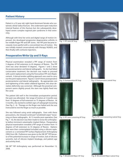
Fig. 1: <strong>PIP</strong> <strong>Joint</strong> AP Xray<br />
Fig. 2: <strong>PIP</strong> <strong>Joint</strong> Lateral Xray<br />
Physical examination revealed a <strong>PIP</strong> range of motion from<br />
5 degrees of full extension to 65 degrees of flexion. The <strong>PIP</strong><br />
<strong>Joint</strong> was ulnar deviated 10 degrees. Figures 1 and 2 show<br />
the posteroanterior and lateral radiographs. As she had failed<br />
conservative treatment, the decision was made to proceed<br />
with a joint replacement using the Pyrocarbon <strong>PIP</strong> <strong>Joint</strong> Replacement.<br />
A dorsal tendon splitting approach was used to carry<br />
out the joint replacement. Figures 3 and 4 show postoperative<br />
posteroanterior and lateral radiographs. By appropriate cuts<br />
and tightening of the radial collateral ligament, the ulnar deviation<br />
deformity was corrected. Although the proximal component<br />
seems slightly proud, the stem was tightly fixed into<br />
the canal.<br />
The patient did well in the immediate postoperative period.<br />
Her pain was alleviated. Her range of motion at 3 months was<br />
from 10 degrees of full extension to 75 degrees of flexion. At<br />
5 months, she started to exhibit signs of radiograph loosening<br />
(See Fig. 5 - 6). Therapy on the finger was halted and she was<br />
instructed to rest the finger as much as possible.<br />
Fig. 3: Postoperative AP Xray<br />
Fig. 4: Postoperative Lateral Xray<br />
She was followed using serial radiographs. Even with these<br />
precautions, she showed continued “windshield wiper” loosening<br />
on these radiographs. At 12 months post-operative, (See<br />
Fig. 7 - 8), the decision was then made to revise the arthroplasty<br />
before imminent catastrophic implant failure. Preoperative<br />
physical examination revealed a <strong>PIP</strong> range of motion from 15<br />
degrees of full extension to 55 degrees of flexion. Options<br />
that were then contemplated included using a silicone replacement<br />
or a cemented <strong>PIP</strong> Surface Replacement <strong>Arthroplasty</strong><br />
(SRA) from SBi. Preference was given to the SBi SR <strong>PIP</strong> because<br />
of the potential increase in range of motion, lack of silicone<br />
synovitis, lack of silicone implant breakage, and patient age<br />
and activity level.<br />
SBi SR <strong>PIP</strong> <strong>Arthroplasty</strong> was performed on November 13,<br />
2008.<br />
Fig. 5: <strong>PIP</strong> <strong>Joint</strong> Loosening AP Xray<br />
Fig. 6: <strong>PIP</strong> <strong>Joint</strong> Loosening Lateral Xray<br />
Fig. 7: Windshield Wiper AP Xray<br />
Fig. 8: Windshield Wiper Lateral Xray