HBM2010 - Organization for Human Brain Mapping
HBM2010 - Organization for Human Brain Mapping
HBM2010 - Organization for Human Brain Mapping

You also want an ePaper? Increase the reach of your titles
YUMPU automatically turns print PDFs into web optimized ePapers that Google loves.
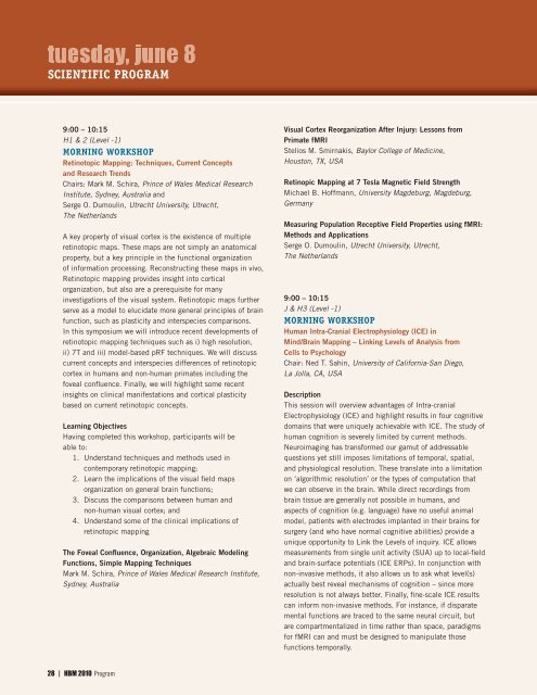
SCIENTIFIC PROGRAM<br />
9:00 – 10:15<br />
H1 & 2 (Level -1)<br />
MORNING WORKSHOP<br />
Retinotopic <strong>Mapping</strong>: Techniques, Current Concepts<br />
and Research Trends<br />
Chairs: Mark M. Schira, Prince of Wales Medical Research<br />
Institute, Sydney, Australia and<br />
Serge O. Dumoulin, Utrecht University, Utrecht,<br />
The Netherlands<br />
A key property of visual cortex is the existence of multiple<br />
retinotopic maps. These maps are not simply an anatomical<br />
property, but a key principle in the functional organization<br />
of in<strong>for</strong>mation processing. Reconstructing these maps in vivo,<br />
Retinotopic mapping provides insight into cortical<br />
organization, but also are a prerequisite <strong>for</strong> many<br />
investigations of the visual system. Retinotopic maps further<br />
serve as a model to elucidate more general principles of brain<br />
function, such as plasticity and interspecies comparisons.<br />
In this symposium we will introduce recent developments of<br />
retinotopic mapping techniques such as i) high resolution,<br />
ii) 7T and iii) model-based pRF techniques. We will discuss<br />
current concepts and interspecies differences of retinotopic<br />
cortex in humans and non-human primates including the<br />
foveal confluence. Finally, we will highlight some recent<br />
insights on clinical manifestations and cortical plasticity<br />
based on current retinotopic concepts.<br />
Learning Objectives<br />
Having completed this workshop, participants will be<br />
able to:<br />
1. Understand techniques and methods used in<br />
contemporary retinotopic mapping;<br />
2. Learn the implications of the visual field maps<br />
organization on general brain functions;<br />
3. Discuss the comparisons between human and<br />
non-human visual cortex; and<br />
4. Understand some of the clinical implications of<br />
retinotopic mapping<br />
The Foveal Confluence, <strong>Organization</strong>, Algebraic Modeling<br />
Functions, Simple <strong>Mapping</strong> Techniques<br />
Mark M. Schira, Prince of Wales Medical Research Institute,<br />
Sydney, Australia<br />
Visual Cortex Reorganization After Injury: Lessons from<br />
Primate fMRI<br />
Stelios M. Smirnakis, Baylor College of Medicine,<br />
Houston, TX, USA<br />
Retinopic <strong>Mapping</strong> at 7 Tesla Magnetic Field Strength<br />
Michael B. Hoffmann, University Magdeburg, Magdeburg,<br />
Germany<br />
Measuring Population Receptive Field Properties using fMRI:<br />
Methods and Applications<br />
Serge O. Dumoulin, Utrecht University, Utrecht,<br />
The Netherlands<br />
9:00 – 10:15<br />
J & H3 (Level -1)<br />
MORNING WORKSHOP<br />
<strong>Human</strong> Intra-Cranial Electrophysiology (ICE) in<br />
Mind/<strong>Brain</strong> <strong>Mapping</strong> – Linking Levels of Analysis from<br />
Cells to Psychology<br />
Chair: Ned T. Sahin, University of Cali<strong>for</strong>nia-San Diego,<br />
La Jolla, CA, USA<br />
Description<br />
This session will overview advantages of Intra-cranial<br />
Electrophysiology (ICE) and highlight results in four cognitive<br />
domains that were uniquely achievable with ICE. The study of<br />
human cognition is severely limited by current methods.<br />
Neuroimaging has trans<strong>for</strong>med our gamut of addressable<br />
questions yet still imposes limitations of temporal, spatial,<br />
and physiological resolution. These translate into a limitation<br />
on ‘algorithmic resolution’ or the types of computation that<br />
we can observe in the brain. While direct recordings from<br />
brain tissue are generally not possible in humans, and<br />
aspects of cognition (e.g. language) have no useful animal<br />
model, patients with electrodes implanted in their brains <strong>for</strong><br />
surgery (and who have normal cognitive abilities) provide a<br />
unique opportunity to Link the Levels of inquiry. ICE allows<br />
measurements from single unit activity (SUA) up to local-field<br />
and brain-surface potentials (ICE ERPs). In conjunction with<br />
non-invasive methods, it also allows us to ask what level(s)<br />
actually best reveal mechanisms of cognition – since more<br />
resolution is not always better. Finally, fine-scale ICE results<br />
can in<strong>for</strong>m non-invasive methods. For instance, if disparate<br />
mental functions are traced to the same neural circuit, but<br />
are compartmentalized in time rather than space, paradigms<br />
<strong>for</strong> fMRI can and must be designed to manipulate those<br />
functions temporally.<br />
28 | HBM 2010 Program