Neoplasms of the Meninges James G. Smirniotopoulos, MD
Neoplasms of the Meninges James G. Smirniotopoulos, MD
Neoplasms of the Meninges James G. Smirniotopoulos, MD

You also want an ePaper? Increase the reach of your titles
YUMPU automatically turns print PDFs into web optimized ePapers that Google loves.
Meningioma - Etiologic Factors<br />
• Trauma<br />
• Radiation<br />
• Viruses<br />
• Familial (Non-NF2) Meningioma<br />
• Neur<strong>of</strong>ibromatosis – Type - 2<br />
‣ MISME Syndrome<br />
Meningioma - Radiation<br />
• Low Dose (2000 cGray = 2000 RADS)<br />
‣ Used for Skull Base Tumors<br />
‣ Pituitary Adenoma<br />
Meningioma - Molecular Biology<br />
• Postulate Tumor suppressor Gene<br />
• Chromosome 22 – deletion in tumor cells<br />
‣ Both sporadic and inherited<br />
‣ W/ or w/o NF-2<br />
• Homozygous for TWO wild-type copies is<br />
normal<br />
• Heterozygous for 22 develops neoplasm<br />
‣ Because <strong>the</strong>re is a subsequent loss <strong>of</strong><br />
<strong>the</strong> OTHER wild-type gene<br />
• Inherited (germline) deletion <strong>of</strong> 22<br />
‣ W/Schwannoma = NF2<br />
Meningiomas<br />
• 1/7 to 1/4 <strong>of</strong> all Intracranial Primary<br />
‣ ~ 6/ 100k / year<br />
‣ Small ones in ~ 1.4% <strong>of</strong> autopsies<br />
• 1/4 – 1/3 <strong>of</strong> all Intraspinal Tumors<br />
• Middle age (40-60)<br />
• Female > Male<br />
‣ Cranial 2-4:1<br />
‣ Spinal 4-8:1<br />
‣ Progesterone receptors in 2/3<br />
‣ Estrogen receptors less common<br />
Meningioma – Sharp Margin [Figure 2]<br />
• Wonton wrapper<br />
• Tortilla<br />
• Arepa (Colombia)<br />
• Sopapilla (Chile)<br />
• Bolo de milho (Brazil)<br />
• Pita (Greece and Middle East)<br />
• Naan (India)<br />
• Lavaash (Farsi/Iranian)<br />
• Injera (Ethiopia)<br />
Meningioma - CT Imaging<br />
• Non-Contrast<br />
‣ Sharply Circumscribed<br />
‣ Homogeneous<br />
‣ Hyperdense (+/- Ca++)<br />
<br />
NOT from psammoma bodies !<br />
‣ Broad Dural Surface<br />
‣ Bone Changes (Hyperostosis)<br />
• Enhanced CT<br />
‣ Homogeneous Enhancement<br />
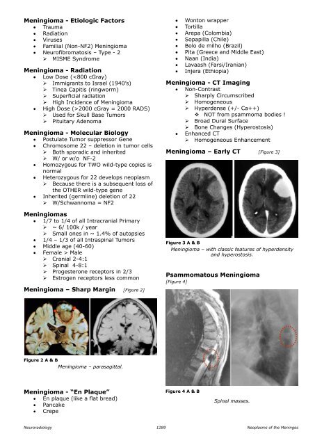
Meningioma – Early CT [Figure 3]<br />
Figure 3 A & B<br />
Meningioma – with classic features <strong>of</strong> hyperdensity<br />
and hyperostosis.<br />
Psammomatous Meningioma<br />
[Figure 4]<br />
Figure 2 A & B<br />
Meningioma – parasagittal.<br />
Meningioma - “En plaque”<br />
• En plaque (like a flat bread)<br />
• Pancake<br />
• Crepe<br />
Figure 4 A & B<br />
Spinal masses.<br />
Neuroradiology<br />
1289<br />
<strong>Neoplasms</strong> <strong>of</strong> <strong>the</strong> <strong>Meninges</strong>