Neoplasms of the Meninges James G. Smirniotopoulos, MD
Neoplasms of the Meninges James G. Smirniotopoulos, MD
Neoplasms of the Meninges James G. Smirniotopoulos, MD

Create successful ePaper yourself
Turn your PDF publications into a flip-book with our unique Google optimized e-Paper software.
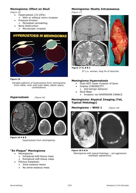
Meningioma: Effect on Skull<br />
[Figure 15]<br />
• Hyperostosis (15-25%)<br />
‣ With or without micro invasion<br />
• Pressure Erosion<br />
‣ Periosteal remodeling<br />
• Bone Destruction<br />
‣ Microscopic invasion<br />
Meningioma: Mostly Intraosseous<br />
[Figure 17]<br />
Figure 17 A, B & C<br />
57 y.o. woman, long Hx <strong>of</strong> seizures.<br />
Figure 15<br />
Variable patterns <strong>of</strong> hyperostosis from meningioma:<br />
Inner table, inner and outer table, diploic space,<br />
combinations.<br />
Hyperostosis [Figure 16]<br />
Meningioma Hyperostosis<br />
• Does NOT mean invasion <strong>of</strong> bone<br />
• Implies CHRONICITY<br />
‣ And benign behavior<br />
• Skull Base<br />
‣ Invasion via HAVERSIAN CANALS<br />
Meningioma: Atypical Imaging (Yet,<br />
typical Histology)<br />
Meningioma – WHO 1 [Figure 18]<br />
Figure 16 A & B<br />
Hyperostosis from meningioma.<br />
“En plaque” Meningioma<br />
• Meningioma:<br />
‣ Intradural s<strong>of</strong>t-tissue mass<br />
‣ Extradural s<strong>of</strong>t-tissue mass<br />
• Fibrous Dysplasia:<br />
‣ Intra-osseous lesion<br />
‣ No extra-osseous mass<br />
Figure 18 A & B<br />
Meningioma with typical histology – yet aggressive<br />
radiologic appearance.<br />
Neuroradiology<br />
1293<br />
<strong>Neoplasms</strong> <strong>of</strong> <strong>the</strong> <strong>Meninges</strong>