Neoplasms of the Meninges James G. Smirniotopoulos, MD
Neoplasms of the Meninges James G. Smirniotopoulos, MD
Neoplasms of the Meninges James G. Smirniotopoulos, MD

You also want an ePaper? Increase the reach of your titles
YUMPU automatically turns print PDFs into web optimized ePapers that Google loves.
MR Signal and Meningioma Types<br />
• Most are Isointense to GM<br />
‣ On both T1W & T2W<br />
• Hyperintense to GM on T1W<br />
‣ “Lipoblastic” (Fatty) Meningioma<br />
‣ Hemorrhage into Meningioma<br />
• Hypointense on T2W<br />
‣ Fibroblastic<br />
‣ Transitional<br />
• Hyperintense on T2W<br />
‣ Meningo<strong>the</strong>lial<br />
‣ “Angioblastic”<br />
• Microcystic (“Humid”)<br />
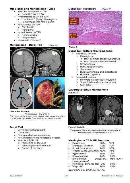
Meningioma - Dural Tail [Figure 8]<br />
Dural tail: Histology [Figure 9]<br />
Figure 9<br />
Dural Tail: Differential Diagnosis<br />
• Extraaxial Lesions<br />
‣ Meningioma<br />
<br />
Most common lesion w/dural tail<br />
<br />
Most common lesions overall<br />
‣ Schwannoma<br />
‣ Hemangiopericytoma<br />
‣ Sarcoidosis<br />
‣ Dural Lymphoma and metastases<br />
‣ Gumma (syphilis)<br />
• Intraaxial Lesions<br />
‣ Pleomorphic Xanthoastrocytoma<br />
‣ Superficial cerebral astrocytoma<br />
‣ GBM (rare)<br />
Cavernous Sinus Meningioma<br />
[Figure 10]<br />
Figure 8 A, B, C & D<br />
Meningioma - Dural Tail.<br />
The upper right image shows branching hypointensities<br />
that may represent flow voids from tumor vessels.<br />
Dural Tail<br />
• Curvilinear enhancement<br />
• “Dural flair”<br />
• First reported w/meningioma<br />
• First reported to be neoplastic invasion<br />
• What is it REALLY?<br />
‣ Thickening <strong>of</strong> <strong>the</strong> dura<br />
‣ Vasocongestion <strong>of</strong> <strong>the</strong> dura<br />
‣ Edema <strong>of</strong> <strong>the</strong> dura<br />
Figure 10 A & B<br />
Cavernous Sinus Meningioma with extensive dural<br />
enhancement along <strong>the</strong> tentorium.<br />
Meningioma CT & MR Features<br />
• Mass effect 88% 90%<br />
• Extraaxial Location 42% 70%<br />
• Broad Dural Attach. 74% 98%<br />
• Typical Dense./Intensity 92% 74%<br />
• Hyperostosis 10% 14%<br />
• Homogeneous 76% 76%<br />
• Enhancement 96%(78%) 96%(80%)<br />
(Homogeneous)<br />
• Meningeal. Enhance (tail) 2% 50%<br />
• “Capsule” 14% 68%<br />
Neuroradiology 1990;32:467-73.<br />
Neuroradiology<br />
1291<br />
<strong>Neoplasms</strong> <strong>of</strong> <strong>the</strong> <strong>Meninges</strong>