o_195egas1b1it2sc6dvd9ii1c7ca.pdf

You also want an ePaper? Increase the reach of your titles
YUMPU automatically turns print PDFs into web optimized ePapers that Google loves.
Case Report<br />
DOI: 10.7241/ourd.20134.155<br />
A CASE OF BULLOUS PEMPHIGOID WITH<br />
IMMUNOREACTIVTY TO BLOOD VESSELS AND SWEAT<br />
GLANDS<br />
Ana Maria Abreu Velez 1 , Juliana Calle-Isaza 2 , Michael S. Howard 1<br />
Source of Support:<br />
Georgia Dermatopathology<br />
Associates, Atlanta, Georgia, USA<br />
(MSH, AMAV).<br />
Competing Interests:<br />
None<br />
1<br />
Georgia Dermatopathology Associates, Atlanta, Georgia, USA<br />
2<br />
Hospital General de Medellin, Medellin, Colombia, S.A<br />
Corresponding author: Ana Maria Abreu Velez, MD PhD<br />
abreuvelez@yahoo.com<br />
Our Dermatol Online. 2013; 4(Suppl. 3): 621-624 Date of submission: 02.10.2013 / acceptance: 25.10.2013<br />
Abstract<br />
Introduction: Bullous pemphigoid (BP) is one of the most prevalent autoimmune blistering diseases, and believed to be mediated by<br />
autoantibodies and complement. The disorder is categorized by the development of urticarial plaques surmounted by subepidermal blisters,<br />
and the deposition of immunoglobulins and complement at the basement membrane zone (BMZ) of the skin.<br />
Case Report: A 70-year-old male Caucasian patient was evaluated for a four day history of multiple itchy, erythematous blisters on his<br />
abdomen. Biopsies for hematoxylin and eosin (H&E) examination, immunohistochemistry (IHC) and direct immunofluorescence (DIF)<br />
analyses were performed.<br />
Results: The H&E biopsy demonstrated a subepidermal blister, with partial re-epithelialization of the blister floor. Within the blister lumen<br />
numerous neutrophils and eosinophils, and occasional lymphocytes were observed. Within the dermis, dilated superficial blood vessels with<br />
a mild, perivascular infiltrate of lymphocytes, histiocytes and eosinophils were seen; mild perivascular leukocytoclastic debris was also<br />
noted. A periodic acid Schiff (PAS) special stain demonstrated positive staining along the BMZ, and around selected dermal blood vessels and<br />
sweat glands. DIF revealed linear deposits of IgG, Complement/C3 and fibrinogen at the BMZ, and around selected dermal blood vessels and<br />
sweat glands. By IHC, positive staining for CD8 and CD45, and occasional CD4 positivity was seen on dermal lymphocytes. These lymphocytes<br />
were present surrounding selected dermal blood vessels and eccrine sweat glands.<br />
Conclusions: The patient displayed immunoreactivity to the BMZ, and also to dermal blood vessels and eccrine glands in his immune<br />
response. Similar immune responses would be of interest in immunologic studies of BP patients.<br />
Key words: bullous pemphigoid; blood vessels; sweat glands; autoantibodies<br />
Abbreviations and acronyms: Bullous pemphigoid (BP), immunohistochemistry (IHC), direct and indirect immunofluorescence (DIF and IIF),<br />
hematoxylin and eosin (H&E), basement membrane zone (BMZ), intercellular staining between epidermal keratinocytes (ICS).<br />
Cite this article:<br />
Ana Maria Abreu Velez, Juliana Calle-Isaza, Michael S. Howard: A case of bullous pemphigoid with immunoreactivty to blood vessels and sweat glands. Our<br />
Dermatol Online. 2013; 4(Suppl.3): 621-624.<br />
Introduction<br />
Patients with bullous pemphigoid (BP) have autoantibodies<br />
binding to specific antigens in the basement membrane zone<br />
(BMZ), causing detachment of the entire epidermis from the<br />
dermis [1]. The autoantibodies directed against the BMZ can<br />
be visualized by both direct and indirect immunofluorescence<br />
(DIF, IIF) [2-4]. Eosinophils, neutrophils, and mast cells<br />
have all been implicated in the pathogenesis of BP [5-7].<br />
It is also believed that the autoantibodies in turn activate<br />
complement and other inflammatory mediators, causing mast<br />
cell degranulation and migration of eosinophils, neutrophils<br />
and antigen presenting cells to the BMZ. These events then<br />
lead to splitting at the dermal/epidermal junctional zone [1].<br />
Specifically, BMZ splitting is thought to occur secondary to<br />
secretion of inflammatory cytokines and proteases.<br />
Clinically, bullous pemphigoid (BP) is a rare skin disorder that<br />
causes tense, fluid-filled blisters on the lower abdomen, upper<br />
thighs, armpits and abdomen. BP is most common in people older<br />
than age 60 [1,2]. Treatment frequently includes corticosteroids<br />
such as prednisone, and other drugs that suppress the immune<br />
system. BP can be life-threatening, especially for older people<br />
who are already in poor health.<br />
www.odermatol.com<br />
© Our Dermatol Online. Suppl. 3.2013 621
Case Report<br />
A 70 year old male patient was referred with a four day<br />
eruption of multiple, severely pruritic, tense bullae with<br />
erythematous bases concentrated on the abdomen (Fig. 1a). Skin<br />
biopsies for hematoxylin and eosin (H&E), direct and indirect<br />
immunofluorescence (DIF and IIF) and immunohistochemistry<br />
(IHC) review were performed. In addition, IIF with 0.1 M<br />
sodium chloride salt split skin was requested.<br />
Methods<br />
DIF<br />
In brief, our DIF was performed utilizing skin cryosections,<br />
incubated with multiple fluorescein isothiocyanate (FITC)-<br />
conjugated secondary antibodies as previously described [8,9].<br />
The secondary antibodies were of rabbit origin, and included a)<br />
anti-human IgG, b) anti-human IgA, c) anti-human IgM, d) antihuman<br />
fibrinogen, and e) anti-human albumin (all with at 1:20 to<br />
1:40, anti-human C3C and C4 FITCI conjugates obtained from<br />
Dako (Carpinteria, California, USA)). We also utilized FITC<br />
conjugated secondary antibodies of goat origin, including a)<br />
anti-human IgE (Vector Laboratories, Bridgeport, New Jersey,<br />
USA) and b) anti-human complement/C1q and IgD FITCI<br />
conjugated (Southern Biotech, Birmingham, Alabama, USA).<br />
The DIF slides were counterstained with 4’,6-diamidino-2-<br />
phenylindole (Dapi) (Pierce, Rockford, Illinois, USA) washed,<br />
coverslipped, and dried overnight at 4oC. The NACl split skin<br />
as performed as previously described [8,9]. IHC staining was<br />
performed using anti-human antibodies to IgG, IgM, IgE,<br />
Complement/C3c, fibrinogen, CD4, CD8, CD45, kappa light<br />
chains, lambda light chains and myeloid/histiocyte antigen<br />
(Clone MAC 387). Our IHC antibodies were all obtained<br />
from Dako. Our IHC staining was performed utilizing a Dako<br />
automatized dual endogenous flex system, and following Dako<br />
technical instructions as previously described [8,9].<br />
Result<br />
Examination of the H&E tissue sections demonstrated a<br />
tense subepidermal blister. The edge of the blister demonstrated<br />
degranulating eosinophils, close to the epidermal BMZ.<br />
Within the dermis, a mild, superficial, perivascular infiltrate<br />
of lymphocytes, histiocytes, eosinophils was identified. The<br />
vessels in the upper dermis were dilated. A PAS special stain<br />
was reviewed; the positive control stained appropriately. The<br />
PAS special stain revealed no fungal organisms, and focally<br />
increased staining around dermal blood vessels. DIF displayed<br />
the following results: IgG (+++, linear BMZ); IgA (+, focal<br />
linear BMZ); IgM (+, Focal dermal perivascular cells); IgD(-<br />
); IgE (-); complement/C1q (+ focal dermal perivascular<br />
cells); complement/C3 (+++, linear BMZ); complement/C4<br />
(-); albumin (+, focal epidermal cell junctions and focal linear<br />
BMZ) and Fibrinogen(+, focal BMZ and diffuse deep dermal).<br />
IIF showed similar results as DIF, and IgG titers of 1:280. Salt<br />
split skin/IIF studies revealed that the primary autoreactivity<br />
was on the blister roof; however, focal staining was also noted<br />
on the blister floor. Finally, by IHC the dermal blood vessels<br />
stained positively for IgG (++), IgM (+++), IgE (+), fibrinogen<br />
(+++) and complement/C3(++). Lymphocytes surrounding<br />
these blood vessels stained positively for CD4(+), CD8(++) and<br />
CD45(+++). The dermal eccrine sweat glands and ducts stained<br />
positive for IgM(+++) and lambda light chains (++). In Figures<br />
1 and 2, we highlight our most significant H&E, DIF and IHC<br />
results.<br />
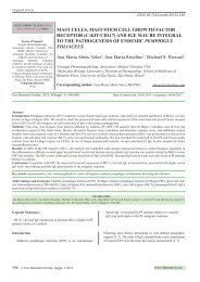
Figure 1. a. A representative clinical blister(black arrow). b. A<br />
representative H&E section, demonstrating a subepidermal<br />
blister (black arrow) (100X). c. DIF positive stain with FITC<br />
conjugated fibrinogen, present in a linear pattern at the BMZ<br />
(green staining; black arrow). Also note the focal positive<br />
staining around upper dermal blood vessels and within the<br />
epidermis. d. Positive IHC staining for IgE around an upper<br />
dermal vessels (red/purple staining; black arrow). e. Positive<br />
IHC staining for IgG around small upper dermal blood<br />
vessels, (brown staining; red arrows). f. Positive IHC staining<br />
for IgM around small upper dermal blood vessels (brown<br />
staining; red arrows). The blue arrow highlights additional<br />
linear staining on the floor of the BP blister.<br />
622 © Our Dermatol Online. Suppl. 3.2013
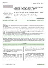
Figure 2. a. Positive DIF staining with FITC conjugated<br />
fibrinogen against a blood vessel in the dermis (green/white<br />
staining; red arrow)(400X). b. Same as a, but adding a Dapi<br />
counterstain to highlight blood vessel endothelial cell nuclei<br />
(blue staining) (400X). c. IHC positive staining for IgM inside<br />
a BP blister lumen, as well as along the BMZ(brown staining;<br />
red arrow) (100x). d. IHC positive staining for IgG on a dermal<br />
blood vessel interior surface (brown staining; red arrow). e.<br />
DIF, showing positive staining with FITC conjugated<br />
Complement/C3c on a dermal eccrine gland coil (green<br />
staining; red arrow). f. IHC positive staining with myeloid/<br />
histiocyte antigen antibody around an eccrine sweat duct<br />
(brown staining; red arrow).<br />
Discussion<br />
Bullous pemphigoid is a subepidermal bullous dermatosis<br />
resulting in a pathologic disruption between basaloid layer of the<br />
epidermis and the dermis, and thus causing formation of tense<br />
clinical blisters [1]. Many previous studies have documented an<br />
increase in blood vessel permeability in BP [5-7]. It is known<br />
that the human cutaneous BMZ contains multiple components,<br />
including BPAGI (230kD) and BPAGII (180 kDa; Collagen<br />
Type XVII) proteins; Type I, IV and VII collagens, alpha6 and<br />
beta4 integrins, laminins 1, 5 and 6, entactin/nidogen, heparan<br />
sulfate proteoglycans and microfibrils. The dermal blood<br />
vessels also contain diverse molecules, including laminin 1,<br />
Type IV collagen, and heparan sulfate proteoglycans. Thus, it<br />
is possible to hypothesize that any of these antigens in the skin<br />
and dermal blood vessels could be immune targets in BP. We<br />
previously reported a different case of BP, having autoantibodies<br />
to dermal blood vessels and sweat glands [10]. It has also been<br />
reported that Collagen XVII is expressed in podocytes of the<br />
renal glomerular barrier [11]. We have also previously observed<br />
strong activity of several proteases and protease inhibitors in<br />
the dermal blood vessels in BP, as well as in other autoimmune<br />
blistering diseases [12].<br />
We were able to demonstrate a direct correlation of our DIF and<br />
IIF/salt split skin positive findings with positive PAS staining<br />
in dermal blood vessels. Notably, vascular dilatation and<br />
perivascular dermal infiltrates have been previously documented<br />
in BP. Recently, we reported that soluble E-selectin (sE-selectin;<br />
an isoform of cell membrane E-selectin, an adhesion molecule<br />
synthesized only by endothelial cells), is significantly increased<br />
in sera of patients with BP and pemphigus vulgaris. We also<br />
reported that collagen XVII (a transmembrane molecule known<br />
to be required for epithelial adhesion) is expressed in podocytes<br />
of normal human and mouse renal tissue, as well as in endothelial<br />
cells of the glomerular filtration barrier. Immunoelectron<br />
microscopy has revealed that the collagen XVII is specifically<br />
localized in the foot processes of the podocytes, and within the<br />
glomerular basement membrane [12].<br />
Multiple authors have previously reported augmentation of<br />
chemokines, cytokines and ICAM/CD54 in BP; in addition,<br />
increased expression of vascular permeability factors including<br />
integrins and selectins in BP has been documented [13-23].<br />
We conclude that our findings of 1) abundant IHC CD45<br />
positive lymphocytes around the dermal blood vessels and<br />
eccrine glands and 2) positive autoantibody staining observed<br />
by DIF and IIF in these areas warrant additional investigation<br />
in BP cases.<br />
Acknowledgement<br />
The study was supported by funding from Georgia<br />
Dermatopathology Associates, Atlanta, Georgia, USA.<br />
REFERENCES<br />
1. Walsh SR, Hogg D, Mydlarski PR: Bullous pemphigoid: From<br />
bench to bedside. Drugs 2005;65:905-26.<br />
2. Jordon RE, Sams WM Jr, Beutner EH: Complement<br />
immunofluorescent staining in bullous pemphigoid. J Lab Clin Med.<br />
1969;74:548-56.<br />
3. Jordon RE, Beutner EH, Witebsky E, Blumental G, Hale WL,<br />
Lever WF: Basement zone antibodies in bullous pemphigoid. JAMA.<br />
1967;29:751-6.<br />
© Our Dermatol Online. Suppl. 3.2013 623
4. Gammon WR, Kowalewski C, Chorzelski TP, Kumar V,<br />
Briggaman RA, Beutner EH: Direct immunofluorescence studies<br />
of sodium chloride-separated skin in the differential diagnosis of<br />
bullous pemphigoid and epidermolysis bullosa acquisita. J Am Acad<br />
Dermatol. 1990;22:664-70.<br />
5. Macvicar DN, Graham JH, Burgoon CF Jr: Dermatitis<br />
herpetiformis, erythema multiforme and bullous pemphigoid: a<br />
comparative histopathological and histochemical study. J Invest<br />
Dermatol. 1963;41:289-300.<br />
6. Lever WF: Pemphigus and pemphigoid. Charles. Thomas,<br />
Springfield 1965.<br />
7. Lever WF: Differential diagnosis of pemphigus vulgaris, bullous<br />
pemphigoid and dermatitis herpetiformis. Med Klin. 1967;62:1173-6.<br />
8. Abreu Velez AM, Howard MS: Neural reactivity detected by<br />
immunofluorescence in a patient with a localized blistering disease.<br />
Our Dermatol Online. 2013;4:91-4.<br />
9. Abreu Velez AM, Brown VM, Howard MS: Cytotoxic and antigen<br />
presenting cells present and non-basement membrane zone pathology<br />
in a case of bullous pemphigoid. Our Dermatol Online. 2012;3:93-9.<br />
10. Abreu Velez AM, Smith JG Jr, Howard MS: IgG/IgE bullous<br />
pemphigoid with CD45 lymphocytic reactivity to dermal blood<br />
vessels, nerves and eccrine sweat glands. North Am J Med Sci.<br />
2010;2:540-43.<br />
11. Hurskainen T, Moilanen J, Sormunen R, Franzke CW, Soininen R,<br />
Loeffek S, et al: Transmembrane collagen XVII is a novel component<br />
of the glomerular filtration barrier. Cell Tissue Res. 2012;348:579-88.<br />
12. Abreu Velez AM, Yepes-Naranjo MM, Avila IC, Londoño<br />
ML, Googe PB, Velásquez-Velez JE, et al: Tissue inhibitor of<br />
metalloproteinase 1, Matrix metalloproteinase 9, αlpha-1 antitrypsin,<br />
metallothionein and urokinase type plasminogen activator receptor<br />
in skin biopsies from patients affected by autoimmune blistering<br />
diseases. Our Dermatol Online. 2013; 4: 275-80.<br />
13. Ameglio F, D’Auria L, Cordiali-Fei P, Mussi A, Valenzano L,<br />
D’Agosto G, et al: Bullous pemphigoid and pemphigus vulgaris:<br />
correlated behaviour of serum VEGF, sE-selectin and TNF-alpha<br />
levels. J Biol Regul Homeost Agents. 1997;11:148-53.<br />
14. Günther C, Wozel G, Meurer M, Pfeiffer C: Up-regulation of<br />
CCL11 and CCL26 is associated with activated eosinophils in bullous<br />
pemphigoid. Clin Exp Immunol. 2011;166:145-53.<br />
15. Miyagaki T, Sugaya M, Kagami S, Nakashima H, Ishiura N,<br />
Watanabe R, et al: Increased CCL1 levels in the sera and blister fluid<br />
of patients with bullous pemphigoid. J Dermatol Sci. 2009;54:45-47.<br />
16. Zebrowska A, Sysa-Jedrzejowska A, Wagrowska-Danilewicz M,<br />
Joss-Wichman E, Erkiert-Polguj A, Waszczykowska E: Expression<br />
of selected integrins and selectins in bullous pemphigoid. Mediators<br />
Inflamm. 2007;2007:31051.<br />
17. Borrego L, Maynard B, Peterson EA, George T, Iglesias L, Peters<br />
MS, et al: Deposition of eosinophil granule proteins precedes blister<br />
formation in bullous pemphigoid. Comparison with neutrophil and<br />
mast cell granule proteins. Am J Pathol. 1996;148:897-909.<br />
18. D’Auria L, Bonifati C, Mussi A, Ameglio F; Monitoring of<br />
sE-selectin serum levels in three different dermatoses. Clin Ter.<br />
1998;149:49-52.<br />
19. Dahlman-Ghozlan K, Ortonne JP, Heilborn JD, Stephansson<br />
E: Altered tissue expression pattern of cell adhesion molecules,<br />
ICAM-1, E-selectin and VCAM-1, in bullous pemphigoid during<br />
methotrexate therapy. Exp Dermatol. 2004;13:65-9.<br />
20. D’Auria L, Cordiali Fei P, Pietravalle M, Ferraro C, Mastroianni<br />
A, Bonifati C, et al: The serum levels of sE-selectin are increased in<br />
patients with bullous pemphigoid or pemphigus vulgaris. Correlation<br />
with the number of skin lesions and recovery after corsticosteroid<br />
therapy. Br J Dermatol.1997;137:59-64.<br />
21. Dahlman-Ghozlan K, Heilborn JD, Stephansson E: Circulating<br />
levels of soluble E-selectin, ICAM-1 and VCAM-1 in bullous<br />
pemphigoid during low-dose methotrexate therapy. A prospective<br />
study. Exp Dermatol. 2000;9:336-40.<br />
22. D’Auria L, Cordiali Fei P, Ameglio F: Cytokines and bullous<br />
pemphigoid. Eur Cytokine Netw. 1999;10:123-34.<br />
23. Brown LF, Harrist TJ, Yeo KT, Ståhle-Bäckdahl M, Jackman<br />
RW, Berse B, et al: Increased expression of vascular permeability<br />
factor (vascular endothelial growth factor) in bullous pemphigoid,<br />
dermatitis herpetiformis, and erythema multiforme. J Invest<br />
Dermatol. 1995;104:744-9.<br />
Copyright by Ana Maria Abreu Velez, et al. This is an open access article distributed under the terms of the Creative Commons Attribution License,<br />
which permits unrestricted use, distribution, and reproduction in any medium, provided the original author and source are credited.<br />
624 © Our Dermatol Online. Suppl. 3.2013