Unveiling the Secrets of Thinking - Carl Zeiss
Unveiling the Secrets of Thinking - Carl Zeiss
Unveiling the Secrets of Thinking - Carl Zeiss

You also want an ePaper? Increase the reach of your titles
YUMPU automatically turns print PDFs into web optimized ePapers that Google loves.
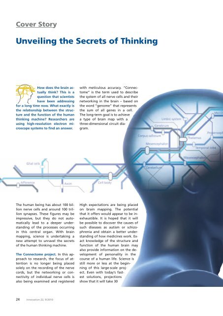
Cover Story<br />
<strong>Unveiling</strong> <strong>the</strong> <strong>Secrets</strong> <strong>of</strong> <strong>Thinking</strong><br />
How does <strong>the</strong> brain actually<br />
think? This is a<br />
question that scientists<br />
have been addressing<br />
for a long time now. What exactly is<br />
<strong>the</strong> relationship between <strong>the</strong> structure<br />
and <strong>the</strong> function <strong>of</strong> <strong>the</strong> human<br />
thinking machine? Researchers are<br />
using high-resolution electron microscope<br />
systems to find an answer.<br />
with meticulous accuracy. “Connectome”<br />
is <strong>the</strong> term used to describe<br />
<strong>the</strong> system <strong>of</strong> all nerve cells and <strong>the</strong>ir<br />
networking in <strong>the</strong> brain – based on<br />
<strong>the</strong> word “genome” that represents<br />
<strong>the</strong> sum <strong>of</strong> all genes in a cell.<br />
The long-term goal is to achieve<br />
a type <strong>of</strong> brain map with a<br />
three-dimensional circuit diagram.<br />
The human being has about 100 billion<br />
nerve cells and around 100 trillion<br />
synapses. These figures may be<br />
impressive, but <strong>the</strong>y do not automatically<br />
lead to a deeper understanding<br />
<strong>of</strong> <strong>the</strong> processes occurring<br />
in this central organ. With brain<br />
mapping, science is undertaking a<br />
new attempt to unravel <strong>the</strong> secrets<br />
<strong>of</strong> <strong>the</strong> human thinking machine.<br />
The Connectome project. In this approach<br />
to research, <strong>the</strong> focus <strong>of</strong> attention<br />
is no longer being placed<br />
solely on <strong>the</strong> recording <strong>of</strong> <strong>the</strong> nerve<br />
cords, but <strong>the</strong> networking or connectivity<br />
<strong>of</strong> individual nerve cells is<br />
also being examined and registered<br />
High expectations are being placed<br />
on brain mapping. The potential<br />
that it <strong>of</strong>fers would appear to be inexhaustible.<br />
It is hoped that it will<br />
be possible to discover <strong>the</strong> causes <strong>of</strong><br />
such diseases as autism or schizophrenia<br />
and obtain a better understanding<br />
<strong>of</strong> how medicines work. Exact<br />
knowledge <strong>of</strong> <strong>the</strong> structure and<br />
function <strong>of</strong> <strong>the</strong> human brain may<br />
also provide information on <strong>the</strong> development<br />
<strong>of</strong> personality in <strong>the</strong><br />
course <strong>of</strong> a human life. Science is<br />
still more or less at <strong>the</strong> beginning<br />
<strong>of</strong> this large-scale project.<br />
Even with today’s fastest<br />
solutions, projections<br />
show that it will take 30<br />
24 Innovation 22, 9 / 2010
Cover Story: (Re)solution<br />
years to record <strong>the</strong> brain<br />
<strong>of</strong> a mouse, not to<br />
mention <strong>the</strong> mapping<br />
<strong>of</strong> <strong>the</strong> human thinking<br />
apparatus.<br />
A puzzle with many facets.<br />
Even <strong>the</strong> initial<br />
preparation <strong>of</strong> a brain<br />
section is no easy matter.<br />
The material is subsequently<br />
embedded in syn<strong>the</strong>tic resin<br />
and cut with a microtome,<br />
a type <strong>of</strong> planing device, into<br />
wafer-thin slices that are applied<br />
to a wafer and imaged in a<br />
scanning electron microscope. Such a<br />
specimen has a volume <strong>of</strong> about one<br />
cubic millimeter; this cube is cut up<br />
into about 20,000 slices. Vast processing<br />
times are required to subsequently<br />
merge <strong>the</strong> resulting huge<br />
quantities <strong>of</strong> image data into a<br />
three-dimensional image in <strong>the</strong> computer<br />
again.<br />
Speed means success. <strong>Carl</strong> <strong>Zeiss</strong> is<br />
supporting this research work with<br />
intelligent solutions. With <strong>the</strong> aid <strong>of</strong><br />
<strong>the</strong> SIGMA TM field emission scanning<br />
electron microscope (FE-SEM), biologist<br />
Jeff Lichtman from Harvard University<br />
in Cambridge, Massachusetts,<br />
is examining pieces <strong>of</strong> a mouse’s<br />
brain. The system is equipped with a<br />
special detector system and s<strong>of</strong>tware<br />
that allow 100 times faster image<br />
generation and storage than with<br />
traditional systems. For John Mendenhall<br />
<strong>of</strong> <strong>the</strong> University <strong>of</strong> Texas in<br />
Austin, speed is an important argument<br />
in favor <strong>of</strong> using a ZEISS FE-<br />
SEM. Toge<strong>the</strong>r with a special application<br />
solution, <strong>the</strong> system has an<br />
image memory with a capacity <strong>of</strong> up<br />
to one gigapixel and <strong>the</strong>refore permits<br />
<strong>the</strong> recording <strong>of</strong> large-area<br />
specimens at maximum resolution.<br />
Dr. Marco Cantoni <strong>of</strong> <strong>the</strong> Ecole Polytechnique<br />
Fédérale de Lausanne in<br />
Switzerland examines mouse brains<br />
with a CrossBeam ® microscope. Instead<br />
<strong>of</strong> a mechanical technique, an<br />
ion beam is used to remove <strong>the</strong> specimen<br />
slice by slice, with <strong>the</strong> result<br />
<strong>the</strong>n being examined in <strong>the</strong> scanning<br />
electron microscope. This process<br />
is largely automated. “The result<br />
was incredible. In 48 hours we<br />
generated 1600 images <strong>of</strong> specimen<br />
slices, each with a thickness <strong>of</strong> just<br />
six nanometers,” explains Dr. Cantoni.<br />
“This gives us an insight into <strong>the</strong><br />
three-dimensional structure <strong>of</strong> <strong>the</strong><br />
tissue to be examined.” The ORION ®<br />
helium ion microscope also <strong>of</strong>fers<br />
possible applications in <strong>the</strong> field <strong>of</strong><br />
brain mapping. The potential <strong>of</strong> this<br />
technology lies, above all, in its extremely<br />
large depth <strong>of</strong> field and <strong>the</strong><br />
innovative contrast mechanisms <strong>of</strong>fered<br />
by imaging with ions.<br />
Brain research as an industry. The<br />
quest for images and data already<br />
shows that this scientific challenge<br />
cannot be mastered without broadbased<br />
research. The opinion is now<br />
increasingly being voiced that Connectome<br />
research should be industrialized,<br />
much like <strong>the</strong> situation with<br />
genome research at <strong>the</strong> end <strong>of</strong> <strong>the</strong><br />
last century. The solution could be<br />
that entire “farms” will emerge<br />
comprising dozens <strong>of</strong> electron microscope<br />
systems in which brain sections<br />
will be imaged day and night.<br />
The computer industry is also faced<br />
with a major challenge. Its job will<br />
be to create gigantic storage media.<br />
Even one cubic millimeter <strong>of</strong> mouse<br />
brain delivers information totaling<br />
1000 terabytes; this would be one<br />
million times 1000 terabytes for <strong>the</strong><br />
human brain.<br />
However, knowing <strong>the</strong> circuit diagram<br />
still does not allow any deductions<br />
to be made about <strong>the</strong> activities<br />
<strong>of</strong> <strong>the</strong> thinking machine. The<br />
question as to <strong>the</strong> effect that a nerve<br />
cell really has on its direct surroundings<br />
remains unresolved for <strong>the</strong><br />
time being.<br />
Monika Etspüler<br />
Innovation 22, 9 / 2010<br />
25